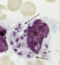
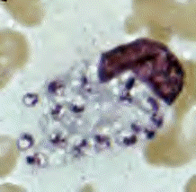
- Leishmania tropica
amastigotes from a skin touch preparation.
- In A, a still intact macrophage is
practically filled with amastigotes, several
of which have clearly visible a nucleus and
a kinetoplast (arrows);
- in B, amastigotes are being freed
from a rupturing macrophage. Patient with
history of travel to Egypt, Africa, and the
Middle East.
- Culture in NNN medium followed by
isoenzyme analysis identified the species as L.
tropica minor.-CDC http://www.dpd.cdc.gov/DPDx/HTML/Leishmaniasis.htm
|

