CHAPTER 8 · ANTIARRHYTHMIC DRUGS

Section 1

The Cardiac Action Potential

Ionic phases, cell-type differences, and the pharmacodynamic map of antiarrhythmic drugs

The cardiac action potential is the electrical event that triggers each heartbeat. Its ionic architecture varies by cell type, and this variation determines where each class of antiarrhythmic drug exerts its primary effect. Before examining any specific drug, a working map of the action potential is essential.

Fast-Response Cells: Working Myocardium and the His-Purkinje System

Fast-response cells have a strongly negative resting membrane potential of approximately negative 90 millivolts, maintained by outward potassium current. Their action potential is divided into five phases, each governed by specific ion movements.

Phase 0, rapid depolarization, occurs when the membrane reaches threshold and voltage-gated sodium channels open rapidly, driving a large inward sodium current. This fast upstroke produces the rapid conduction velocity characteristic of working ventricular muscle and the His-Purkinje system. Class I antiarrhythmic drugs act here by blocking sodium channels and slowing conduction.

Phase 1, early repolarization, begins as sodium channels inactivate within milliseconds. A brief outward potassium current drives the membrane toward the plateau. Phase 2, the plateau, is sustained by a balance between inward calcium current through L-type calcium channels and outward potassium currents. This calcium entry triggers contraction. Class IV agents — the non-dihydropyridine calcium channel blockers — and amiodarone act at the calcium channel during this phase.

Phase 3, rapid repolarization, occurs as calcium channel current declines and outward potassium currents dominate, restoring the resting potential. Class III agents prolong this phase by blocking the repolarizing potassium channels, which is the basis for their action potential duration prolongation and the mechanism of drug-induced QT interval prolongation. Phase 4 is electrically stable in non-pacemaker cells; the sodium-potassium pump restores ionic gradients, and an inward-rectifying potassium current holds the membrane at rest.
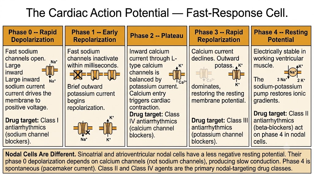
Diagram of the five phases of the cardiac action potential in a fast-response ventricular muscle cell, showing the ion movements responsible for each phase and the Vaughan Williams drug classes that act at each phase, with a note box describing how nodal cells differ.
Source: AI-generated figure (Gemini). Educational use.

Slow-Response Cells: The Sinoatrial and Atrioventricular Nodes

Nodal cells operate on fundamentally different ionic machinery. Their resting membrane potential is less negative — approximately negative 60 to negative 70 millivolts — which keeps fast sodium channels largely inactivated. As a result, nodal cell depolarization depends on calcium current through L-type calcium channels rather than on sodium channels. This produces a slow upstroke and slow conduction velocity through the nodes.

The clinically defining feature of nodal cells is spontaneous phase 4 depolarization, the pacemaker potential. This gradual drift toward threshold is driven by inward current through channels that open on hyperpolarization (the funny current, or If), combined with declining outward potassium current and late-activating calcium current. The sinoatrial node fires fastest and drives the heart rate under normal conditions; the atrioventricular node follows as a subsidiary pacemaker.

Because nodal automaticity depends on calcium current and adrenergic input to the If channel, these cells are the primary targets for rate control: beta-blockers reduce the slope of phase 4 by decreasing adrenergic stimulation of If, while non-dihydropyridine calcium channel blockers (verapamil, diltiazem) reduce calcium-dependent upstroke and atrioventricular nodal conduction. Class II (beta-blockers) and Class IV agents are the nodal-targeting drug classes.

Drug Class to Action Potential Phase — Quick Reference

Class I (sodium channel blockers) — Phase 0. Reduce upstroke velocity and slow conduction in fast-response tissue.

Class II (beta-blockers) — Phase 4 in nodal cells. Reduce slope of spontaneous depolarization; slow atrioventricular nodal conduction.

Class III (potassium channel blockers) — Phase 3. Prolong repolarization and action potential duration; lengthen QT interval.

Class IV (calcium channel blockers) — Phase 2 in fast-response cells; phase 0 and phase 4 in nodal cells. Reduce calcium-dependent upstroke and automaticity in the sinoatrial and atrioventricular nodes.
Section 2

Mechanisms of Arrhythmogenesis

The three electrophysiologic pathways through which arrhythmias arise

Arrhythmias arise through three distinct electrophysiologic mechanisms: abnormal automaticity, triggered activity, and re-entry. Recognizing the mechanism matters clinically because drugs that terminate one type of arrhythmia may worsen another.

Abnormal Automaticity

Normal automaticity is confined to the sinoatrial node and subsidiary pacemakers in the atrioventricular node and His-Purkinje system. The sinoatrial node dominates because its intrinsic rate exceeds that of subordinate pacemakers, which it suppresses by repeated excitation (overdrive suppression).

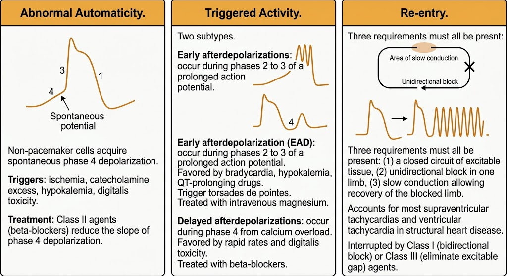
Abnormal automaticity occurs when non-pacemaker cells acquire spontaneous phase 4 depolarization. This happens in the setting of ischemia, catecholamine excess, hypokalemia, or digitalis toxicity — conditions that partially depolarize the resting membrane potential, shifting it toward the threshold for spontaneous discharge. Beta-blockers suppress both normal and abnormal automaticity by reducing adrenergic drive to the phase 4 pacemaker current.

Triggered Activity

Triggered activity refers to membrane depolarizations that arise from a preceding action potential rather than spontaneously. Two subtypes are recognized based on when they occur in the action potential cycle.

Early afterdepolarizations (EADs) occur during phases 2 or 3 of the action potential, while the membrane is still depolarized. They are favored by anything that prolongs the action potential duration: bradycardia, hypokalemia, hypomagnesemia, and Class Ia or Class III antiarrhythmic drugs. Early afterdepolarizations are the proximate trigger for torsades de pointes, a potentially fatal polymorphic ventricular tachycardia. Intravenous magnesium is the treatment of choice, as it suppresses the calcium-dependent oscillations underlying early afterdepolarization formation.

Delayed afterdepolarizations (DADs) occur during phase 4, after full repolarization, when intracellular calcium overload triggers spontaneous calcium release and a transient inward current that depolarizes the membrane. They are favored by high heart rates (the opposite of early afterdepolarizations) and by conditions that increase intracellular calcium, including digitalis toxicity and catecholamine excess. Beta-blockers are effective against delayed afterdepolarization-driven arrhythmias by reducing adrenergic calcium loading.

Three-panel diagram showing the three mechanisms of arrhythmogenesis: abnormal automaticity with spontaneous phase 4 depolarization, triggered activity showing early and delayed afterdepolarizations, and re-entry showing the three required conditions of closed circuit, unidirectional block, and slow conduction.
Source: AI-generated figure (Gemini). Educational use.

Re-entry

Re-entry is responsible for the majority of clinically significant tachyarrhythmias, including atrial flutter, most supraventricular tachycardias, and sustained ventricular tachycardia in the setting of structural heart disease. It occurs when an electrical impulse circulates continuously through a loop of tissue rather than dying out after a single depolarization.

Three conditions must be present simultaneously for re-entry to occur. First, there must be a circuit — a closed loop of excitable tissue, which may be anatomic (a scar border zone, an accessory pathway) or functional (tissue with heterogeneous refractoriness). Second, one limb of the circuit must have unidirectional block at the moment of the initiating impulse, forcing the wavefront to travel only in one direction. Third, conduction through the circuit must be slow enough that the blocked limb has time to recover excitability before the impulse arrives from the other direction.

Pharmacologic interruption of re-entry exploits these requirements. Class I agents can slow conduction to the point of bidirectional block, terminating the circuit. Class III agents prolong refractoriness, eliminating the excitable gap through which the re-entrant wavefront travels. The choice of approach depends on the substrate and whether structural heart disease is present.


Section 3

The Vaughan Williams Classification

The organizing framework for antiarrhythmic drug pharmacology

Proposed by Vaughan Williams in 1970, this classification groups antiarrhythmic drugs by their primary electrophysiologic action on the cardiac action potential. Despite acknowledged limitations, it remains the dominant clinical and educational framework and governs the organization of this chapter series.

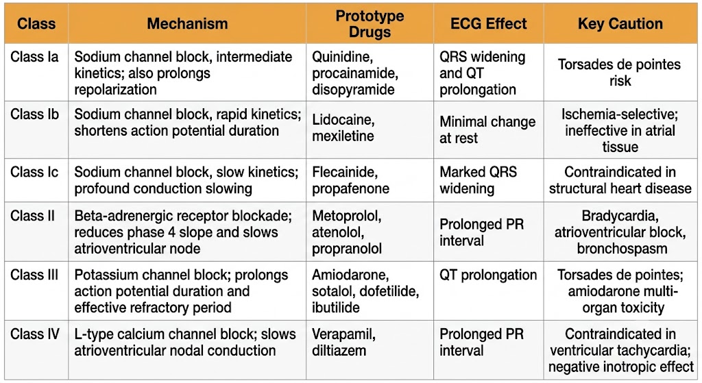
Reference table summarizing the Vaughan Williams classification of antiarrhythmic drugs with six rows covering Class Ia, Class Ib, Class Ic, Class II, Class III, and Class IV, showing mechanism, prototype drugs, electrocardiogram effect, and key caution for each class.
Source: AI-generated figure (Gemini). Educational use.

Class I — Sodium Channel Blockers

Class I agents block voltage-gated fast sodium channels in fast-response tissue, slowing the rate of phase 0 depolarization and reducing conduction velocity. The three subclasses differ in their speed of channel binding and unbinding, which produces distinct clinical profiles.

Class Ia agents — quinidine, procainamide, and disopyramide — have intermediate binding kinetics. They slow conduction, prolong action potential duration, and produce QT interval prolongation on the electrocardiogram. They carry risk of torsades de pointes because of this action potential duration prolongation.

Class Ib agents — lidocaine and mexiletine — bind and dissociate rapidly. They shorten action potential duration and are selectively active in diseased or ischemic tissue that fires rapidly. Because they dissociate so quickly, they have minimal effect on normal tissue at resting heart rates, making them relatively safe for acute ventricular arrhythmia management in ischemic settings.

Class Ic agents — flecainide and propafenone — bind slowly and dissociate very slowly, producing marked slowing of conduction with QRS widening on the electrocardiogram but minimal change in action potential duration. This profound conduction slowing is effective in atrial arrhythmias in structurally normal hearts but carries serious risk in the presence of structural heart disease.

Use-Dependence and the Structural Heart Disease Contraindication

Class I agents exhibit use-dependence: their sodium channel blockade intensifies at higher heart rates because more channels cycle through the open and inactivated states per unit time, increasing drug-channel interaction. This property makes Class Ic agents particularly dangerous during rapid tachycardias in scarred myocardium, where slowed conduction can convert a non-sustained arrhythmia into a sustained or fatal one. The Cardiac Arrhythmia Suppression Trial demonstrated that encainide and flecainide increased mortality approximately 2.5-fold in patients with prior myocardial infarction, despite suppressing ambient ventricular ectopy. Class Ic agents are contraindicated in structural heart disease.

Class II — Beta-Adrenergic Receptor Antagonists

Class II agents competitively block beta-adrenergic receptors, reducing sympathetic drive to the heart. Their antiarrhythmic effects are predominantly on nodal tissue and on arrhythmias driven by catecholamine excess. They decrease the slope of spontaneous phase 4 depolarization in pacemaker cells, prolong atrioventricular nodal conduction and refractoriness (increasing the PR interval), and suppress delayed afterdepolarization-mediated triggered activity in catecholamine-sensitive arrhythmias. Beta-blockers are also anti-ischemic, reducing arrhythmia substrate by decreasing myocardial oxygen demand.

Class III — Potassium Channel Blockers

Class III agents block the repolarizing potassium channels responsible for phase 3, prolonging action potential duration and the effective refractory period. The net electrocardiographic effect is QT interval prolongation. By prolonging refractoriness, these agents eliminate the excitable gap in re-entrant circuits — the pharmacologic rationale for rhythm control in both atrial and ventricular arrhythmias.

Amiodarone is the most widely used Class III agent but is mechanistically unusual: it blocks sodium channels, calcium channels, beta-adrenergic receptors, and potassium channels — effectively spanning all four Vaughan Williams classes. This multi-channel activity makes it the broadest-spectrum antiarrhythmic drug available. Its extraordinary volume of distribution and elimination half-life of 40 to 55 days necessitate loading protocols and produce toxicities that develop over months to years.

Sotalol has both Class III potassium channel blocking activity and significant beta-adrenergic receptor antagonism. Dofetilide and ibutilide are more selective potassium channel blockers used for rhythm control in atrial fibrillation. A critical property of Class III agents is reverse use-dependence: their action potential duration prolongation is paradoxically greatest at slow heart rates, which is also the condition most favorable for early afterdepolarization formation — meaning their proarrhythmic risk is highest precisely when they provide the least protection at rapid rates.

Class IV — Calcium Channel Blockers

Class IV agents block L-type calcium channels. Their primary antiarrhythmic targets are the sinoatrial and atrioventricular nodes, where calcium current drives the upstroke and contributes to the pacemaker potential. Verapamil and diltiazem — the two antiarrhythmic agents in this class — reduce atrioventricular nodal conduction velocity and prolong atrioventricular nodal refractoriness, making them effective for ventricular rate control in atrial fibrillation and for terminating atrioventricular nodal-dependent re-entrant tachycardias.

Class IV agents have minimal effect on fast-response ventricular tissue at therapeutic concentrations. This explains both their utility for supraventricular arrhythmias and a critical contraindication: verapamil and diltiazem must not be used in wide-complex tachycardias presumed to be ventricular tachycardia, where their negative inotropic and vasodilating effects can cause hemodynamic collapse without providing antiarrhythmic benefit.

Class I — Sodium Channel Block

Three Subclasses by Kinetics

  • Ia — quinidine, procainamide, disopyramide — intermediate kinetics, QT prolongation
  • Ib — lidocaine, mexiletine — rapid kinetics, shorten action potential duration, ischemic tissue selective
  • Ic — flecainide, propafenone — slow kinetics, marked QRS widening, contraindicated in structural heart disease

Classes II, III, IV

Nodal, Repolarization, and Calcium Targets

  • II — beta-blockers — decrease automaticity, slow atrioventricular node
  • III — amiodarone, sotalol, dofetilide, ibutilide — prolong action potential duration, QT prolongation
  • IV — verapamil, diltiazem — slow atrioventricular nodal conduction, rate control

Section 4

Agents Outside the Vaughan Williams Framework

Three clinically important antiarrhythmics that do not fit the four-class schema

Several antiarrhythmic agents are in routine clinical use but do not fit cleanly into the four Vaughan Williams classes. Understanding their mechanisms independently is essential because they address specific arrhythmia situations that the class agents do not cover.

Adenosine

Adenosine acts on adenosine receptors in the atrioventricular node, activating a potassium channel that hyperpolarizes nodal cells and transiently blocks atrioventricular nodal conduction. Its elimination half-life is less than ten seconds, making it ultrashort in duration. This brevity is both a safety feature and a diagnostic tool: a bolus of adenosine can transiently block the atrioventricular node, terminating atrioventricular node-dependent re-entrant tachycardias (including atrioventricular nodal reentrant tachycardia and atrioventricular reentrant tachycardia) within seconds. It is first-line therapy for these supraventricular tachycardias in the acute setting and also reveals underlying atrial activity (atrial flutter waves, for example) during the brief period of nodal block. Adenosine must not be used in patients with Wolff-Parkinson-White syndrome and atrial fibrillation, where blocking the atrioventricular node can redirect conduction exclusively through the accessory pathway at dangerous rates.

Digoxin

Digoxin enhances vagal tone to the atrioventricular node and additionally inhibits the sodium-potassium pump, leading to indirect increases in intracellular calcium. Its antiarrhythmic utility in contemporary practice is limited to ventricular rate control in atrial fibrillation, particularly in patients with concomitant heart failure where its mild positive inotropic effect is beneficial. A narrow therapeutic index, multiple drug interactions, and dose adjustment requirements in renal impairment constrain its use. At toxic concentrations, digoxin produces enhanced automaticity (through intracellular calcium overload and delayed afterdepolarizations) and enhanced vagal tone, causing a characteristic pattern of increased ectopy combined with atrioventricular block.

Magnesium

Intravenous magnesium sulfate is the first-line treatment for torsades de pointes regardless of the measured serum magnesium level — even patients with normal magnesium levels respond. Magnesium suppresses the calcium-dependent oscillatory currents responsible for early afterdepolarization formation, interrupting the mechanism that drives the polymorphic ventricular tachycardia. It is also used in certain cases of digoxin toxicity and refractory ventricular fibrillation.


Section 5

Applying the Classification: Clinical Considerations

Limitations of the framework and the clinical principles that govern antiarrhythmic prescribing

The Vaughan Williams classification has organized antiarrhythmic pharmacology for five decades. It is useful as an educational framework but has recognized limitations that matter clinically. Understanding both the classification and its limits is part of using these drugs safely.

Limitations of the Vaughan Williams Classification

Several agents do not fit neatly into a single class. Amiodarone has Class I, II, III, and IV properties simultaneously. Sotalol has Class II and Class III activity. Assigning a single class label to these agents misrepresents their pharmacology and can create a false sense of predictability.

Reverse use-dependence is a particularly important limitation for Class III agents. Their action potential prolongation is paradoxically greatest at slow heart rates — the condition most conducive to early afterdepolarization formation and torsades de pointes. This means the proarrhythmic risk of Class III drugs is highest at the heart rates where they provide the least anti-tachycardia protection.

The classification is based on drug mechanism, not arrhythmia mechanism. Knowing that a drug is Class Ic does not indicate which arrhythmias it should or should not treat. Clinical decision-making requires knowledge of both the drug class and the arrhythmia substrate.

Clinical Principles That Apply Across Drug Classes

Structural heart disease constrains drug selection more than any other single factor. Class I agents that slow conduction can convert a non-sustained arrhythmia into a sustained or fatal one in scarred myocardium by facilitating stable re-entrant circuits. This is why Class Ic agents are contraindicated in structural heart disease, a restriction established by the Cardiac Arrhythmia Suppression Trial and still fundamental to antiarrhythmic prescribing today.

Proarrhythmia is class-specific and predictable from mechanism. QT interval prolongation and torsades de pointes risk arise from Class Ia and Class III agents. Negative inotropic effects are most pronounced with Class Ic agents and verapamil. Atrioventricular block is a risk with Class II, Class IV, adenosine, and digoxin. Anticipating these effects based on mechanism — rather than encountering them as surprises — is the goal of mechanistic pharmacology training.

Combination antiarrhythmic therapy does not reliably produce additive benefit but does produce additive toxicity. Combining a Class Ia agent with a Class III agent, for example, compounds QT prolongation risk substantially. Polypharmacy in this drug class requires explicit attention to the cumulative proarrhythmic burden.

The Core Principle Across All Antiarrhythmic Drug Classes

Every antiarrhythmic drug can also cause arrhythmias — proarrhythmia is an inherent risk of altering cardiac electrophysiology. The decision to treat an arrhythmia pharmacologically always requires weighing the risk of the arrhythmia itself against the proarrhythmic and other adverse effects of the drug. This risk-benefit calculus, not the classification alone, drives rational antiarrhythmic prescribing.


Suggested References
Author / Organization Title Source
Nerbonne JM, Kass RS Molecular physiology of cardiac repolarization Physiol Rev. 2005;85(4):1205–1253
Grant AO Cardiac ion channels Circ Arrhythm Electrophysiol. 2009;2(2):185–194
Bers DM Cardiac excitation-contraction coupling Nature. 2002;415(6868):198–205
Wit AL, Rosen MR Pathophysiologic mechanisms of cardiac arrhythmias Am Heart J. 1983;106(4 Pt 2):798–811
Antzelevitch C, Burashnikov A Overview of basic mechanisms of cardiac arrhythmia Card Electrophysiol Clin. 2011;3(1):23–45
Vaughan Williams EM A classification of antiarrhythmic actions reassessed after a decade of new drugs J Clin Pharmacol. 1984;24(4):129–147
Echt DS, Liebson PR, Mitchell LB, et al Mortality and morbidity in patients receiving encainide, flecainide, or placebo: the Cardiac Arrhythmia Suppression Trial N Engl J Med. 1991;324(12):781–788
Siddoway LA Amiodarone: guidelines for use and monitoring Am Fam Physician. 2003;68(11):2189–2196
Bigger JT Jr, Hoffman BF Antiarrhythmic drugs. In: Hardman JG, Limbird LE, eds. Goodman and Gilman's The Pharmacological Basis of Therapeutics. 9th ed. McGraw-Hill; 1996:839–874
Task Force of the Working Group on Arrhythmias of the European Society of Cardiology The Sicilian Gambit: a new approach to the classification of antiarrhythmic drugs based on their actions on arrhythmogenic mechanisms Circulation. 1991;84(4):1831–1851