CHAPTER 8 ยท ANTIARRHYTHMIC DRUGS

Section 1

Supraventricular Tachycardia: Classification and Acute Management Principles

Anatomic classification, the diagnostic value of adenosine, and the Wolff-Parkinson-White contraindication

Supraventricular tachycardia encompasses all tachyarrhythmias originating above the bundle of His. In clinical practice the term most often refers to the paroxysmal regular narrow-complex tachycardias -- primarily atrioventricular nodal reentrant tachycardia and atrioventricular reentrant tachycardia -- that present acutely and respond to atrioventricular nodal blockade. Atrial fibrillation and atrial flutter, though supraventricular by origin, are managed under their own frameworks covered in Module 6.

The Key Anatomic Distinction

The most clinically important classification divides supraventricular tachycardias by whether the atrioventricular node is a required component of the circuit.

Atrioventricular nodal-dependent tachycardias -- atrioventricular nodal reentrant tachycardia and atrioventricular reentrant tachycardia -- require the atrioventricular node as an obligate limb of the re-entrant circuit. Blocking the atrioventricular node with adenosine or calcium channel blockers terminates these arrhythmias rather than merely slowing the ventricular rate.

Atrial tachycardias -- focal atrial tachycardia, atrial flutter, atrial fibrillation -- originate entirely within atrial tissue and do not require the atrioventricular node for circuit maintenance. Atrioventricular nodal blockade slows the ventricular rate but does not terminate the arrhythmia; it continues in the atria and resumes its ventricular conduction when the drug wears off.

Adenosine as a Diagnostic and Therapeutic Tool

Adenosine is first-line for acute management of regular narrow-complex tachycardia and is also the most powerful bedside diagnostic tool. When given as a rapid intravenous bolus with continuous rhythm recording, three response patterns are possible:

Termination of the tachycardia confirms atrioventricular nodal dependence -- the arrhythmia was atrioventricular nodal reentrant tachycardia or orthodromic atrioventricular reentrant tachycardia. This is both therapeutic and diagnostic in one step.

Transient slowing of the ventricular rate without termination reveals an atrial origin -- the atrioventricular node is slowed but the atrial circuit continues. This pattern diagnoses atrial tachycardia, atrial flutter, or atrial fibrillation.

No response is less diagnostically useful but can occur with inadequate dose, peripheral vein delivery, or prior caffeine consumption blocking the adenosine receptor.

Acute Management Sequence

For a hemodynamically stable regular narrow-complex tachycardia, the sequence is: vagal maneuvers first (Valsalva, carotid sinus massage); if unsuccessful, adenosine rapid intravenous bolus; if unsuccessful or contraindicated, intravenous verapamil or diltiazem as second-line. Beta-blockers (intravenous metoprolol or esmolol) are an alternative second-line option. For any hemodynamic instability -- hypotension, altered consciousness, chest pain -- immediate synchronized direct-current cardioversion without waiting for drug therapy.


Section 2

Atrioventricular Nodal Reentrant Tachycardia and Atrioventricular Reentrant Tachycardia

The two most common paroxysmal supraventricular tachycardias -- and the Wolff-Parkinson-White emergency

Atrioventricular nodal reentrant tachycardia accounts for approximately 60 percent of paroxysmal supraventricular tachycardia presentations. Atrioventricular reentrant tachycardia accounts for most of the remainder. Both are atrioventricular nodal-dependent and respond to the same acute pharmacologic management -- with one critical exception: when Wolff-Parkinson-White syndrome is complicated by atrial fibrillation.

Atrioventricular Nodal Reentrant Tachycardia

Atrioventricular nodal reentrant tachycardia arises from a re-entrant circuit within or immediately adjacent to the atrioventricular node, using two functionally distinct conduction pathways -- one fast, one slow. A premature atrial beat triggers the circuit when one pathway is refractory. In typical atrioventricular nodal reentrant tachycardia, atrial and ventricular activation occur nearly simultaneously, producing a P wave that is buried within or immediately after the QRS complex. A characteristic finding is a pseudo-R prime wave in lead V1 or a pseudo-S wave in inferior leads that is absent on the baseline electrocardiogram -- this represents retrograde atrial activation coinciding with ventricular repolarization.

Acute termination: adenosine first-line (90 to 95% success). Chronic suppression: beta-blockers or non-dihydropyridine calcium channel blockers first-line. Catheter ablation of the slow pathway is curative in over 95% of cases and is preferred for frequent symptomatic episodes.

Atrioventricular Reentrant Tachycardia and Wolff-Parkinson-White Syndrome

Atrioventricular reentrant tachycardia uses an accessory pathway -- an anomalous muscular connection between atrium and ventricle that bypasses the atrioventricular node -- as one limb of the re-entrant circuit. Wolff-Parkinson-White syndrome is the combination of electrocardiographic pre-excitation (delta wave, short PR interval, widened QRS during sinus rhythm) and symptomatic tachyarrhythmias.

In the most common form (orthodromic atrioventricular reentrant tachycardia), conduction goes down through the normal atrioventricular node and retrograde up through the accessory pathway, producing a regular narrow-complex tachycardia. Because the atrioventricular node is the antegrade limb, adenosine and calcium channel blockers terminate it -- the same as for atrioventricular nodal reentrant tachycardia.
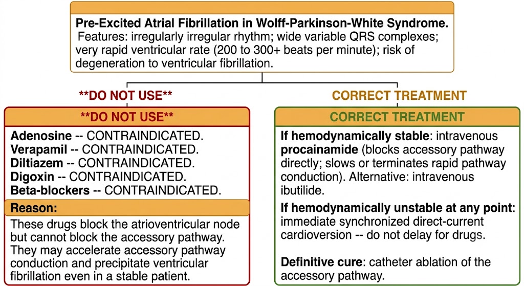
Emergency management flow diagram for pre-excited atrial fibrillation in Wolff-Parkinson-White showing contraindicated drugs on the left and correct treatment with procainamide or cardioversion on the right.
Source: AI-generated figure (Gemini). Educational use.

Pre-Excited Atrial Fibrillation in Wolff-Parkinson-White -- The Emergency

The most dangerous arrhythmia in Wolff-Parkinson-White syndrome is atrial fibrillation with rapid conduction through the accessory pathway. Unlike the atrioventricular node -- which slows conduction adaptively -- accessory pathways conduct at a fixed rapid rate. In pre-excited atrial fibrillation, atrial impulses can be conducted to the ventricle at rates of 200 to 300 beats per minute or faster, producing an irregularly irregular wide-complex tachycardia that can degenerate to ventricular fibrillation within seconds. This is a life-threatening emergency.

The electrocardiogram shows an irregularly irregular rhythm with wide, variable QRS complexes at a very rapid ventricular rate -- the irregular RR interval distinguishes pre-excited atrial fibrillation from the regular wide-complex tachycardia of antidromic atrioventricular reentrant tachycardia.

Absolutely contraindicated: adenosine, verapamil, diltiazem, and digoxin. These drugs block the atrioventricular node but have no effect on accessory pathway conduction. By eliminating competing wavefronts from atrioventricular nodal conduction, they can paradoxically accelerate accessory pathway conduction and precipitate ventricular fibrillation -- even in a hemodynamically stable patient.

Correct treatment: intravenous procainamide, which directly blocks accessory pathway conduction and slows or terminates rapid pathway conduction. Intravenous ibutilide is an alternative. If the patient is hemodynamically unstable at any point, immediate synchronized direct-current cardioversion without delay for drugs.

Wolff-Parkinson-White Safety Rule -- Memorize This

In pre-excited atrial fibrillation (Wolff-Parkinson-White with atrial fibrillation): adenosine, verapamil, diltiazem, digoxin, and beta-blockers are ALL contraindicated. Any atrioventricular nodal blocker can accelerate accessory pathway conduction and cause ventricular fibrillation. Treat with intravenous procainamide or electrical cardioversion.
Section 3

Atrial Flutter and Focal Atrial Tachycardia

Macro-re-entrant and focal atrial arrhythmias -- and why rate control is harder in flutter than in atrial fibrillation

Atrial Flutter

Atrial flutter is a macro-re-entrant atrial tachycardia with a characteristic sawtooth electrocardiogram pattern. The typical circuit circulates around the tricuspid valve annulus, with conduction through the cavotricuspid isthmus -- the narrow channel between the tricuspid annulus and the inferior vena cava -- serving as the critical slow zone. The atrial rate is approximately 300 beats per minute. Atrioventricular nodal conduction typically occurs at 2:1, producing a regular ventricular rate of approximately 150 beats per minute. A regular tachycardia at 150 beats per minute should always prompt consideration of atrial flutter.

Rate control is considerably more difficult in atrial flutter than in atrial fibrillation. The organized, high-amplitude flutter impulses penetrate the atrioventricular node more effectively than disorganized fibrillatory activity, requiring higher doses of atrioventricular nodal blocking agents and often producing unpredictable swings between 2:1 and 4:1 conduction rather than smooth rate control. This pharmacologic limitation makes cardioversion or catheter ablation the preferred approach for most patients with symptomatic flutter rather than long-term rate control.

For pharmacologic cardioversion of recent-onset atrial flutter, ibutilide is the most effective agent (cardioversion rates approximately 65 to 70%, higher than for atrial fibrillation). Dofetilide is an effective oral option. Direct-current cardioversion is highly effective and requires lower energy than for atrial fibrillation. Cavotricuspid isthmus catheter ablation is curative in over 95 percent of cases and is the preferred definitive treatment for symptomatic typical flutter.

Focal Atrial Tachycardia

Focal atrial tachycardia arises from a discrete atrial site and spreads centrifugally from that point. It accounts for approximately 10 to 15 percent of supraventricular tachycardia presentations. The electrocardiogram shows a P wave that differs in morphology from the sinus P wave, preceding the QRS with a normal PR interval. The tachycardia is typically regular at 130 to 250 beats per minute. A gradual onset with progressive rate acceleration at initiation suggests an automatic mechanism driven by enhanced automaticity of the focus.

Beta-blockers and non-dihydropyridine calcium channel blockers are first-line, providing rate control and partial suppression of catecholamine-driven automatic foci. Class Ic agents (flecainide, propafenone) suppress triggered and re-entrant focal atrial tachycardia in patients without structural heart disease. Adenosine transiently suppresses or slows automatic focal atrial tachycardia -- useful diagnostically (termination rules in atrioventricular nodal reentrant tachycardia/atrioventricular reentrant tachycardia; slowing or brief suppression with resumption suggests atrial origin). Catheter ablation is curative when the focus is localized and accessible, with success rates varying by anatomic location.


Section 4

Wide-Complex Tachycardia: Differential Diagnosis and Management

Treat as ventricular tachycardia until proven otherwise -- and why verapamil is dangerous

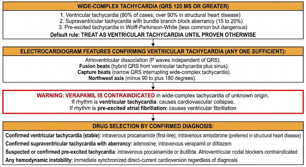
A wide-complex tachycardia is any tachycardia with QRS duration of 120 milliseconds or greater. The differential includes three possibilities: ventricular tachycardia, supraventricular tachycardia with aberrant conduction (bundle branch block), and pre-excited tachycardia (antidromic atrioventricular reentrant tachycardia or pre-excited atrial fibrillation). Getting this distinction wrong -- especially giving verapamil to a patient in ventricular tachycardia -- can be fatal.

Wide-complex tachycardia management flow diagram showing default ventricular tachycardia diagnosis, electrocardiogram confirmation features, verapamil contraindication warning, and drug selection by confirmed diagnosis.
Source: AI-generated figure (Gemini). Educational use.

The Frequency Problem: Ventricular Tachycardia is the Default Diagnosis

Ventricular tachycardia accounts for approximately 80 percent of all wide-complex tachycardias presenting to the emergency department, and over 90 percent of cases in patients with structural heart disease or prior myocardial infarction. Supraventricular tachycardia with aberrancy accounts for most of the remainder. The clinical rule follows directly: in any patient with structural heart disease presenting with wide-complex tachycardia, ventricular tachycardia is the presumptive diagnosis until proven otherwise.

Electrocardiogram Features That Confirm Ventricular Tachycardia

When present, the following features effectively confirm ventricular tachycardia without requiring further analysis:

Atrioventricular dissociation -- P waves marching independently of QRS complexes at a different rate, visible as notching or deformation of the QRS baseline in some leads. This is pathognomonic of ventricular tachycardia because any supraventricular tachycardia would conduct to the ventricle with maintained atrioventricular relationship.

Fusion beats -- a QRS morphology that is a hybrid between the ventricular tachycardia QRS and the sinus QRS, occurring when a sinus impulse partially activates the ventricle simultaneously with the ventricular tachycardia focus.

Capture beats -- a narrow QRS interrupting the wide-complex tachycardia, representing a sinus impulse that fully captures the ventricle during a brief gap in ventricular tachycardia refractoriness.

Northwest axis -- a QRS axis between minus 90 and plus 180 degrees (no man's land axis), which cannot be produced by any supraventricular conduction pathway and is highly specific for ventricular tachycardia.

Why Verapamil Is Dangerous in Wide-Complex Tachycardia of Unknown Origin

Intravenous verapamil is absolutely contraindicated in wide-complex tachycardia of unknown origin. It causes harm through two distinct mechanisms depending on the actual rhythm. If the rhythm is ventricular tachycardia: verapamil's negative inotropic and vasodilatory effects precipitate hemodynamic collapse without any antiarrhythmic benefit, because ventricular tachycardia does not depend on atrioventricular nodal conduction. Cardiovascular collapse and death from verapamil given to ventricular tachycardia misidentified as supraventricular tachycardia with bundle branch block are well documented. If the rhythm is pre-excited atrial fibrillation: verapamil blocks the atrioventricular node but accelerates accessory pathway conduction, potentially precipitating ventricular fibrillation.

The safe approach is to treat any wide-complex tachycardia of uncertain origin as ventricular tachycardia. Procainamide and amiodarone are effective for ventricular tachycardia and are also effective for supraventricular tachycardia with aberrancy, so treating as ventricular tachycardia carries no harm in the minority of cases that turn out to be supraventricular in origin.

Drug Selection by Confirmed Diagnosis

Confirmed ventricular tachycardia (hemodynamically stable): intravenous procainamide first-line; intravenous amiodarone preferred in structural heart disease. Synchronized cardioversion immediately if any hemodynamic instability.

Confirmed supraventricular tachycardia with bundle branch block aberrancy: adenosine (diagnostic and therapeutic); intravenous verapamil or diltiazem.

Confirmed or suspected pre-excited tachycardia: intravenous procainamide or intravenous ibutilide. Atrioventricular nodal blocking agents specifically contraindicated.

Uncertain diagnosis in a stable patient with high pre-test probability of ventricular tachycardia: treat as ventricular tachycardia with procainamide or amiodarone. Adenosine may be used cautiously to clarify the diagnosis given its ultrashort half-life, but only after pre-excitation has been excluded.


Section 5

Catheter Ablation: Indications and Outcomes

When to prefer ablation over long-term drug therapy -- and what cure rates to expect

Catheter ablation has transformed supraventricular tachycardia management over the past three decades, offering curative therapy for arrhythmias that previously required lifelong pharmacologic suppression. Understanding which arrhythmias are best treated by ablation, what success rates are realistic, and what the major complications are enables appropriate patient counseling and clinical decision-making.

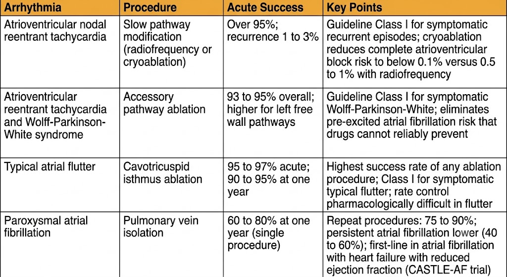
Reference table comparing catheter ablation outcomes for four arrhythmia types showing procedure, acute success rate, and key points for atrioventricular nodal reentrant tachycardia, atrioventricular reentrant tachycardia, typical atrial flutter, and paroxysmal atrial fibrillation.
Source: AI-generated figure (Gemini). Educational use.

Energy Sources

Radiofrequency ablation delivers energy through the catheter tip, generating heat at the contact point that creates a permanent coagulation lesion. It is the dominant modality for most supraventricular tachycardia procedures. Cryoablation freezes the contact tissue to very low temperatures. Its key advantage is cryomapping -- at an intermediate temperature the tissue is temporarily rendered non-conductive without permanent injury, allowing the operator to confirm that atrioventricular conduction is preserved before committing to a permanent freeze. This makes cryoablation the preferred approach for atrioventricular nodal reentrant tachycardia ablation near the His bundle, where the risk of inadvertent complete atrioventricular block is highest, reducing that complication rate to below 0.1 percent compared to 0.5 to 1 percent with radiofrequency ablation at the same site.

Outcomes by Arrhythmia Type

Atrioventricular nodal reentrant tachycardia (slow pathway modification): acute success over 95 percent; recurrence 1 to 3 percent at one year. Guideline Class I for symptomatic recurrent atrioventricular nodal reentrant tachycardia. Major risk: complete atrioventricular block requiring pacemaker (0.5 to 1 percent with radiofrequency; below 0.1 percent with cryoablation).

Atrioventricular reentrant tachycardia and Wolff-Parkinson-White: acute success 93 to 95 percent overall; higher for left free wall pathways (95 to 97 percent), lower for septal pathways near the His bundle. Guideline Class I for symptomatic Wolff-Parkinson-White. Ablation eliminates the risk of pre-excited atrial fibrillation, which drug therapy cannot reliably achieve.

Typical atrial flutter (cavotricuspid isthmus ablation): highest success rate of any ablation procedure -- 95 to 97 percent acute success; 90 to 95 percent long-term freedom from flutter. Guideline Class I for symptomatic typical flutter. Complication rates low.

Paroxysmal atrial fibrillation (pulmonary vein isolation): 60 to 80 percent freedom from atrial fibrillation at one year after a single procedure in paroxysmal atrial fibrillation without significant structural disease. Repeat procedures improve success to 75 to 90 percent. Persistent atrial fibrillation has lower single-procedure success (40 to 60 percent). The CASTLE-AF trial established ablation as first-line rhythm control in atrial fibrillation with heart failure with reduced ejection fraction.

Ablation vs. Drug Therapy: The Decision Framework

For atrioventricular nodal reentrant tachycardia and atrioventricular reentrant tachycardia in structurally normal hearts, ablation is preferred when symptoms are frequent or poorly tolerated, drugs have failed or produced unacceptable adverse effects, the patient prefers a potentially curative procedure over lifelong medication, or high-risk features are present (Wolff-Parkinson-White with short accessory pathway refractory period). Drug therapy is reasonable for infrequent well-tolerated episodes or patient preference for medical management.

For typical atrial flutter, the high ablation success rate and the pharmacologic difficulty of rate control in flutter favor ablation as the preferred strategy in most symptomatic patients. For atrial fibrillation, the decision integrates symptom burden, heart failure status, patient preference, and whether early rhythm control is indicated -- with ablation increasingly favored as first-line in younger symptomatic patients with paroxysmal atrial fibrillation and in atrial fibrillation with heart failure with reduced ejection fraction.


Suggested References
Author / Organization Title Source
Blomstrom-Lundqvist C, Scheinman MM, Aliot EM, et al ACC/AHA/ESC guidelines for the management of patients with supraventricular arrhythmias J Am Coll Cardiol. 2003;42(8):1493–1531
Delacretaz E Clinical practice: supraventricular tachycardia N Engl J Med. 2006;354(10):1039–1051
Ferguson JD, DiMarco JP Contemporary management of paroxysmal supraventricular tachycardia Circulation. 2003;107(8):1096–1099
Brugada J, Katritsis DG, Arbelo E, et al 2019 ESC Guidelines for the management of patients with supraventricular tachycardia Eur Heart J. 2020;41(5):655–720
Pappone C, Santinelli V, Manguso F, et al A randomized study of prophylactic catheter ablation in asymptomatic patients with the Wolff-Parkinson-White syndrome N Engl J Med. 2003;349(19):1803–1811
Timmermans C, Smeets JL, Rodriguez LM, et al Aborted sudden death in the Wolff-Parkinson-White syndrome Am J Cardiol. 1995;76(7):492–494
Schmieder S, Ndrepepa G, Dong J, et al Acute and long-term results of radiofrequency ablation of common atrial flutter and the influence of the right atrial isthmus ablation on the occurrence of atrial fibrillation Eur Heart J. 2003;24(10):956–962
Kistler PM, Roberts-Thomson KC, Haqqani HM, et al P-wave morphology in focal atrial tachycardia: development of an algorithm to predict the anatomic site of origin J Am Coll Cardiol. 2006;48(5):1010–1017
Calkins H, Hindricks G, Cappato R, et al 2017 HRS/EHRA/ECAS/APHRS/SOLAECE expert consensus statement on catheter and surgical ablation of atrial fibrillation Heart Rhythm. 2017;14(10):e275–e444
Brugada P, Brugada J, Mont L, Smeets J, Andries EW A new approach to the differential diagnosis of a regular tachycardia with a wide QRS complex Circulation. 1991;83(5):1649–1659
Akhtar M, Shenasa M, Jazayeri M, Caceres J, Tchou PJ Wide QRS complex tachycardia: reappraisal of a common clinical problem Ann Intern Med. 1988;109(11):905–912
Marrouche NF, Brachmann J, Andresen D, et al Catheter ablation for atrial fibrillation with heart failure (CASTLE-AF) N Engl J Med. 2018;378(5):417–427
Lerman BB, Belardinelli L Cardiac electrophysiology of adenosine: basic and clinical concepts Circulation. 1991;83(5):1499–1509