Introduction to Medical Pharmacology
Module 3 — Autonomic Receptors: Classification, Signaling, and Tissue Distribution
ANSI · Module 3 of 4Section 1
G-protein coupling, second messengers, tissue locations, and clinical drug targets
Muscarinic acetylcholine receptors are G-protein-coupled receptors activated by acetylcholine at parasympathetic postganglionic neuroeffector junctions and at a small number of sympathetically innervated tissues (notably sweat glands). Five subtypes — M1 through M5 — have been identified, each encoded by a distinct gene and distinguished by its G-protein coupling partner, tissue distribution, and functional role. The clinical pharmacology of muscarinic drugs is best understood by knowing which subtypes predominate at each clinically relevant target organ.
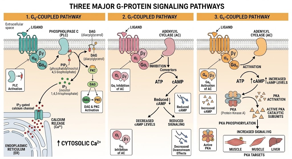
The five subtypes divide into two functional groups based on G-protein coupling. M1, M3, and M5 couple to Gq, activating phospholipase C, which generates inositol trisphosphate and diacylglycerol, ultimately increasing intracellular calcium and activating protein kinase C. M2 and M4 couple to Gi, inhibiting adenylyl cyclase, reducing cyclic adenosine monophosphate, and in the heart opening potassium channels that slow sinoatrial node firing and conduction through the atrioventricular node.
The clinical consequence of this subtype division is that most available muscarinic antagonists (atropine, scopolamine, glycopyrrolate, oxybutynin) are non-selective — they block all five subtypes. Their organ-level effects therefore reflect the muscarinic subtype predominating at each target: atropine accelerates heart rate (blocking M2 at the sinoatrial node), dries secretions (blocking M3 at salivary and lacrimal glands), dilates pupils (blocking M3 at the iris sphincter), and relaxes bronchial smooth muscle (blocking M3). Selective M3 antagonists such as tiotropium and ipratropium are used in pulmonary disease precisely because M3 mediates bronchoconstriction.
Gq-coupled (M1, M3, M5)
Phospholipase C Pathway
Gi-coupled (M2, M4)
Adenylyl Cyclase Inhibition
Muscarinic Agonist vs Antagonist — Predicting Organ Effects
To predict the effect of a muscarinic drug at any organ: identify which subtype predominates there, then determine what the drug does to that receptor. Atropine blocks M2 at the sinoatrial node → removes vagal braking → heart rate rises. Atropine blocks M3 at the bladder detrusor → relaxes detrusor → urinary retention. The same drug, different organs, different effects — all from knowing which subtype is dominant where.
Section 2
Ligand-gated ion channels, two subtypes, and drugs targeting each location
Nicotinic acetylcholine receptors are ligand-gated ion channels — a structurally and mechanistically distinct receptor class from the muscarinic receptors. When two molecules of acetylcholine bind to the receptor, a central sodium and calcium permeable ion channel opens, generating a rapid depolarizing potential in the postsynaptic cell. This ionotropic mechanism produces fast synaptic responses — milliseconds versus the seconds-to-minutes timescale of G-protein-coupled muscarinic signaling.
Two pharmacologically distinct nicotinic receptor subtypes are present in the autonomic nervous system and neuromuscular junction. The ganglionic subtype (designated N-N or NN) is found at all autonomic ganglia in both the sympathetic and parasympathetic divisions, as well as in the adrenal medulla. The neuromuscular subtype (designated N-M or NM) is found at the skeletal neuromuscular junction. Although both subtypes are activated by acetylcholine and nicotine, they differ in their subunit composition, pharmacological sensitivity, and the drugs that selectively target them.
This subtype distinction is clinically fundamental. Neuromuscular blocking drugs — succinylcholine (depolarizing) and rocuronium, vecuronium, cisatracurium (non-depolarizing) — act selectively at NM receptors to produce skeletal muscle paralysis for anesthesia and intubation without disrupting ganglionic transmission. Ganglionic blockers such as trimethaphan and mecamylamine act at NN receptors and block both sympathetic and parasympathetic ganglia, producing the broad autonomic suppression described in Module 1. Nicotine itself acts primarily at NN receptors at low doses, explaining its cardiovascular stimulant effects via sympathetic ganglionic activation.
The Critical Clinical Distinction: N-N vs N-M
N-N (ganglionic): autonomic ganglia and adrenal medulla. Blocked by trimethaphan, mecamylamine. Also activated by nicotine and by acetylcholinesterase inhibitor overdose (excess acetylcholine). Blockade produces bilateral autonomic suppression.
N-M (neuromuscular): skeletal muscle motor endplate only. Blocked by non-depolarizing agents (rocuronium, vecuronium — competitive antagonists, reversed by acetylcholinesterase inhibitors). Activated then desensitized by succinylcholine (depolarizing blocker, not reversible with neostigmine). No autonomic effects at clinical doses.
Section 3
G-protein coupling, tissue distribution, signaling cascades, and drug selectivity
Adrenergic receptors mediate the actions of norepinephrine (released from sympathetic postganglionic nerve terminals) and epinephrine (released from the adrenal medulla into the bloodstream). All adrenergic receptors are G-protein-coupled. They are classified into two major families — alpha and beta — each with clinically important subtypes. The subtype present at a given tissue determines the nature and direction of the sympathetic response at that organ.
Alpha-1 receptors couple to Gq, activating phospholipase C and raising intracellular calcium. They are located predominantly on vascular smooth muscle, the iris dilator muscle, the prostate and urethral sphincter, and the liver. Activation produces vasoconstriction, mydriasis, and increased urethral resistance. Alpha-1 antagonists (prazosin, tamsulosin, terazosin) lower blood pressure and relax the prostate — the dual utility that makes them useful for both hypertension and benign prostatic hyperplasia.
Alpha-2 receptors couple to Gi, inhibiting adenylyl cyclase and reducing cyclic adenosine monophosphate. They are found in two functionally distinct locations: presynaptically on sympathetic nerve terminals (where activation reduces norepinephrine release — a negative feedback autoreceptor mechanism) and postsynaptically in the central nervous system (where activation reduces sympathetic outflow). Clonidine and alpha-methyldopa exploit central alpha-2 receptors to lower blood pressure. Alpha-2 receptors in the pancreas inhibit insulin secretion, and in platelets promote aggregation.
Beta-1 receptors couple to Gs, stimulating adenylyl cyclase and raising cyclic adenosine monophosphate. They predominate in the heart (sinoatrial node, atrioventricular node, ventricular myocardium) and kidney (juxtaglomerular cells). Activation increases heart rate, conduction velocity, and contractility, and stimulates renin release. Beta-1 selective antagonists (metoprolol, atenolol, bisoprolol) are used for hypertension, heart failure, and arrhythmias, with the selectivity advantage of avoiding bronchoconstriction at therapeutic doses.
Beta-2 receptors also couple to Gs but are concentrated in bronchial smooth muscle, vascular smooth muscle of skeletal muscle beds, the uterus, and the liver. Activation causes bronchodilation, vasodilation in skeletal muscle, uterine relaxation, and glycogenolysis. Selective beta-2 agonists (albuterol, salmeterol, formoterol) are the primary bronchodilators in asthma and chronic obstructive pulmonary disease. Beta-3 receptors in adipose tissue mediate lipolysis; mirabegron targets beta-3 receptors in the bladder detrusor for overactive bladder.
Alpha Receptors
Alpha-1 and Alpha-2
Beta Receptors
Beta-1, Beta-2, Beta-3
Section 4
D1 and D2 subtypes, renal vasodilation, and peripheral dopaminergic pharmacology
Dopamine receptors (D1 through D5) are G-protein-coupled receptors that mediate the actions of dopamine in both the central nervous system and the periphery. While their central nervous system roles — in movement, reward, and psychosis — are covered in dedicated chapters on antipsychotic and antiparkinsonian drugs, the peripheral dopamine receptors have direct autonomic pharmacology relevance.
D1-like receptors (D1 and D5) couple to Gs, stimulating adenylyl cyclase and increasing cyclic adenosine monophosphate. In the periphery, D1 receptors in the renal and mesenteric vasculature mediate vasodilation — the basis for the renal-selective vasodilatory effect of low-dose dopamine infusion in critical care. D2-like receptors (D2, D3, D4) couple to Gi, inhibiting adenylyl cyclase. D2 receptors on presynaptic terminals in the sympathetic nervous system function as autoreceptors, inhibiting norepinephrine release in a manner analogous to alpha-2 autoreceptors.
The dopamine dose-response relationship in clinical use illustrates receptor selectivity in action. At low infusion rates, dopamine acts primarily on D1 receptors, causing renal and splanchnic vasodilation. At intermediate doses, it activates beta-1 receptors, increasing cardiac output. At high doses, it activates alpha-1 receptors, producing vasoconstriction. This dose-dependent receptor recruitment makes dopamine infusion a useful hemodynamic tool but also a pharmacologically complex one — the net effect shifts substantially with dose. Note that dopamine's role in central nervous system pharmacology (Parkinson disease, antipsychotics, reward pathways) is addressed in the dedicated central nervous system chapters.
Section 5
How prolonged agonist exposure reduces receptor responsiveness and the clinical consequences
Prolonged or repeated exposure to an agonist reduces the receptor's ability to respond — a phenomenon called desensitization. Two distinct processes underlie this reduction. Rapid desensitization (minutes) results from phosphorylation of the receptor by G-protein-coupled receptor kinases, which recruit arrestin proteins that uncouple the receptor from its G-protein. The receptor remains on the cell surface but no longer signals. This process is reversible once the agonist is removed.
Downregulation (hours to days) involves internalization of receptors from the cell surface into endosomes, followed by either recycling back to the membrane or lysosomal degradation. The result is a net reduction in the number of functional receptors available, producing a sustained decrease in drug response. Clinically, this is the mechanism underlying beta-2 agonist tolerance in asthma with overuse of short-acting rescue inhalers — the bronchodilator response diminishes with chronic over-reliance.
The converse process — upregulation — occurs when receptors are chronically blocked by an antagonist. The cell compensates by increasing receptor number and sensitivity. This is why abrupt withdrawal of beta-blockers can trigger rebound tachycardia and hypertension: the now-upregulated beta-1 receptors respond to endogenous catecholamines with exaggerated effect. The clinical rule is that beta-blockers should always be tapered, never stopped abruptly, in patients with coronary artery disease or hypertension.
Beta-Blocker Withdrawal — A Receptor Upregulation Hazard
Chronic beta-1 receptor blockade causes compensatory upregulation of beta-1 receptors. Abrupt discontinuation exposes these upregulated receptors to circulating catecholamines, producing rebound tachycardia, hypertension, and potentially angina or myocardial infarction in susceptible patients. Always taper beta-blockers over one to two weeks when discontinuing. This is a clinical consequence of receptor regulation, not of the drug itself.
| Author / Organization | Title | Source |
|---|---|---|
| Caulfield MP, Birdsall NJM | International Union of Pharmacology. XVII. Classification of muscarinic acetylcholine receptors | Pharmacological Reviews. 1998;50(2):279-290. |
| Harvey RD, Belevych AE | Muscarinic regulation of cardiac ion channels | British Journal of Pharmacology. 2003;139(6):1074-1084. |
| Eglen RM | Muscarinic receptor subtype pharmacology and physiology | Progress in Medicinal Chemistry. 2005;43:105-136. |
| Wess J, Eglen RM, Gautam D | Muscarinic acetylcholine receptors: mutant mice provide new insights for drug development | Nature Reviews Drug Discovery. 2007;6(9):721-733. |
| Sine SM | End-plate acetylcholine receptor: structure, mechanism, pharmacology, and disease | Physiological Reviews. 2012;92(3):1189-1234. |
| Naguib M, Lien CA, Aker J | Neuromuscular physiology and pharmacology. In: Miller RD, ed. Miller's Anesthesia. 8th ed. | Elsevier Saunders; 2015:423-468. |
| Benowitz NL | Nicotine addiction | New England Journal of Medicine. 2010;362(24):2295-2303. |
| Michel MC, Vrydag W | Alpha1-, alpha2- and beta-adrenoceptors in the urinary bladder, urethra and prostate | British Journal of Pharmacology. 2006;147(Suppl 2):S88-S119. |
| Brede M, Philipp M, Knaus A, Muthig V, Hein L | Alpha2-adrenergic receptor subtypes — novel functions uncovered in gene-targeted mouse models | Biology of the Cell. 2004;96(5):343-348. |
| Bristow MR | Beta-adrenergic receptor blockade in chronic heart failure | Circulation. 2000;101(5):558-569. |
| Johnson M | Molecular mechanisms of beta2-adrenergic receptor function, response, and regulation | Journal of Allergy and Clinical Immunology. 2006;117(1):18-24. |
| Andersson KE | Beta-adrenoceptors and their role in the regulation of lower urinary tract function | Acta Pharmacologica Sinica. 2022;43(11):2741-2751. |
| Vallone D, Picetti R, Borrelli E | Structure and function of dopamine receptors | Neuroscience and Biobehavioral Reviews. 2000;24(1):125-132. |
| Seeman P | Atypical antipsychotics: mechanism of action | Canadian Journal of Psychiatry. 2002;47(1):27-38. |
| Lohse MJ, Benovic JL, Caron MG, Lefkowitz RJ | Multiple pathways of rapid beta2-adrenergic receptor desensitization | Journal of Biological Chemistry. 1990;265(6):3202-3211. |
| Wehrwein EA, Orer HS, Barman SM | Overview of the anatomy, physiology, and pharmacology of the autonomic nervous system | Comprehensive Physiology. 2016;6(3):1239-1278. |