Estrogens act through two nuclear receptor subtypes: estrogen receptor alpha and estrogen receptor beta. Both are ligand-activated transcription factors that, upon estrogen binding, move into the nucleus and regulate gene expression. The two subtypes differ in their tissue distribution, and this difference is the pharmacological foundation for selective estrogen receptor modulators.
Estrogen receptor alpha predominates in the uterus, breast, liver, bone, and the hypothalamic-pituitary axis. It mediates the negative feedback of estradiol on gonadotropin secretion. Estrogen receptor beta is expressed preferentially in the ovary, colon, lung, and central nervous system, and its activation tends to oppose the proliferative effects driven by estrogen receptor alpha in tissues where both receptors are present.
Estrogen enters cells and binds the estrogen receptor in the cytoplasm or nucleus. The receptor-ligand complex dimerizes, binds to specific deoxyribonucleic acid sequences in the promoter regions of target genes, and activates or represses transcription. This genomic pathway takes hours to produce effects because it requires new messenger ribonucleic acid synthesis and protein production. Rapid membrane-associated estrogen effects (within minutes) also exist but are less relevant to Step 1 pharmacology.
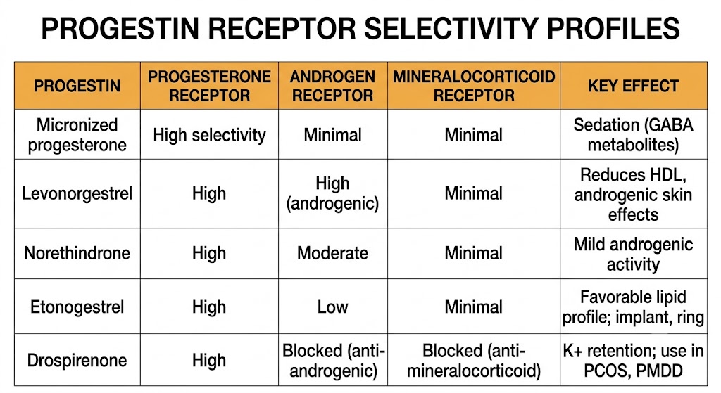
Natural progesterone binds the progesterone receptor selectively with minimal activity at the androgen, glucocorticoid, or mineralocorticoid receptors. Synthetic progestins were designed to improve oral bioavailability but retained structural features that produce off-target receptor binding. This cross-reactivity explains the major adverse effect differences between progestin generations: progestins derived from testosterone (such as levonorgestrel) bind the androgen receptor and can worsen lipid profiles and cause acne. Progestins derived from spironolactone (such as drospirenone) block both the androgen receptor and the mineralocorticoid receptor, producing anti-androgenic and potassium-retaining effects respectively.
Selective estrogen receptor modulators exploit the tissue-selective consequences of estrogen receptor alpha versus estrogen receptor beta distribution. Tamoxifen blocks estrogen receptor alpha in breast tissue (therapeutic in breast cancer) while acting as a partial agonist at estrogen receptor alpha in uterus and bone. Raloxifene blocks estrogen receptor alpha in both breast and uterus while retaining bone agonism. Understanding which receptor subtype predominates in which tissue predicts these tissue-selective drug effects throughout this chapter.
Ovarian estrogen production requires cooperative action between two cell types. Luteinizing hormone stimulates theca cells to produce androgens (androstenedione and testosterone) from cholesterol. Follicle-stimulating hormone stimulates granulosa cells to express aromatase (the enzyme encoded by CYP19A1), which converts those theca-derived androgens into estrogens. Androstenedione becomes estrone; testosterone becomes estradiol. Neither cell type can produce estradiol alone.
This two-cell model is a high-yield concept because it explains several pharmacological interventions: aromatase inhibitors block the final conversion step and suppress estrogen production in both premenopausal and postmenopausal women; in polycystic ovary syndrome, excess luteinizing hormone-driven theca androgen production with impaired granulosa aromatization creates the androgen excess characteristic of the syndrome.
Three endogenous estrogens exist in women. Estradiol is the most potent and is the primary secretory product of the premenopausal ovary. Estrone is weaker than estradiol and is produced mainly by peripheral aromatization of adrenal androstenedione in adipose tissue, liver, and muscle; it becomes the dominant circulating estrogen after menopause. Estriol is the weakest of the three and is produced in large amounts by the placenta during pregnancy; it is used in low-potency local vaginal preparations.
After menopause, the ovary stops producing estradiol and circulating estrogen falls dramatically. The dominant postmenopausal estrogen is estrone, produced by aromatization of adrenal androstenedione in adipose tissue. Because adipose aromatase is not regulated by gonadotropins, postmenopausal estrone production is proportional to body fat mass. This explains why aromatase inhibitors are highly effective in postmenopausal women with estrogen receptor-positive breast cancer: adipose tissue is the only remaining significant estrogen source, and blocking aromatase eliminates it.
During the reproductive years, estradiol exerts negative feedback on follicle-stimulating hormone and luteinizing hormone secretion from the pituitary. The unique mid-cycle switch from negative to positive feedback — triggered by sustained high estradiol concentrations — generates the luteinizing hormone surge that causes ovulation.
Theca cell makes androgens (luteinizing hormone-driven). Granulosa cell converts androgens to estrogens (follicle-stimulating hormone-driven, aromatase-dependent). Aromatase inhibitors work in postmenopausal women because adipose tissue is the remaining estrogen source. Clomiphene and letrozole both ultimately increase follicle-stimulating hormone to drive granulosa aromatase activity. This model recurs throughout this chapter series.
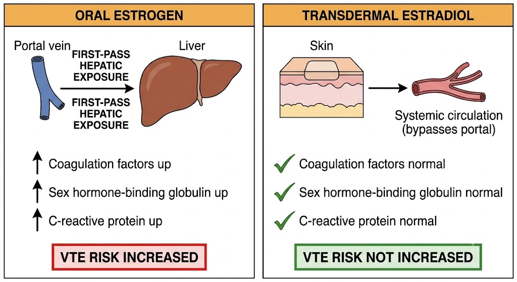
Estradiol is the bioidentical form of the primary ovarian estrogen. When taken orally, it undergoes extensive first-pass metabolism in the intestinal mucosa and liver, where it is converted to the weaker estrone and then to inactive sulfate conjugates. The portal circulation delivers high estrogen concentrations directly to the liver, stimulating hepatic synthesis of sex hormone-binding globulin, coagulation factors, angiotensinogen, and inflammatory proteins — effects that increase venous thromboembolism risk.
Transdermal estradiol (patches, gels, sprays) bypasses this first-pass hepatic exposure. Estradiol absorbed through the skin enters the systemic circulation directly, reaching the liver at normal physiological concentrations rather than as a concentrated portal bolus. The result is minimal stimulation of hepatic coagulation and inflammatory proteins. Observational data consistently show that transdermal estradiol does not increase venous thromboembolism risk even at higher doses, making it the preferred route in women with risk factors for clotting. The rule is straightforward: oral estrogen increases venous thromboembolism risk; transdermal estradiol does not.
Ethinyl estradiol is a synthetic estrogen designed for oral use. A chemical modification at the C-17 position blocks the hepatic enzyme that rapidly degrades natural estradiol, giving ethinyl estradiol a much higher oral bioavailability than estradiol. Despite this improved stability, ethinyl estradiol is still a potent hepatic stimulant: even at the low doses used in modern combined oral contraceptives (20 to 35 micrograms per day), it produces substantially greater hepatic estrogenic stimulation than any transdermal preparation. This hepatic potency is the reason combined oral contraceptive use increases venous thromboembolism risk approximately three to four times above baseline.
Conjugated equine estrogens (Premarin) are a mixture of water-soluble estrogen sulfates extracted from pregnant mares' urine. The mixture includes several estrogens not naturally present in humans, including equilin and equilenin, which have longer biological activity than estradiol. Conjugated equine estrogens were the estrogen used in the Women's Health Initiative trial — they are not interchangeable with transdermal estradiol in interpreting trial risk data.
Estetrol is a native fetal estrogen now available in a combined oral contraceptive paired with drospirenone. It has lower hepatic estrogenic stimulation than ethinyl estradiol, with early data suggesting a more favorable venous thromboembolism and metabolic profile, though long-term comparative outcome data are still accumulating.
Oral estrogen (ethinyl estradiol in contraceptives; oral estradiol or conjugated equine estrogens in hormone therapy) increases venous thromboembolism risk by stimulating hepatic coagulation factor production through first-pass portal exposure. Transdermal estradiol does not. In women with venous thromboembolism risk factors, the route of administration matters as much as the dose.
Natural progesterone has poor oral bioavailability due to extensive first-pass hepatic metabolism. Micronized progesterone (Prometrium) — progesterone formulated as very small particles in oil-filled capsules — achieves sufficient oral absorption for clinical use. It is metabolized to compounds that potentiate the gamma-aminobutyric acid type A receptor, producing the sedation that is a characteristic side effect of oral micronized progesterone. Vaginal and intramuscular progesterone formulations bypass first-pass metabolism and are used for luteal phase support in assisted reproduction.
Synthetic progestins were developed to overcome the bioavailability limitations of natural progesterone. Their side effect profiles are determined by which non-progesterone receptors they also bind.
Androgenic progestins (levonorgestrel, norethindrone acetate) worsen lipid profiles and may cause androgenic skin effects. Anti-androgenic progestins (drospirenone, dienogest) are preferred in androgen-excess conditions such as polycystic ovary syndrome and hirsutism. Drospirenone requires serum potassium monitoring when used with potassium-retaining drugs. Medroxyprogesterone acetate and micronized progesterone are pharmacologically distinct and should not be considered interchangeable, particularly when counseling about long-term hormone therapy.
The most pharmacologically important kinetic distinction for estrogens is the presence or absence of first-pass hepatic exposure. Oral estradiol and ethinyl estradiol both expose the liver to high concentrations via the portal circulation, stimulating hepatic production of sex hormone-binding globulin, coagulation factors, C-reactive protein, and angiotensinogen. Transdermal estradiol bypasses this exposure and does not produce these hepatic effects at therapeutic doses. This single difference determines the venous thromboembolism risk difference between oral and transdermal estrogen formulations.
Vaginal estrogen preparations (creams, suppositories, low-dose rings) are used for genitourinary syndrome of menopause and act primarily locally with minimal systemic absorption at standard doses, making them generally suitable even for women with contraindications to systemic estrogen.
Ethinyl estradiol in combined oral contraceptives potently induces hepatic sex hormone-binding globulin production, raising circulating levels two to four times above baseline. Because testosterone is transported bound to sex hormone-binding globulin, this induction reduces the free (biologically active) testosterone fraction. This pharmacodynamic effect is the basis for prescribing ethinyl estradiol-containing combined oral contraceptives in women with androgen-excess conditions such as polycystic ovary syndrome, acne, and hirsutism.
Androgenic progestins such as levonorgestrel compete with testosterone for sex hormone-binding globulin binding, partially countering this effect and increasing free testosterone. Anti-androgenic progestins such as drospirenone do not, making drospirenone-containing pills preferable in androgen-excess conditions.
Depot medroxyprogesterone acetate (150 mg intramuscular every 12 weeks) achieves contraceptive serum concentrations within 24 hours of injection and maintains them for 3 months. A high-yield counseling point: the average time to return of fertility after discontinuing depot medroxyprogesterone acetate is 9 to 10 months, substantially longer than for any other reversible contraceptive method. Patients planning pregnancy in the near term should be counseled about this delay before starting the injection.
The etonogestrel subdermal implant (Nexplanon) provides contraceptive concentrations for 3 years from a single insertion and is the most effective reversible contraceptive available. Fertility returns within 3 to 4 weeks of implant removal.
Both ethinyl estradiol and most synthetic progestins are metabolized by cytochrome P450 3A4. Drugs that induce this enzyme accelerate estrogen and progestin metabolism, lower plasma concentrations, and can cause contraceptive failure or reduced therapeutic efficacy of hormone therapy. The clinical consequence ranges from breakthrough bleeding to complete loss of ovulation suppression.
Rifampin is the most potent cytochrome P450 3A4 inducer in clinical use and renders hormonal contraception unreliable. Even a short course of rifampin produces enzyme induction that persists for approximately 4 weeks after the last dose. Patients on combined hormonal contraceptives who require rifampin must use a non-hormonal backup method (copper intrauterine device or barrier method) during rifampin therapy and for 4 weeks after stopping.
Among antiepileptic drugs, the major enzyme-inducing agents that reduce hormonal contraceptive efficacy are carbamazepine, phenytoin, phenobarbital, primidone, and oxcarbazepine. Non-enzyme-inducing antiepileptic drugs — including valproate, levetiracetam, gabapentin, and pregabalin — do not affect hormonal contraceptive efficacy.
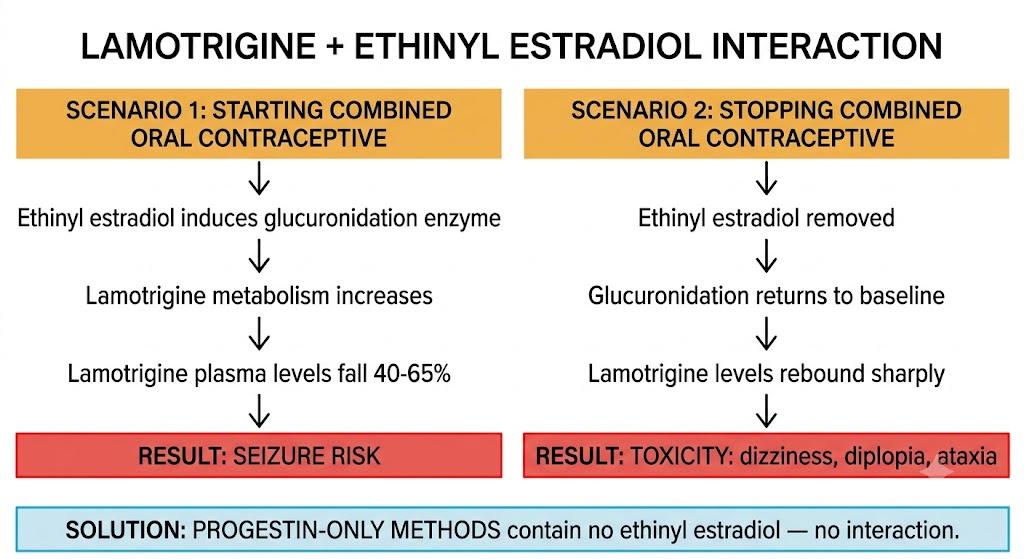
This bidirectional interaction is high-yield. Ethinyl estradiol induces the enzyme responsible for lamotrigine glucuronidation and inactivation. When a woman taking lamotrigine for epilepsy starts a combined oral contraceptive containing ethinyl estradiol, lamotrigine plasma levels fall by up to 40 to 65 percent, substantially increasing the risk of breakthrough seizures. When the combined oral contraceptive is stopped — or during the pill-free week of cyclic regimens — lamotrigine levels rebound sharply and can cause lamotrigine toxicity (dizziness, diplopia, ataxia).
This interaction does not occur with progestin-only methods, because they contain no ethinyl estradiol and do not induce lamotrigine metabolism. For women with epilepsy who are stabilized on lamotrigine, progestin-only contraception or a non-hormonal method is strongly preferred over combined oral contraceptives.
Several classes of antiretroviral drugs interact with hormonal contraceptives. Non-nucleoside reverse transcriptase inhibitors vary: some (including efavirenz) are potent cytochrome P450 3A4 inducers that reduce hormonal contraceptive efficacy, while others (including rilpivirine) do not. Ritonavir-boosted protease inhibitor regimens also reduce ethinyl estradiol concentrations. Integrase strand-transfer inhibitors (such as dolutegravir and raltegravir) do not have clinically significant interactions with hormonal contraceptives and are preferred antiretroviral agents when reliable hormonal contraception is required.
No oral, patch, or ring hormonal contraceptive is reliable during rifampin therapy or for 4 weeks after stopping. The copper intrauterine device is the preferred backup option. The lamotrigine interaction is the most common clinically encountered bidirectional drug interaction in hormonal contraception: adding ethinyl estradiol causes seizures (lamotrigine levels fall); stopping it causes toxicity (levels rebound). Progestin-only methods avoid this interaction entirely.
| Author / Organization | Title | Source |
|---|---|---|
| Heldring N, Pike A, Andersson S, et al. | Estrogen receptors: how do they signal and what are their targets | Physiol Rev. 2007;87(3):905–931 |
| Nilsson S, Makela S, Treuter E, et al. | Mechanisms of estrogen action | Physiol Rev. 2001;81(4):1535–1565 |
| Levin ER. | Membrane oestrogen receptor alpha signalling to cell functions | J Physiol. 2009;587(21):5019–5023 |
| Schindler AE, Campagnoli C, Druckmann R, et al. | Classification and pharmacology of progestins | Maturitas. 2008;61(1–2):171–180 |
| Drummond AE, Fuller PJ. | The importance of ERbeta signalling in the ovary | J Endocrinol. 2010;205(1):15–23 |
| Simpson ER, Davis SR. | Minireview: aromatase and the regulation of estrogen biosynthesis | Endocrinology. 2001;142(11):4589–4594 |
| Kuhnz W, Blode H, Zimmermann H. | Pharmacokinetics of exogenous natural and synthetic estrogens and antiestrogens | In: Estrogens and Antiestrogens II. Springer; 1999:261–322 |
| Canonico M, Oger E, Plu-Bureau G, et al; ESTHER Study Group. | Hormone therapy and venous thromboembolism among postmenopausal women: impact of the route of estrogen administration and progestogens | Circulation. 2007;115(7):840–845 |
| Stanczyk FZ, Hapgood JP, Winer S, Mishell DR Jr. | Progestogens used in postmenopausal hormone therapy: differences in their pharmacological properties, intracellular actions, and clinical effects | Endocr Rev. 2013;34(2):171–208 |
| Tavaniotou A, Smitz J, Bourgain C, Devroey P. | Comparison between different routes of progesterone administration as luteal phase and early pregnancy support | Hum Reprod Update. 2000;6(2):139–148 |
| van den Heuvel MW, van Bragt AJ, Alnabawy AK, Kaptein MC. | Comparison of ethinylestradiol pharmacokinetics in three hormonal contraceptive formulations: the vaginal ring, the transdermal patch and an oral contraceptive | Contraception. 2005;72(3):168–174 |
| Simmons KB, Haddad LB, Nanda K, Curtis KM. | Drug interactions between non-rifamycin antibiotics and hormonal contraception: a systematic review | Am J Obstet Gynecol. 2018;218(1):88–97 |
| Reimers A, Brodtkorb E, Sabers A. | Interactions between hormonal contraception and antiepileptic drugs: clinical and mechanistic considerations | Seizure. 2015;28:66–70 |