Gonadotropin-releasing hormone (GnRH), also called luteinizing hormone-releasing hormone, is a decapeptide produced by approximately 1,000 to 3,000 neurons in the medial preoptic area and arcuate nucleus of the hypothalamus. It is released in discrete pulses into the hypothalamic-pituitary portal circulation approximately once every 60 to 90 minutes during the follicular phase of the menstrual cycle and once every 2 to 4 hours during the luteal phase.
The pulsatile pattern is not incidental: it is the pharmacological signal that determines the response of pituitary gonadotrophs. Pulsatile gonadotropin-releasing hormone stimulates the synthesis and secretion of both luteinizing hormone and follicle-stimulating hormone. Continuous gonadotropin-releasing hormone receptor occupancy, as produced by long-acting agonist depot formulations, paradoxically suppresses both hormones through receptor downregulation. This is the mechanism underlying medical castration.
The gonadotropin-releasing hormone receptor is a seven-transmembrane G protein-coupled receptor that uniquely lacks an intracellular carboxyl-terminal tail. This structural feature slows receptor internalization compared to other G protein-coupled receptors, making the receptor more susceptible to sustained-stimulation-induced downregulation. Receptor coupling is through Gq/11 proteins, which activate phospholipase C beta, generating inositol trisphosphate and diacylglycerol. Inositol trisphosphate mobilizes intracellular calcium and diacylglycerol activates protein kinase C, together driving luteinizing hormone and follicle-stimulating hormone exocytosis.
With continuous agonist exposure, the receptor undergoes two sequential events: rapid desensitization through protein kinase C-mediated uncoupling from Gq, followed by slower internalization via a clathrin-independent pathway. Surface receptor density falls by 80 to 95%, producing the pharmacodynamic basis for medical castration. Testosterone reaches castrate levels within 3 to 4 weeks of initiating a depot gonadotropin-releasing hormone agonist.
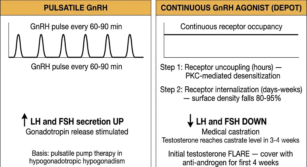
Pulsatile gonadotropin-releasing hormone stimulates luteinizing hormone and follicle-stimulating hormone. Continuous gonadotropin-releasing hormone receptor activation suppresses both hormones via receptor downregulation. This distinction explains why depot agonists (leuprolide, goserelin) produce medical castration, while a single injection can trigger an ovulatory luteinizing hormone surge in assisted reproduction. The dosing interval and formulation determine the pharmacodynamic outcome — not the drug structure.
Thyrotropin-releasing hormone is a tripeptide produced in the paraventricular nucleus of the hypothalamus. It stimulates thyroid-stimulating hormone secretion from pituitary thyrotrophs and also stimulates prolactin secretion from lactotrophs. The thyrotropin-releasing hormone receptor is a Gq-coupled G protein-coupled receptor, activating phospholipase C beta through the same inositol trisphosphate-diacylglycerol-calcium-protein kinase C pathway used by the gonadotropin-releasing hormone receptor. This shared pathway explains why primary hypothyroidism with chronically elevated thyrotropin-releasing hormone can produce hyperprolactinemia and galactorrhea.
The clinical pharmacology of thyrotropin-releasing hormone centers on the thyrotropin-releasing hormone stimulation test. Synthetic thyrotropin-releasing hormone (protirelin) administered intravenously produces a thyroid-stimulating hormone rise within 30 minutes in normal individuals. A blunted or absent response indicates pituitary insufficiency; an exaggerated or delayed response suggests hypothalamic disease with an intact but understimulated pituitary. With sensitive thyroid-stimulating hormone assays now available, the test is rarely performed but remains conceptually useful for understanding feedback regulation of the hypothalamic-pituitary-thyroid axis.
Corticotropin-releasing hormone is a 41-amino-acid peptide produced predominantly in the paraventricular nucleus. It drives adrenocorticotropic hormone secretion from corticotroph cells in the anterior pituitary. Two receptor subtypes exist: corticotropin-releasing hormone receptor type 1 and type 2, both Gs-coupled G protein-coupled receptors that activate adenylyl cyclase, raise intracellular cyclic adenosine monophosphate, and activate protein kinase A to stimulate pro-opiomelanocortin gene transcription and adrenocorticotropic hormone release. Corticotropin-releasing hormone receptor type 1 is the dominant mediator of pituitary adrenocorticotropic hormone secretion.
The corticotropin-releasing hormone stimulation test is used to differentiate the causes of Cushing syndrome. Intravenous corticotropin-releasing hormone produces an exaggerated adrenocorticotropic hormone and cortisol rise in pituitary-dependent Cushing disease, a blunted or absent rise in adrenal Cushing syndrome, and typically no rise with ectopic adrenocorticotropic hormone syndrome because ectopic tumors lack corticotropin-releasing hormone receptor type 1. The test is most useful when combined with inferior petrosal sinus sampling, where a central-to-peripheral adrenocorticotropic hormone gradient after corticotropin-releasing hormone stimulation confirms pituitary origin of adrenocorticotropic hormone excess.
Pituitary Cushing disease: adrenocorticotropic hormone rises by more than 35% above baseline because corticotroph adenoma cells retain corticotropin-releasing hormone receptor type 1 responsiveness. Adrenal Cushing syndrome: no adrenocorticotropic hormone rise; autonomous adrenal cortisol suppresses pituitary corticotroph function. Ectopic adrenocorticotropic hormone: usually no rise because ectopic tumors lack corticotropin-releasing hormone receptor type 1. A blunted response does not definitively exclude pituitary disease; inferior petrosal sinus sampling is required for definitive localization.
Growth hormone-releasing hormone is a 44-amino-acid peptide produced in the arcuate nucleus. It binds a Gs-coupled G protein-coupled receptor on pituitary somatotroph cells, raising intracellular cyclic adenosine monophosphate and activating protein kinase A to stimulate growth hormone gene transcription and secretion. Growth hormone pulsatility results from alternating episodic growth hormone-releasing hormone stimulation and tonic somatostatin inhibitory tone, not from an intrinsic growth hormone secretory pulse generator. The largest growth hormone secretory burst normally occurs during slow-wave sleep.
Somatostatin exists in two bioactive forms: somatostatin-14 (predominant in hypothalamic neurons) and somatostatin-28 (predominant in the gastrointestinal tract and pancreas). Both forms have plasma half-lives of 1 to 3 minutes, making native somatostatin pharmacologically impractical and driving the development of synthetic analogs.
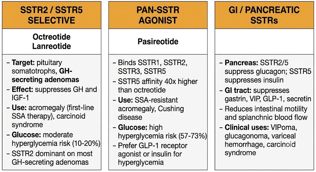
Somatostatin acts through five receptor subtypes (somatostatin receptor subtypes 1 through 5), all of which are Gi-coupled G protein-coupled receptors. Gi activation inhibits adenylyl cyclase, opens inwardly rectifying potassium channels (hyperpolarizing the cell), and inhibits voltage-gated calcium channels — together suppressing hormone secretion. The subtype distribution determines both physiological function and drug selectivity. Somatostatin receptor subtype 2 and somatostatin receptor subtype 5 predominate on pituitary somatotrophs and are the primary targets for growth hormone suppression. Somatostatin receptor subtype 2 is the dominant subtype on most growth hormone-secreting pituitary adenomas. Somatostatin receptor subtype 3 mediates antiproliferative signaling in tumor cells.
In the gastrointestinal tract and pancreas, somatostatin receptor activation suppresses multiple secretory hormones including gastrin, secretin, vasoactive intestinal peptide, glucagon, and insulin, and reduces splanchnic blood flow. These peripheral effects underlie the clinical applications of somatostatin analogs in carcinoid syndrome, variceal hemorrhage, and pancreatic tumors.
Somatostatin receptor subtype 2 and subtype 5-selective agents (octreotide, lanreotide): effective growth hormone suppression in most acromegaly; moderate hyperglycemia risk. Pan-somatostatin receptor agonist (pasireotide): additional somatostatin receptor subtype 1, 3, and 5 activity provides superior growth hormone and insulin-like growth factor-1 control in somatostatin receptor subtype 2-poor tumors and Cushing disease (corticotroph adenomas express somatostatin receptor subtype 5); substantially higher hyperglycemia incidence (57 to 73%) because somatostatin receptor subtype 5-mediated insulin suppression is more pronounced. Subtype profiling of pituitary adenomas can predict analog response before treatment.
Dopamine functions as a hypothalamic inhibiting hormone through the tuberoinfundibular dopaminergic pathway: dopaminergic neurons in the arcuate nucleus project to the median eminence, releasing dopamine into the portal circulation. At the anterior pituitary, dopamine binds dopamine type 2 receptors on lactotroph cells, activating Gi proteins that inhibit adenylyl cyclase and suppress prolactin gene transcription and secretion. Dopamine is therefore the primary physiological inhibitor of prolactin — the only major anterior pituitary hormone under predominantly inhibitory rather than stimulatory hypothalamic control.
This explains drug-induced hyperprolactinemia as a class effect of dopamine type 2 receptor antagonists. Antipsychotics (first-generation agents such as haloperidol; most second-generation agents such as risperidone) block pituitary dopamine type 2 receptors and reliably elevate prolactin, causing galactorrhea, amenorrhea, and hypogonadism in women, and gynecomastia and sexual dysfunction in men. Among second-generation antipsychotics, clozapine, quetiapine, and aripiprazole are prolactin-sparing; aripiprazole is a partial dopamine type 2 receptor agonist at the pituitary level and can normalize prolactin when added to a prolactin-elevating antipsychotic regimen. Metoclopramide and domperidone elevate prolactin through the same mechanism. Chronic opioid use suppresses tuberoinfundibular dopaminergic neuron firing through mu-opioid receptor activation, contributing to opioid-induced endocrinopathy.
Oxytocin and vasopressin (antidiuretic hormone) are nonapeptides synthesized in magnocellular neurons of the supraoptic and paraventricular nuclei, transported axonally to the posterior pituitary (neurohypophysis), and released directly into the systemic circulation. Oxytocin acts at the oxytocin receptor, a Gq-coupled G protein-coupled receptor on uterine myometrial cells and mammary myoepithelial cells. Myometrial oxytocin receptor density increases markedly at term gestation due to estrogen-driven upregulation, explaining the sharp rise in uterine sensitivity to oxytocin near delivery. Synthetic oxytocin (Pitocin) is used for labor induction; carbetocin is a long-acting oxytocin analog; atosiban is an oxytocin receptor antagonist used as a tocolytic in preterm labor in Europe.
Vasopressin acts through three receptor subtypes. Vasopressin receptor 1a receptors are Gq-coupled and mediate vascular smooth muscle contraction and platelet aggregation. Vasopressin receptor 1b receptors are Gq-coupled and mediate adrenocorticotropic hormone release from the anterior pituitary. Vasopressin receptor 2 receptors are Gs-coupled and mediate antidiuresis by promoting aquaporin-2 insertion into renal collecting duct cells. Desmopressin is a selective vasopressin receptor 2 agonist with negligible vasopressin receptor 1 activity, providing antidiuresis without vasoconstriction. Tolvaptan and conivaptan are vasopressin receptor 2 and combined vasopressin receptor 1/2 antagonists, respectively, used for hyponatremia in syndrome of inappropriate antidiuretic hormone secretion.
Causes: dopamine type 2 receptor antagonists (antipsychotics, metoclopramide, domperidone); verapamil (interferes with dopamine release); chronic opioids (suppress tuberoinfundibular dopaminergic neuron activity via mu receptors). Prolactin-sparing antipsychotics: clozapine, quetiapine, aripiprazole. Management: switch to prolactin-sparing agent if clinically feasible; dopamine agonist (cabergoline) can lower prolactin but risks worsening underlying psychosis if the patient is on antipsychotics for that indication.
Native hypothalamic peptides have three pharmacological liabilities that preclude direct therapeutic use: extremely short plasma half-lives (seconds to minutes) from rapid peptidase degradation, negligible oral bioavailability because peptide bonds are hydrolyzed in the gastrointestinal tract before absorption, and rapid receptor desensitization at the concentrations achievable with exogenous native peptide. Analog design strategies address each liability through targeted structural modifications.
Substitution of D-amino acids at peptidase-susceptible cleavage sites prevents enzymatic recognition and degradation. For gonadotropin-releasing hormone analogs, D-amino acid substitution at position 6 of the decapeptide extends the half-life from 2 to 4 minutes (native gonadotropin-releasing hormone) to 3 to 8 hours (leuprolide, buserelin). C-terminal amidation further protects against carboxypeptidase attack. For octreotide, a cyclic octapeptide structure incorporating D-phenylalanine and D-tryptophan substitutions produces a half-life of approximately 1.7 to 2 hours compared to 1 to 3 minutes for native somatostatin.
Depot formulation technology converts short-acting peptides into once-monthly or once-quarterly clinical agents. Poly(lactic-co-glycolic acid) microsphere technology encapsulates leuprolide acetate or octreotide in biodegradable polymer microspheres injected intramuscularly; slow hydrolysis releases drug over 28 to 90 days. Lanreotide uses a self-assembling deep subcutaneous autogel that releases drug over approximately 28 days. Goserelin is formulated as a rod-shaped biodegradable subcutaneous implant releasing drug over 28 or 84 days.
Oral bioavailability was achieved by departing entirely from the peptide backbone. The oral gonadotropin-releasing hormone antagonists elagolix, relugolix, and linzagolix are non-peptide small molecules that competitively block the gonadotropin-releasing hormone receptor without the peptide bonds that confer gastrointestinal degradability. Elagolix has oral bioavailability of approximately 57% and is metabolized primarily by cytochrome P450 3A4. Relugolix has oral bioavailability of approximately 12% and is a substrate and moderate inhibitor of P-glycoprotein and breast cancer resistance protein. Both achieve immediate testosterone suppression without the initial luteinizing hormone surge (flare) that characterizes depot gonadotropin-releasing hormone agonist initiation.
D-amino acid substitution at peptidase cleavage sites extends half-life (leuprolide, buserelin, octreotide). C-terminal amidation prevents carboxypeptidase degradation. Poly(lactic-co-glycolic acid) microsphere depot converts hours half-life into monthly dosing (leuprolide long-acting release, octreotide long-acting release). Non-peptide small molecule design achieves oral bioavailability by eliminating the peptide backbone (elagolix, relugolix, macimorelin). Somatostatin receptor subtype selectivity is engineered by varying amino acid residues at receptor-contact positions: somatostatin receptor subtype 2 and subtype 5-selective (octreotide, lanreotide) versus pan-receptor agonism (pasireotide).
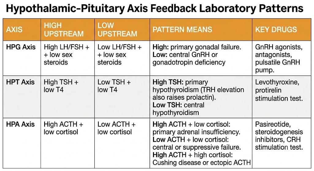
Each hypothalamic-pituitary-peripheral axis provides a predictable laboratory pattern when the axis is disrupted at a specific level. Understanding these patterns is essential for interpreting endocrine laboratory tests and predicting the effects of drugs that act on these axes.
In the hypothalamic-pituitary-gonadal axis: low luteinizing hormone and follicle-stimulating hormone with low sex steroids indicates central (gonadotropin-releasing hormone or gonadotropin) failure. High luteinizing hormone and follicle-stimulating hormone with low sex steroids indicates primary gonadal failure. This distinction guides both diagnostic workup and treatment: central failure responds to exogenous pulsatile gonadotropin-releasing hormone or gonadotropin administration; primary gonadal failure does not.
In the hypothalamic-pituitary-thyroid axis: high thyroid-stimulating hormone with low thyroxine indicates primary hypothyroidism. Low thyroid-stimulating hormone with low thyroxine indicates central hypothyroidism (thyrotropin-releasing hormone or thyroid-stimulating hormone deficiency). In patients on levothyroxine replacement, a thyroid-stimulating hormone that fails to suppress suggests either non-compliance, malabsorption, or drug interactions increasing thyroid hormone clearance (most commonly rifampin or other cytochrome P450-inducing drugs).
In the hypothalamic-pituitary-adrenal axis: low adrenocorticotropic hormone with low cortisol identifies either primary adrenal failure after exogenous glucocorticoid suppression or primary pituitary-hypothalamic failure. High adrenocorticotropic hormone with low cortisol identifies primary adrenal insufficiency (Addison disease). High adrenocorticotropic hormone with high cortisol identifies either Cushing disease or ectopic adrenocorticotropic hormone secretion, distinguishable by corticotropin-releasing hormone stimulation test and imaging.
Hypothalamic-pituitary-gonadal axis: low luteinizing hormone/follicle-stimulating hormone + low sex steroids = central failure; high luteinizing hormone/follicle-stimulating hormone + low sex steroids = primary gonadal failure. Hypothalamic-pituitary-thyroid axis: high thyroid-stimulating hormone + low thyroxine = primary hypothyroidism; low thyroid-stimulating hormone + low thyroxine = central hypothyroidism. Hypothalamic-pituitary-adrenal axis: low adrenocorticotropic hormone + low cortisol = central or suppressive failure; high adrenocorticotropic hormone + low cortisol = primary adrenal insufficiency; high adrenocorticotropic hormone + high cortisol = Cushing disease or ectopic adrenocorticotropic hormone. Each pattern maps to a specific level of axis dysfunction and guides pharmacological intervention.
| Author / Organization | Title | Source |
|---|---|---|
| Millar RP, Lu ZL, Pawson AJ, Flanagan CA, Morgan K, Maudsley SR | Gonadotropin-releasing hormone receptors | Endocr Rev. 2004;25(2):235–275 |
| Conn PM, Crowley WF Jr | Gonadotropin-releasing hormone and its analogs | N Engl J Med. 1991;324(2):93–103 |
| Shore ND, Saad F, Cookson MS, et al; HERO Study Investigators | Oral relugolix for androgen-deprivation therapy in advanced prostate cancer | N Engl J Med. 2020;382(23):2187–2196 |
| Vale W, Spiess J, Rivier C, Rivier J | Characterization of a 41-residue ovine hypothalamic peptide that stimulates secretion of corticotropin and beta-endorphin | Science. 1981;213(4514):1394–1397 |
| Nieman LK, Biller BM, Findling JW, et al | The diagnosis of Cushing syndrome: an Endocrine Society clinical practice guideline | J Clin Endocrinol Metab. 2008;93(5):1526–1540 |
| Muller EE, Locatelli V, Cocchi D | Neuroendocrine control of growth hormone secretion | Physiol Rev. 1999;79(2):511–607 |
| Patel YC | Somatostatin and its receptor family | Front Neuroendocrinol. 1999;20(3):157–198 |
| Gadelha MR, Bronstein MD, Brue T, et al | Pasireotide versus continued treatment with octreotide or lanreotide in patients with inadequately controlled acromegaly (PAOLA): a randomised, phase 3 trial | Lancet Diabetes Endocrinol. 2014;2(11):875–884 |
| Molitch ME | Drugs and prolactin | Pituitary. 2008;11(2):209–218 |
| Gimpl G, Fahrenholz F | The oxytocin receptor system: structure, function, and regulation | Physiol Rev. 2001;81(2):629–683 |
| Veber DF, Holly FW, Nutt RF, et al | Highly active cyclic and bicyclic somatostatin analogues of reduced ring size | Nature. 1979;280(5722):512–514 |