Reversible, localized nerve blockade without loss of consciousness
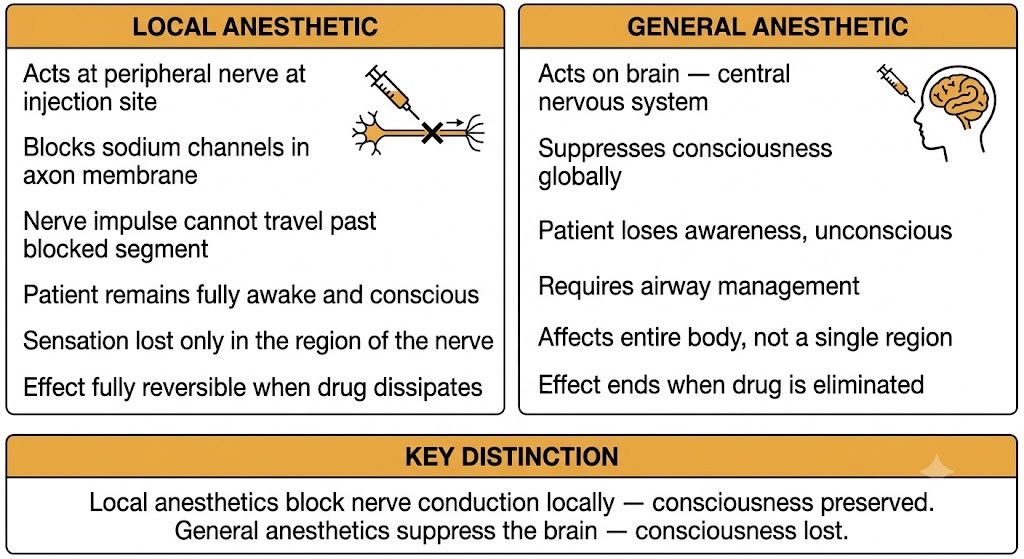
Local anesthetics are a class of drugs that reversibly block nerve conduction in a defined anatomical region, producing loss of sensation — and at higher concentrations, loss of motor function — without causing unconsciousness. This distinguishes them fundamentally from general anesthetics, which act on the brain to produce a global loss of consciousness.
When a dentist injects local anesthetic into the gum before a procedure, the patient remains awake and conversational while the entire lower jaw is numb. That clinical picture captures the defining feature of local anesthetics: the drug acts at the site of injection, blocks the nerves at that location, and the effect is entirely confined to the region supplied by those nerves. No drug reaches the brain in significant concentration at clinical doses, so consciousness is preserved.
General anesthetics work in the opposite direction. They are delivered systemically — inhaled or administered intravenously — and act directly on the central nervous system to suppress consciousness. A patient under general anesthesia feels nothing and cannot be roused. A patient under local anesthesia is awake, can communicate, and can feel sensations outside the blocked region.
All clinically used local anesthetics share a single primary mechanism: blockade of voltage-gated sodium channels in nerve cell membranes. Sodium influx through these channels is the electrical event that generates and propagates the action potential. By blocking sodium entry, local anesthetics raise the threshold required for an action potential to fire and prevent the nerve impulse from traveling along the axon. The nerve is still physically intact — no damage occurs — and when the drug dissipates, full function returns. This reversibility is a defining property of local anesthetics.
The sodium channel mechanism operates the same way in every tissue where sodium channels exist. This creates both the therapeutic effect in peripheral nerves and the basis for the most serious toxicity: systemic local anesthetic toxicity that affects the central nervous system and the heart, both of which also depend on sodium channels for their electrical activity.
Local anesthetics block nerve conduction at the site of administration — patient remains awake. General anesthetics suppress central nervous system function — patient loses consciousness. This distinction governs their clinical use: local anesthetics are used for regional procedures where the patient needs to remain conscious or where a systemic anesthetic would be unnecessary or risky.
Binding to the inner face of the channel prevents sodium influx and arrests action potential propagation
The voltage-gated sodium channel is the molecular target of all local anesthetics. Understanding how and where local anesthetics bind to this channel — and the critical fact that they act from the inside of the cell — explains both the clinical pharmacology and the pH-dependent variation in efficacy.
Voltage-gated sodium channels are transmembrane proteins that open in response to membrane depolarization, allowing sodium ions to rush into the cell down their electrochemical gradient. This rapid sodium influx is the inward current that generates the rising phase of the action potential. Once opened, the channel rapidly inactivates — it closes and cannot reopen immediately — producing the refractory period.
For a nerve impulse to travel from one point to another, each segment of membrane must depolarize enough to trigger opening of sodium channels in the adjacent segment. This propagation continues all the way from the site of stimulation to the nerve terminal. Block the sodium channels in any segment of the axon, and the impulse fails to propagate beyond that point.
Local anesthetics bind to a receptor site located on the intracellular face of the sodium channel — the side facing the inside of the cell, not the extracellular surface. This is a high-yield anatomical fact: the drug must reach the inside of the axon to reach its binding site on the channel.
Once bound, the local anesthetic physically occludes the inner pore of the channel. Sodium ions cannot pass through the blocked channel. The threshold for action potential generation rises, and if enough channels are blocked, no action potential can be generated or propagated in that nerve segment. The sensory information — a pain signal, a touch signal, a temperature signal — cannot reach the central nervous system. The patient feels nothing in the distribution of that nerve.
Local anesthetics block voltage-gated sodium channels from the intracellular side. Sodium influx is prevented. Without sodium influx, the action potential cannot be generated or propagated. Nerve conduction fails in the blocked segment. Sensation is lost in the territory of that nerve. Effect is reversible when drug dissipates.
The voltage-gated sodium channel exists in three functional states: resting (closed but able to open), open (conducting sodium), and inactivated (closed and unable to open until the membrane repolarizes). Local anesthetics have very different affinities for these states — they bind the open and inactivated states with much higher affinity than the resting state.
This state preference has a direct clinical consequence. Nerves that are firing frequently — such as pain fibers transmitting a barrage of nociceptive signals — spend more time in the open and inactivated states than nerves at rest. More time in these states means more drug binding, more channels blocked, and greater sensitivity to local anesthetic blockade. A nerve that is actively signaling is more susceptible than a nerve at rest. This concept is called state-dependent (or use-dependent) blockade and is addressed further in Section 4.
Two molecular forms of the drug serve two different roles — and only one can cross the nerve membrane
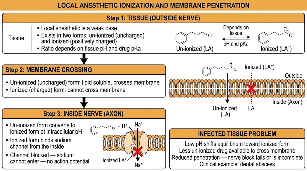
Local anesthetics are weak bases. In solution, they exist in two interconvertible forms: an uncharged (un-ionized) form and a positively charged (ionized) form. The ratio of these two forms depends on the tissue pH and the drug's acid dissociation constant (pKa). This equilibrium is pharmacologically important because the two forms have completely different jobs: one form crosses the nerve membrane, the other blocks the channel.
The un-ionized (uncharged) form of the local anesthetic is lipid-soluble and membrane-permeant. It can diffuse across the phospholipid bilayer of the axon membrane and enter the interior of the nerve cell. This membrane-crossing step is essential — because the binding site on the sodium channel is on the intracellular face of the channel, the drug must first get inside the cell.
Once inside the axon, some of the un-ionized drug converts to the ionized (positively charged) form at the lower intracellular pH. The ionized form is the active form — it is the species that binds to the receptor site on the inner face of the sodium channel and produces blockade. The ionized form cannot easily cross back through the membrane, so it becomes trapped inside the cell where it exerts its effect.
Step 1 — Crossing the membrane: Un-ionized (uncharged) form diffuses through the lipid bilayer into the axon interior.
Step 2 — Binding the channel: Inside the axon, the ionized (charged) form binds to the intracellular receptor site on the voltage-gated sodium channel and blocks sodium conduction.
Both steps are required. Neither form alone is sufficient.
Normal tissue has a pH near 7.4. Local anesthetics with typical pKa values of 7.6 to 8.9 exist in a mixture of ionized and un-ionized forms at this pH, with a significant fraction in the un-ionized (membrane-crossing) form. This provides adequate drug penetration into the nerve for reliable blockade under normal conditions.
Infected or inflamed tissue is acidic — bacterial metabolism, tissue hypoxia, and inflammatory mediators lower local pH, sometimes to values as low as 6 or below in an abscess cavity. At lower pH, the equilibrium of the weak base shifts toward the ionized form. A larger proportion of the drug molecules carry a positive charge and cannot cross the axon membrane. Less un-ionized drug is available to penetrate the nerve, and less drug reaches the intracellular receptor site.
The clinical consequence is well known to dental practitioners: local anesthetics often fail to produce adequate anesthesia in the vicinity of a dental abscess or area of acute cellulitis. The patient may feel the injection but the nerve block never develops properly. No amount of additional injection fixes the problem as long as the tissue pH remains acidic. The pharmacological explanation is this ionization shift — the drug cannot get into the nerve in sufficient concentration.
In infected or inflamed tissue, local pH is low (acidic). The local anesthetic weak base equilibrium shifts toward the ionized form. Less un-ionized drug crosses the nerve membrane. Blockade is incomplete or fails entirely. This is a pharmacokinetic problem with no simple solution at the injection site — dental abscesses often require incision and drainage, systemic antibiotics, or alternative approaches to achieve adequate pain control for procedures.
Rapidly firing neurons accumulate more drug in their channels with each successive action potential
Because local anesthetics bind preferentially to sodium channels in the open and inactivated states — not the resting state — neurons that are actively firing are more susceptible to blockade than neurons at rest. Each action potential opens more channels, and open channels are better drug targets. This is called state-dependent or use-dependent blockade, and it is the biophysical basis for the clinical observation that pain fibers are blocked at lower drug concentrations than motor fibers.
Consider what happens during repetitive nerve firing. Each time an action potential fires, the sodium channels open (high-affinity state), then inactivate (still high-affinity), then return to the resting state (low-affinity). If local anesthetic drug is present in the axon, drug molecules bind during the open and inactivated phases. When the channel recovers to the resting state, the drug dissociates partially — but not completely, especially with slower-dissociating drugs. With each successive action potential, more channels accumulate bound drug. The blockade deepens with increasing use — hence the name "use-dependent."
A nerve firing at high frequency spends proportionally more time in the open and inactivated states than a nerve at rest or firing slowly. High-frequency firing means more drug binding per unit time, which means faster onset of blockade and more complete blockade at any given drug concentration.
Pain fibers — the fibers transmitting nociceptive signals in the setting of tissue damage or inflammation — are among the most rapidly firing neurons in the peripheral nervous system during a pain state. They discharge at high frequency to signal injury. Motor neurons, by contrast, have lower baseline firing frequencies during normal activity. Proprioceptive fibers and touch fibers also fire at lower frequencies than acutely activated pain pathways.
The consequence of this firing rate difference is that at clinical doses, pain fibers accumulate more local anesthetic blockade than motor fibers or large sensory fibers. The patient loses pain sensation while retaining significant motor function and some touch and pressure sensation. This is desirable: a patient undergoing an epidural block for labor can be largely free of pain while still able to bear weight and move the legs. State-dependent blockade is one reason this differential effect is achievable.
Use-dependent blockade means the drug preferentially silences the most active neurons. Pain neurons firing at high frequency during tissue injury are blocked at lower drug concentrations than resting motor neurons. At appropriate epidural anesthetic concentrations, patients achieve effective pain relief while maintaining motor function — a practical benefit of state-dependent sodium channel pharmacology.
Fiber size and myelination determine the order in which nerve functions are lost — and recovered
Not all nerve fibers are equally sensitive to local anesthetic blockade. Two anatomical properties — fiber diameter and myelination — determine how readily a fiber is blocked. Smaller fibers and unmyelinated fibers are blocked at lower drug concentrations than larger myelinated fibers. This size-dependent sensitivity produces a predictable sequence of functional loss called differential block, which has important clinical applications.
In myelinated nerves, the action potential jumps between nodes of Ranvier — the small exposed gaps in the myelin sheath where sodium channels are concentrated. A drug must block a minimum number of consecutive nodes (typically three) to stop propagation completely. In large, thickly myelinated fibers, the nodes are widely spaced and the internodal distances are long. A drug molecule penetrating from the injection site has to diffuse farther between nodes to block enough of them in sequence. This geometry makes large myelinated fibers relatively resistant to blockade.
Small fibers have shorter internodal distances, closer together nodes, and in the case of unmyelinated type C fibers (which carry pain and temperature signals), no myelin at all. Local anesthetic can act across a greater fraction of the total fiber length at lower drug concentrations. The result is that small fibers are blocked first and with lower drug concentrations.
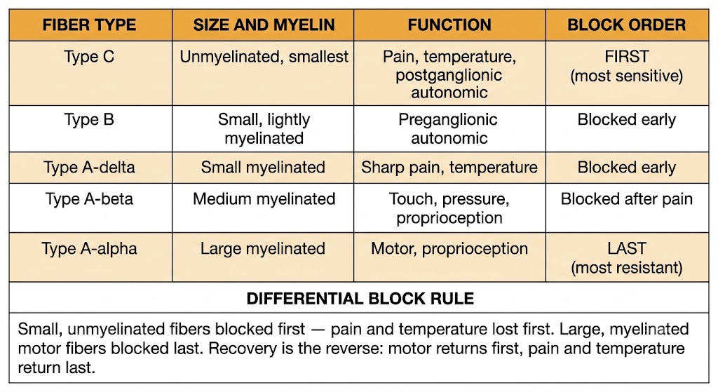
The approximate sequence in which nerve functions are lost as local anesthetic concentration builds at a nerve trunk follows fiber size and myelination. Small unmyelinated and small myelinated fibers are blocked earliest; large myelinated motor and proprioceptive fibers are blocked last.
| Fiber Type | Size / Myelination | Function | Sensitivity to Block |
|---|---|---|---|
| Type C | Unmyelinated, smallest | Pain, temperature, postganglionic autonomic | Blocked first (most sensitive) |
| Type B | Small, lightly myelinated | Preganglionic autonomic | Blocked early |
| Type A-delta | Small myelinated | Sharp pain, temperature, pressure | Blocked early |
| Type A-beta | Medium myelinated | Touch, pressure, proprioception | Blocked after pain fibers |
| Type A-alpha | Large myelinated | Motor, proprioception | Blocked last (most resistant) |
As a regional nerve block develops, the patient notices a predictable sequence of sensory and motor changes. Pain and temperature sensation are lost first — the patient can no longer feel sharp or thermal stimuli. At this stage, touch and pressure may still be detectable. Motor power is the last function to be lost as drug concentration builds.
Recovery follows the reverse sequence. As the drug wears off, motor function returns first (the most resistant fibers recover first), followed by touch and pressure, and finally pain and temperature sensation are restored last. A patient emerging from a spinal anesthetic who can move the legs but still cannot feel pinprick in the feet is experiencing this differential recovery — motor function before pain sensation.
This gradient is clinically useful. At lower drug concentrations — for example, in a dilute epidural infusion for labor pain management — it is possible to achieve near-complete pain blockade while preserving significant motor function. The patient can remain ambulatory or can bear weight while experiencing little or no pain from contractions. Selecting the drug concentration and volume for the target level of differential block is a key skill in regional anesthesia practice.
Order of blockade (first to last): Pain and temperature → autonomic → touch and pressure → motor.
Order of recovery (first to last): Motor → touch and pressure → autonomic → pain and temperature.
Rule: Small fibers (pain, temperature, autonomic) are blocked first and recover last. Large motor fibers are blocked last and recover first.
| Author / Organization | Title | Source |
|---|---|---|
| Butterworth JF 4th, Strichartz GR | Molecular mechanisms of local anesthesia: a review | Anesthesiology. 1990;72(4):711–734 |
| Catterall WA | From ionic currents to molecular mechanisms: the structure and function of voltage-gated sodium channels | Neuron. 2000;26(1):13–25 |
| Hille B | Ion Channels of Excitable Membranes. 3rd ed. | Sunderland, MA: Sinauer Associates; 2001:385–420 |
| Ragsdale DS, McPhee JC, Scheuer T, Catterall WA | Molecular determinants of state-dependent block of Na+ channels by local anesthetics | Science. 1994;265(5179):1724–1728 |
| Strichartz GR | The inhibition of sodium currents in myelinated nerve by quaternary derivatives of lidocaine | J Gen Physiol. 1973;62(1):37–57 |
| Tucker GT | Pharmacokinetics of local anesthetics | Br J Anaesth. 1986;58(7):717–731 |
| Becker DE, Reed KL | Local anesthetics: review of pharmacological considerations | Anesth Prog. 2012;59(2):90–102 |
| Routledge PA | The plasma protein binding of basic drugs | Br J Clin Pharmacol. 1986;22(5):499–506 |
| Fink BR | Mechanisms of differential axial blockade in epidural and subarachnoid anesthesia | Anesthesiology. 1989;70(5):851–858 |
| Wong CA, Scavone BM, Peaceman AM, et al. | The risk of cesarean delivery with neuraxial analgesia given early versus late in labor | N Engl J Med. 2005;352(7):655–665 |
| Rosenberg PH, Veering BT, Urmey WF | Maximum recommended doses of local anesthetics: a multifactorial concept | Reg Anesth Pain Med. 2004;29(6):564–575 |