Introduction to Medical Pharmacology
Volume of distribution, plasma protein binding, tissue barriers, and loading dose
Chapter 2 · Module 2 of 5 · PKIN-02Section 1
The apparent volume concept, determinants of distribution, and the clinical spectrum from plasma-confined to extensively tissue-distributed drugs
Once a drug enters the systemic circulation, it distributes throughout the body into various fluid compartments and tissues. The volume of distribution is the pharmacokinetic parameter that quantifies this distribution behavior and links the total amount of drug in the body to the concentration measurable in plasma.
The volume of distribution (abbreviated Vd) is defined as the apparent volume into which a drug appears to distribute, calculated by dividing the total amount of drug in the body by the plasma concentration. The word "apparent" is essential: Vd is not a real anatomical space. It is a mathematical construct that can substantially exceed the total body volume — and for some drugs, reaches values of hundreds of liters per kilogram — when drug accumulates avidly in tissues, keeping plasma concentrations very low relative to the amount of drug present in the body.
To interpret a Vd value intuitively: plasma volume in a 70-kilogram person is approximately 3 to 4 liters. Extracellular fluid volume is approximately 14 to 15 liters. Total body water is approximately 42 liters. A drug with Vd near 4 liters is confined almost entirely to the intravascular compartment. A drug with Vd near 15 liters distributes into plasma and interstitial fluid. A drug with Vd near 40 liters distributes throughout total body water. Any Vd well above 40 liters indicates that the drug is concentrating in tissues — often because it is highly lipophilic or binds extensively to tissue proteins, phospholipids, or cellular membranes.
Two properties compete to determine where a drug distributes: plasma protein binding and tissue affinity. A drug that binds extensively to plasma proteins (albumin, alpha-1-acid glycoprotein) is retained in the intravascular compartment, resulting in a small Vd. A drug with low plasma protein binding and high affinity for tissue proteins, phospholipids, or adipose tissue distributes rapidly out of plasma into tissue reservoirs, producing a large Vd.
Lipophilicity is the single most powerful predictor of large Vd: highly lipophilic drugs accumulate in adipose tissue and lipid-rich cell membranes throughout the body, keeping plasma concentrations low relative to total body drug burden. This is also why lipophilic drugs take longer to clear from the body — the tissue reservoir slowly releases drug back into plasma for elimination.
Warfarin has a Vd of approximately 0.14 liters per kilogram (roughly 10 liters in a 70-kilogram patient), reflecting its approximately 99 percent plasma protein binding to albumin and low tissue penetration. Gentamicin, a polar aminoglycoside, has a Vd of approximately 0.25 liters per kilogram, confined largely to extracellular fluid because it cannot penetrate cell membranes effectively. Both drugs are dialyzable because their relatively small Vd means most of the body's drug burden is present in plasma and accessible to the dialysis membrane.
Digoxin has a Vd of approximately 7 liters per kilogram, reflecting extensive binding to sodium-potassium-adenosine triphosphatase pumps in cardiac and skeletal muscle. This enormous tissue reservoir means that plasma digoxin concentrations are very low relative to the total amount of drug in the body, making plasma level interpretation timing-sensitive — a digoxin level drawn immediately after an intravenous dose will be falsely elevated because redistribution to tissue is still occurring. Digoxin toxicity cannot be treated by dialysis because only a tiny fraction of the body's digoxin burden is in the plasma at any time.
Amiodarone has a Vd of approximately 60 liters per kilogram due to its extreme lipophilicity and accumulation in adipose tissue, liver, lung, and thyroid. This enormous tissue reservoir explains its exceptionally long half-life — measured in weeks to months — and the persistence of its pharmacological effects and toxicities (thyroid dysfunction, pulmonary toxicity, corneal microdeposits) long after the drug is discontinued.
The volume of distribution and clearance together determine a drug's elimination half-life according to the relationship: half-life equals 0.693 multiplied by Vd, divided by clearance. This relationship has several important clinical implications. A large Vd prolongs the half-life even if clearance is normal, because the extensive tissue reservoir slowly releases drug back into plasma for elimination. This is why amiodarone, despite being hepatically cleared, has a half-life of weeks. Conversely, a small Vd allows even modest clearance to produce a short half-life.
When Vd increases — as in fluid overload, edema, or ascites for water-soluble drugs — the half-life prolongs even with unchanged clearance. This has direct dosing implications: a patient with massive ascites may require a higher loading dose of a water-soluble antibiotic such as gentamicin to achieve the target peak concentration, because the drug distributes into a larger apparent volume.
Volume of Distribution — Clinical Reference Points
Small Vd (plasma-confined, ~3–5 L): heparin, insulin — dialyzable. Moderate Vd (extracellular fluid, ~15 L): warfarin, gentamicin, most penicillins — partially dialyzable. Larger Vd (total body water, ~40 L): ethanol, lithium. Extensive tissue distribution (Vd >100 L): digoxin (~490 L), amiodarone (~4,200 L in 70 kg) — not dialyzable. Half-life = (0.693 × Vd) / clearance — large Vd extends half-life independent of clearance.
Section 2
Albumin and alpha-1-acid glycoprotein, the free drug hypothesis, displacement interactions, and disease states that alter binding
Most drugs circulate in plasma as a mixture of protein-bound and unbound (free) drug. Only the free fraction is pharmacologically active, able to cross membranes, and available for elimination. Understanding plasma protein binding is essential for interpreting drug levels in patients with hypoalbuminemia or acute illness.
Albumin is the most abundant plasma protein, with a normal concentration of 3.5 to 5.0 grams per deciliter. It is the principal binding protein for acidic drugs: warfarin, phenytoin, valproic acid, furosemide, and most nonsteroidal anti-inflammatory drugs bind extensively to albumin. Alpha-1-acid glycoprotein is an acute-phase reactant protein present at lower concentrations than albumin and is the primary binding protein for basic drugs: lidocaine, propranolol, methadone, tricyclic antidepressants, and many opioids bind preferentially to alpha-1-acid glycoprotein.
This protein-class pairing matters clinically because albumin and alpha-1-acid glycoprotein concentrations change in opposite directions during acute illness. Albumin falls in critical illness, surgery, hepatic disease, and nephrotic syndrome. Alpha-1-acid glycoprotein rises as an acute-phase reactant during the same acute illnesses. The net effect on drug binding depends on which protein binds the drug in question.
Only the unbound (free) drug fraction produces pharmacological effects, crosses membranes, and is available for hepatic metabolism and renal filtration. This principle — the free drug hypothesis — has a direct and commonly misunderstood consequence for therapeutic drug monitoring: standard drug assays measure total plasma drug concentration (bound plus free). If protein binding changes, the same total concentration corresponds to a different free concentration and a different pharmacological effect.
Phenytoin is the paradigmatic clinical example. The standard therapeutic range of 10 to 20 micrograms per milliliter applies to total phenytoin in patients with normal albumin (approximately 4.4 grams per deciliter). In a patient with hypoalbuminemia — as occurs in hepatic cirrhosis, nephrotic syndrome, malnutrition, or critical illness — the free fraction of phenytoin increases substantially, and a measured total phenytoin level of 8 micrograms per milliliter may represent fully adequate free drug exposure despite appearing subtherapeutic. Treating to the standard total concentration target in a hypoalbuminemic patient risks phenytoin toxicity. The corrected phenytoin concentration can be estimated using a formula that adjusts the measured total level for the patient's albumin, though the corrected value is an estimate and measurement of free phenytoin concentration is preferable when clinically important decisions are being made.
Valproic acid presents the same challenge, but with an additional complication: valproic acid binding to albumin is saturable at higher drug concentrations, meaning the free fraction increases disproportionately at high total concentrations. This non-linear relationship between total and free valproic acid is another reason total levels can be misleading in clinical practice.
When two drugs compete for the same albumin binding site, the drug with lower affinity may be displaced into the free fraction, transiently raising its unbound drug concentration. Classic pharmacology teaching attributed many clinically significant interactions to displacement; the current understanding is more nuanced.
For most drugs, displacement interactions are self-limiting and clinically minor. The displaced free drug is simultaneously more available for distribution into tissues (increasing Vd) and more available for elimination (increasing clearance). The net result is a transient rise in free drug concentration that returns toward the original steady state as distribution and elimination compensate. Displacement interactions are clinically meaningful only for drugs with all three of these characteristics simultaneously: high protein binding (above approximately 90 percent), narrow therapeutic index, and small Vd (so that the displaced fraction represents a large change in total body free drug). Warfarin meets all three criteria, which is why displacing agents such as amiodarone — combined with concurrent inhibition of warfarin's cytochrome P450 2C9-mediated metabolism — can significantly potentiate anticoagulation.
Several clinical conditions alter plasma protein concentrations enough to affect drug pharmacokinetics in a clinically important way. Hypoalbuminemia (albumin below 3.0 grams per deciliter) increases the free fraction of highly albumin-bound drugs including phenytoin, valproic acid, warfarin, and diazepam. This occurs in hepatic cirrhosis (reduced albumin synthesis), nephrotic syndrome (urinary albumin loss), severe malnutrition, and critical illness.
Alpha-1-acid glycoprotein rises up to threefold during inflammation, surgery, myocardial infarction, and other acute illnesses, increasing the bound fraction of basic drugs such as lidocaine and propranolol. In the acute postoperative state, total lidocaine or propranolol levels may appear elevated while free drug concentrations — and pharmacological effects — are actually unchanged or even reduced. Alpha-1-acid glycoprotein concentrations fall in hepatic insufficiency and are low in neonates, giving newborns higher free fractions of basic drugs at standard weight-based doses.
Protein Binding in Clinical Practice — High-Yield Traps
Phenytoin in hypoalbuminemia: standard total level (10–20 mcg/mL) does not apply — correct for albumin or measure free phenytoin. Valproic acid at high concentrations: free fraction increases non-linearly; total level understates toxicity risk. Warfarin displacement: clinically relevant due to narrow therapeutic index, small Vd, and high protein binding — watch with amiodarone, fluconazole, and other protein binding displacers that also inhibit metabolism. Alpha-1-acid glycoprotein rises in acute illness: total levels of lidocaine and propranolol may rise without change in free drug or clinical effect.
Section 3
The blood-brain barrier, placenta, and selective tissue distribution
Not all body compartments are equally accessible to drugs. Specialized anatomical barriers limit drug distribution to certain tissues, with the blood-brain barrier being the most pharmacologically and clinically consequential.
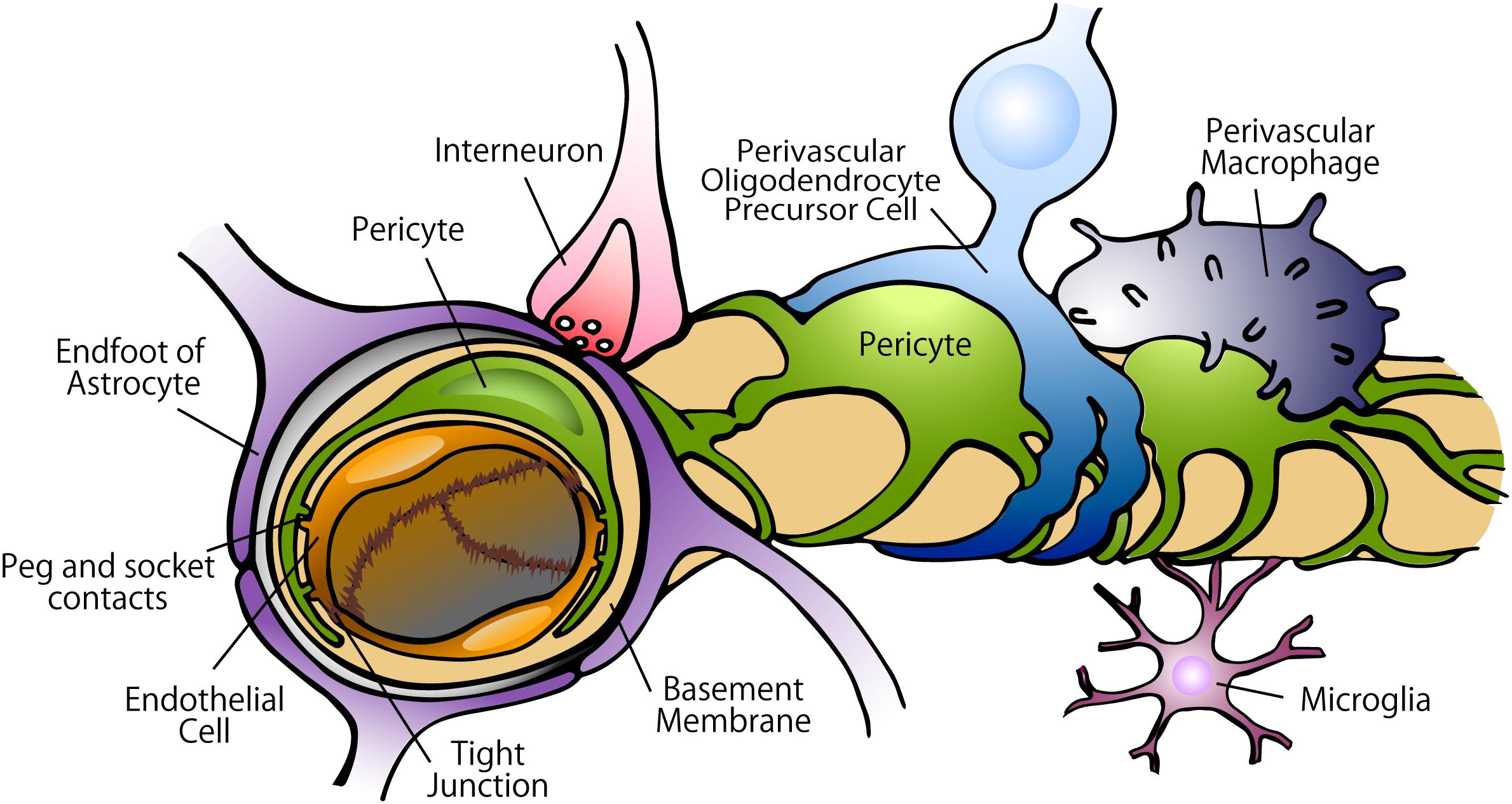
The blood-brain barrier is formed by specialized endothelial cells lining the capillaries of the central nervous system. Unlike peripheral capillaries, which have gaps between cells allowing relatively free passage of solutes, blood-brain barrier endothelial cells are joined by extensive tight junctions that seal the paracellular pathway almost completely. Astrocyte foot processes envelop the outer surface of these capillaries, and pericytes embedded in the basement membrane contribute to barrier maintenance. The result is one of the most selective barriers in the body.
For a drug to cross the blood-brain barrier by passive diffusion, it must be lipophilic, small (generally below approximately 400 to 500 daltons), and largely un-ionized at physiological pH. Drugs that fail these criteria — large hydrophilic molecules, highly ionized drugs, drugs that are substrates for efflux transporters including P-glycoprotein expressed at the blood-brain barrier — are effectively excluded from the central nervous system. This is pharmacologically useful (protecting the brain from most blood-borne toxins and drugs) but also a challenge for central nervous system drug delivery (requiring special strategies to deliver large therapeutic molecules such as antibiotics into the cerebrospinal fluid).
P-glycoprotein at the blood-brain barrier actively pumps drug molecules that have entered endothelial cells back into the blood, providing a second line of defense against central nervous system drug penetration beyond the tight junctions. Many of the same drugs that are P-glycoprotein substrates at the intestinal brush border — including loperamide, certain antiretrovirals, and many chemotherapeutic agents — are excluded from the central nervous system by blood-brain barrier P-glycoprotein. Loperamide is an instructive example: it is a potent opioid receptor agonist that cannot cross the blood-brain barrier at therapeutic doses (due to P-glycoprotein efflux), making it effective for diarrhea without central nervous system opioid effects.
Meningitis and encephalitis inflame the blood-brain barrier, increasing permeability to drugs that would normally be excluded. This is why hydrophilic antibiotics such as penicillin, ampicillin, and vancomycin — which cross the blood-brain barrier poorly under normal circumstances — achieve therapeutically adequate cerebrospinal fluid concentrations in bacterial meningitis. As inflammation resolves with treatment, barrier integrity is restored, which may paradoxically reduce antibiotic penetration during the later phases of treatment when it is still needed.
The placenta was historically described as a barrier protecting the fetus from drugs. This framing is misleading: the placenta is a partial, selective, and often ineffective barrier to drug transfer. Lipophilic, small, un-ionized drugs cross the placenta readily by passive diffusion. The clinical implication is that most small-molecule drugs administered to a pregnant patient reach the fetus to some degree, and fetal drug exposure must be assumed for any drug with lipophilic and small-molecule characteristics.
Ion trapping affects drug distribution to the fetus. Fetal blood is slightly more acidic than maternal blood. Weak bases such as local anesthetics (lidocaine, bupivacaine) are more ionized in the acidic fetal compartment than in maternal plasma, trapping the ionized form on the fetal side. This can lead to fetal drug accumulation — "ion trapping" — if the maternal-fetal pH gradient persists or worsens, as in fetal acidosis.
Blood-Brain Barrier Drug Penetration — Requirements and Exceptions
Crosses blood-brain barrier (lipophilic, small, un-ionized): diazepam, morphine, most general anesthetics, ethanol, most CNS-active drugs by design. Poor blood-brain barrier penetration (hydrophilic or P-glycoprotein substrate): penicillin, vancomycin, gentamicin, loperamide. Blood-brain barrier disruption in meningitis: allows hydrophilic antibiotics to achieve therapeutic cerebrospinal fluid levels during active inflammation. Loperamide: P-glycoprotein efflux at blood-brain barrier prevents central opioid effects at therapeutic doses — accounts for its peripheral-only antidiarrheal action.
Section 4
Loading dose calculation, dialysis, and toxicological implications of drug distribution
The volume of distribution is not merely a pharmacokinetic abstraction. It directly governs three important clinical decisions: whether a loading dose is needed, whether hemodialysis can remove a drug in overdose, and why some drugs are nearly impossible to remove from the body once distributed.
A loading dose is a larger-than-maintenance initial dose intended to rapidly achieve a target plasma concentration rather than waiting for the drug to accumulate to steady state through repeated maintenance dosing. The loading dose is calculated as the target plasma concentration multiplied by the volume of distribution: loading dose equals target concentration multiplied by Vd. A drug with a large Vd requires a large loading dose to fill the tissue reservoir before the desired plasma concentration is achieved.
Digoxin illustrates this principle: its Vd of approximately 7 liters per kilogram means that the tissue reservoir is enormous. To achieve a therapeutic plasma digoxin concentration rapidly (as in atrial fibrillation with rapid ventricular response), multiple intravenous loading doses are required to fill the tissue compartment before plasma concentrations reach the therapeutic range. Vancomycin loading doses in critically ill patients with expanded extracellular fluid volumes — sepsis, fluid resuscitation, edema — must be higher than for standard patients because the effective Vd for this hydrophilic drug is substantially larger. Similarly, weight-based loading doses for water-soluble drugs in obese patients should use lean body weight, not total body weight, because adipose tissue does not appreciably distribute hydrophilic drugs.
Hemodialysis and other extracorporeal removal techniques can only remove drug that is present in plasma at the time of dialysis. A drug with a small Vd, where most of the body's drug burden is in plasma, will be effectively removed by dialysis. A drug with a large Vd, where most of the body's drug burden is sequestered in tissues, will not be effectively removed by dialysis even if plasma concentrations transiently fall during the session — because drug rapidly redistributes from tissue back into plasma as soon as the session ends.
This principle is clinically important in overdose management. Lithium has a Vd of approximately 0.8 liters per kilogram and is efficiently removed by dialysis — a mainstay of management in severe lithium toxicity. Digoxin has a Vd of approximately 7 liters per kilogram and is not meaningfully removed by dialysis; treatment of digoxin toxicity relies on digoxin-specific antibody fragments, not dialysis. Tricyclic antidepressants have Vd values exceeding 20 liters per kilogram and are essentially undialyzable; management of overdose focuses on supportive care rather than drug removal.
Large Vd drugs present unique challenges in toxicology because the drug has distributed into tissues before clinical toxicity becomes apparent, and reversal of toxicity does not correlate with plasma drug level reduction. Amiodarone accumulates over months to years in tissues at concentrations far exceeding plasma, explaining why pulmonary and thyroid toxicity can persist for months after the drug is stopped. When a clinician says a drug is "out of the system" based on plasma levels being undetectable, this reasoning fails for drugs with enormous Vd where tissue concentrations remain high despite undetectable plasma levels.
Loading Dose Calculation
LD = Target Cp × Vd
Dialyzability
Small Vd = dialyzable; Large Vd = not
Section 5
How edema, obesity, liver disease, renal failure, and critical illness change drug distribution and require dose adjustment
Distribution is not a fixed property of a drug in isolation. It changes with the patient's physiological state, and many common clinical conditions substantially alter where drugs distribute and how much is needed to achieve a therapeutic plasma concentration.
Water-soluble (hydrophilic) drugs distribute into extracellular fluid. When extracellular fluid volume expands — in edema from heart failure or hypoalbuminemia, in ascites from hepatic cirrhosis, in third-spacing from surgery or burns — the effective Vd of hydrophilic drugs increases substantially. A standard weight-based dose will yield a lower-than-expected peak plasma concentration because the drug has distributed into a larger fluid volume.
This is clinically relevant for aminoglycoside antibiotics, beta-lactams at high doses, and vancomycin in fluid-overloaded patients. In severe sepsis with aggressive fluid resuscitation, patients may accumulate 10 to 20 liters of extracellular fluid above their baseline, effectively doubling or tripling the distribution volume for hydrophilic antibiotics. Standard weight-based dosing in this setting often produces subtherapeutic peak concentrations. As the patient diureses and fluid is mobilized, the Vd contracts and drug doses may need to be reduced to avoid toxicity.
Lipophilic drugs accumulate in adipose tissue. In obese patients, lipophilic drugs have a substantially larger Vd than in patients of normal weight, requiring higher total doses to achieve equivalent plasma concentrations. Loading doses for lipophilic drugs — including certain benzodiazepines, lipophilic opioids such as fentanyl, and lipophilic antibiotics — should generally be based on total body weight in obese patients, because adipose tissue is a significant part of the distribution volume.
Hydrophilic drugs behave differently in obesity: since adipose tissue does not meaningfully distribute hydrophilic drugs, the Vd of hydrophilic drugs correlates with lean body mass rather than total body weight. Dosing hydrophilic drugs by total body weight in obese patients risks overdosing and toxicity. The practical approach is to use lean body weight (or adjusted body weight for some drugs) for hydrophilic drugs and total body weight (or actual body weight) for lipophilic drugs, though drug-specific guidance should always be consulted.
Hepatic cirrhosis affects distribution through two mechanisms: reduced albumin synthesis (increasing free fraction of albumin-bound drugs) and altered body fluid distribution (ascites increasing Vd of hydrophilic drugs). These effects compound each other: a patient with cirrhosis and ascites may need a different total dose and frequency of dosing compared with a healthy patient, with the direction of adjustment depending on which effect predominates for the specific drug.
Renal failure generates endogenous organic acids that compete with drugs for albumin binding sites, increasing the free fraction of albumin-bound drugs. This is clinically most important for phenytoin, which can have a free fraction of 20 to 30 percent in severe renal failure compared to the normal 10 percent. A patient in renal failure with a measured total phenytoin of 8 micrograms per milliliter may have a free phenytoin concentration that corresponds to a total level of 14 to 16 micrograms per milliliter in a patient with normal renal function — well within or even above the therapeutic range.
Distribution Changes in Pathological States — Summary
Edema / ascites: Vd increases for hydrophilic drugs — higher loading doses needed; reduce dose as fluid mobilizes. Obesity: lipophilic drugs use total body weight for dosing; hydrophilic drugs use lean body weight. Hepatic cirrhosis: reduced albumin synthesis — increased free fraction of acidic drugs; ascites — increased Vd of hydrophilic drugs. Renal failure: endogenous albumin displacers — increased free phenytoin; correct total phenytoin for renal failure or measure free level directly.
Section 6
One-compartment versus two-compartment behavior and why drug level timing matters after intravenous administration
Compartment models are mathematical frameworks used to describe how drug concentrations change over time after administration. For clinicians, the most practical implication is understanding why drug levels drawn immediately after an intravenous dose may be misleading and why a distribution phase must complete before concentrations are interpretable.
In a one-compartment model, the drug is assumed to distribute instantaneously and uniformly throughout the body after administration. Following an intravenous bolus, the plasma concentration declines in a single exponential phase reflecting only elimination. Drugs that behave this way tend to be hydrophilic and distribute primarily within the extracellular fluid, reaching distribution equilibrium rapidly. For these drugs, any blood sample drawn after administration gives a concentration that reflects the drug's true distribution state, and the concentration-time curve forms a single straight line on a semi-logarithmic plot.
Many drugs, particularly lipophilic ones, exhibit two-compartment behavior. After an intravenous bolus, the concentration-time curve shows two distinct phases on a semi-logarithmic plot. The initial rapid decline — the distribution phase, also called the alpha phase — represents the drug moving rapidly from plasma into a peripheral tissue compartment. Plasma concentrations fall steeply during this phase not because the drug is being eliminated, but because it is redistributing into tissues. The subsequent slower decline — the elimination phase, also called the beta phase — represents true elimination from the body and runs at a pace governed by clearance and the equilibrium volume of distribution.
This two-phase behavior has a critically important practical implication: drug levels drawn during the distribution phase are artifactually high and do not reflect the equilibrium concentration at the site of action. Drawing a digoxin level within two hours of an intravenous dose yields a falsely elevated result because digoxin is still distributing into myocardium and other tissue; the plasma level will be far higher than what the tissue is actually experiencing. The recommended approach is to wait at least six to eight hours after an intravenous digoxin dose — until distribution is essentially complete — before drawing a clinically interpretable level.
Lidocaine is another classical example of two-compartment behavior with important clinical consequences. After an intravenous bolus for cardiac arrhythmia, plasma lidocaine concentrations fall rapidly during the distribution phase. If a repeat bolus is given based on the falling plasma concentration without recognizing that this fall reflects redistribution rather than elimination, successive boluses can lead to lidocaine accumulation and toxicity — the "flash and crash" phenomenon. This is why lidocaine is given as a bolus followed by a maintenance infusion, rather than repeated boluses, for arrhythmia management.
The concept also applies to interpreting vancomycin trough levels. Vancomycin exhibits two-compartment kinetics, and trough levels drawn at the appropriate time (30 to 60 minutes before the next dose, after distribution is complete) reflect true equilibrium concentrations and are interpretable. Vancomycin area under the curve-to-minimum inhibitory concentration ratio-guided dosing increasingly replaces simple trough monitoring, but the underlying principle — that drug levels must be drawn at a time that reflects true distribution equilibrium — remains the same regardless of the monitoring strategy.
One-Compartment
Single exponential decline after IV bolus
Two-Compartment
Biexponential decline — distribution then elimination
| Author / Organization | Title | Source |
|---|---|---|
| Rowland M, Tozer TN | Clinical Pharmacokinetics and Pharmacodynamics: Concepts and Applications, 4th ed. | Lippincott Williams & Wilkins, 2011 |
| Shargel L, Wu-Pong S, Yu ABC | Applied Biopharmaceutics and Pharmacokinetics, 7th ed. | McGraw-Hill, 2016 |
| Brunton LL, Hilal-Dandan R, Knollmann BC, eds. | Goodman & Gilman’s: The Pharmacological Basis of Therapeutics, 13th ed. | McGraw-Hill, 2018 |
| Benet LZ, Hoener BA | Changes in plasma protein binding have little clinical relevance | Clin Pharmacol Ther. 2002;71(3):115-121 |
| Winter ME | Basic Clinical Pharmacokinetics, 5th ed. | Lippincott Williams & Wilkins, 2010 |
| Rolan PE | Plasma protein binding displacement interactions — why are they still regarded as clinically important? | Br J Clin Pharmacol. 1994;37(2):125-128 |
| Ogu CC, Maxa JL | Drug interactions due to cytochrome P450 | Proc (Bayl Univ Med Cent). 2000;13(4):421-423 |
| Joukhadar C, Muller M | Microdialysis: current applications in clinical pharmacokinetic studies and its potential role in the future | Clin Pharmacokinet. 2005;44(9):895-913 |