CHAPTER 25  ·  PULMONARY PHARMACOLOGY
Section 1

Acute Respiratory Distress Syndrome: Berlin Definition, Pathophysiology, and Dexamethasone

Severity stratification by PaO2/FiO2 ratio, diffuse alveolar damage as the pathological substrate, lung-protective ventilation, and the DEXA-ARDS corticosteroid evidence

Acute respiratory distress syndrome is a diffuse inflammatory lung injury whose pharmacological management is largely supportive, but two interventions — lung-protective ventilation and adjunctive corticosteroids — have demonstrated mortality benefit in appropriately selected patients and constitute the pharmacological core of management.

Berlin Definition and Severity Stratification

The Berlin definition categorizes acute respiratory distress syndrome by severity based on the ratio of partial pressure of arterial oxygen to fraction of inspired oxygen, measured with at least 5 cmH2O of positive end-expiratory pressure. Mild acute respiratory distress syndrome is a ratio of 201 to 300 mmHg; moderate is 101 to 200 mmHg; and severe is 100 mmHg or below. The bilateral chest imaging infiltrates must not be fully explained by effusions, lobar collapse, or nodules, and respiratory failure must not be primarily attributable to cardiac failure or fluid overload. Onset must occur within one week of a known clinical insult. Severity stratifies the risk of death and the threshold for escalating pharmacological and non-pharmacological interventions including neuromuscular blockade and prone positioning.

Pathophysiology: Diffuse Alveolar Damage

The pathological substrate is diffuse alveolar damage, characterized by flooding of alveoli with protein-rich edema fluid from damaged alveolar-capillary membranes, formation of hyaline membranes from fibrin and plasma proteins, loss of surfactant function producing alveolar collapse and reduced compliance, and recruitment of neutrophils whose proteases, reactive oxygen species, and inflammatory cytokines perpetuate the injury. The resulting lung is heterogeneous: some alveoli are flooded and collapsed, some are recruitable, and some remain aerated. Tidal volumes sized for the full lung weight preferentially overdistend the still-aerated regions — compounding injury through a mechanism called volutrauma, while repetitive opening and closing of recruitable regions produces atelectrauma.

The ARDSNet trial established that tidal volume limitation to 6 milliliters per kilogram of predicted body weight, with plateau pressures kept below 30 cmH2O, reduces mortality by approximately 22% compared with traditional tidal volumes of 12 milliliters per kilogram. This lung-protective ventilation strategy is now the standard of care for acute respiratory distress syndrome. Although it is primarily a ventilator management decision rather than a pharmacological one, second-year students are expected to know the tidal volume target and its rationale as USMLE Step 1 content.

Dexamethasone in ARDS

The DEXA-ARDS trial demonstrated that early dexamethasone (20 mg intravenously daily for 5 days, then 10 mg daily for 5 days) reduced the number of ventilator-free days and 60-day mortality in patients with moderate-to-severe acute respiratory distress syndrome. The mechanistic rationale is that the sustained cytokine storm driving diffuse alveolar damage is glucocorticoid-sensitive in a meaningful proportion of patients, and early anti-inflammatory intervention can attenuate the inflammatory phase before irreversible fibroproliferation occurs. Dexamethasone is the preferred glucocorticoid in this setting due to its favorable potency, duration of action, and absence of mineralocorticoid activity. The timing matters — late corticosteroid administration (after 14 days) in the fibroproliferative phase has not shown consistent benefit and may be harmful.
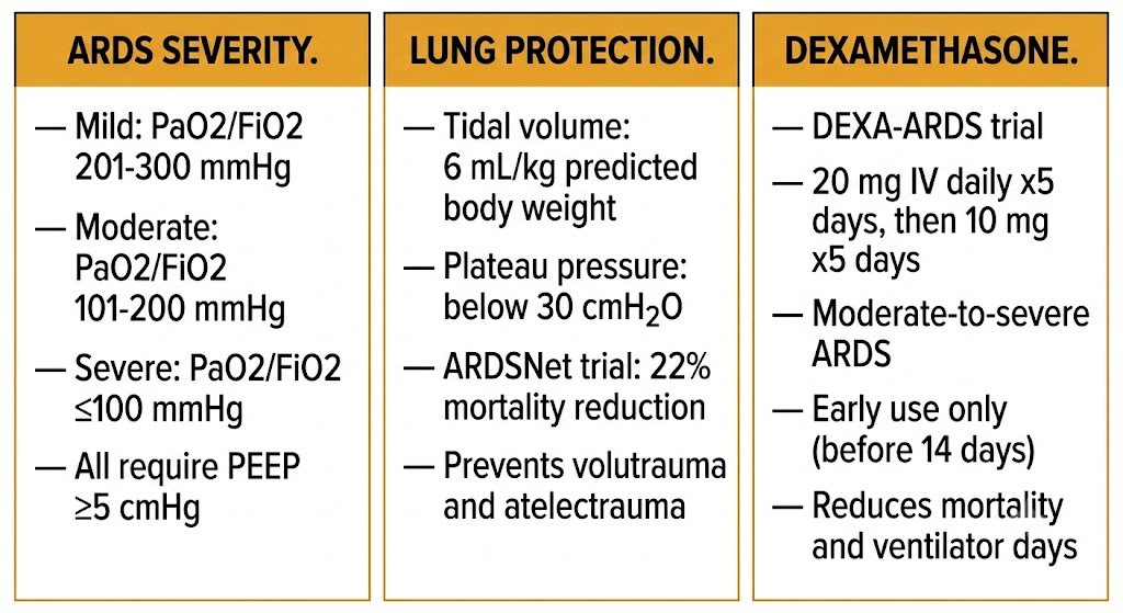
Three-panel ARDS summary: left panel shows Berlin severity thresholds by PaO2/FiO2 ratio (mild 201-300, moderate 101-200, severe 100 or below, all with PEEP at least 5 cmH2O); center panel shows ARDSNet lung-protective ventilation with tidal volume 6 mL/kg predicted body weight and plateau pressure below 30 cmH2O achieving 22 percent mortality reduction; right panel shows dexamethasone DEXA-ARDS protocol with 20 mg IV daily times 5 days then 10 mg times 5 days for moderate-to-severe ARDS before 14 days.
ARDS Berlin severity stratification, ARDSNet lung-protective ventilation, and dexamethasone (DEXA-ARDS trial). Generated with Gemini AI for educational use.
ARDS: High-Yield Pharmacology Points

Berlin severity: mild (PaO2/FiO2 201–300), moderate (101–200), severe (≤100) — all measured with positive end-expiratory pressure ≥5 cmH2O.

Lung-protective ventilation: tidal volume 6 mL/kg predicted body weight; plateau pressure below 30 cmH2O. ARDSNet trial: 22% mortality reduction.

Dexamethasone (DEXA-ARDS): 20 mg intravenously daily ×5 days then 10 mg daily ×5 days. Reduces ventilator days and 60-day mortality in moderate-to-severe ARDS. Early use only — not beneficial after 14 days.

Section 2

ICU Sedation and Analgesia: Propofol, Dexmedetomidine, Midazolam, and Fentanyl

Mechanisms and adverse effect profiles of the major ICU sedative agents, the analgesia-first approach, and propofol infusion syndrome

Mechanically ventilated patients require sedation and analgesia for comfort, safety, and tolerance of the endotracheal tube. Contemporary practice emphasizes an analgesia-first approach — treating pain before adding sedation — because uncontrolled pain is itself a major driver of agitation, and adequate analgesia often reduces the sedative requirement substantially.

Propofol

Propofol is a lipid-soluble phenol that potentiates gamma-aminobutyric acid type A receptor activity, producing rapid sedation with an onset of 30 to 60 seconds. Its very short context-sensitive half-time after infusion cessation — a consequence of rapid redistribution — makes it ideal for daily awakening trials, where the goal is temporary cessation of sedation to assess neurological status and readiness for extubation. Propofol infusion syndrome is the most serious adverse effect: a rare but potentially fatal syndrome occurring with prolonged high-dose infusions (typically above 5 mg/kg/hour for more than 48 hours) characterized by metabolic acidosis, rhabdomyolysis, renal failure, cardiac arrhythmias, and lipemic plasma. The mechanism involves impairment of mitochondrial electron transport. Propofol is formulated in a lipid emulsion, which provides significant caloric load and can contribute to hypertriglyceridemia; triglyceride levels should be monitored in patients receiving prolonged infusions.

Dexmedetomidine

Dexmedetomidine is a selective alpha-2 adrenergic receptor agonist that produces sedation, anxiolysis, and analgesia through central alpha-2 receptor activation in the locus coeruleus — the same pathway involved in natural non-rapid eye movement sleep. Unlike propofol and benzodiazepines, dexmedetomidine produces a state of cooperative sedation: patients are calm and easily arousable but return to sedation when unstimulated, can follow commands, and can participate in ventilator weaning assessments. It does not suppress respiratory drive, making it uniquely useful for non-invasive ventilation support and for facilitating extubation readiness. Bradycardia and hypotension are the primary adverse effects, resulting from reduced sympathetic outflow. The MIDEX and PRODEX trials demonstrated non-inferiority of dexmedetomidine to midazolam and propofol respectively for sedation maintenance, with shorter time to extubation in the dexmedetomidine arms.

Midazolam and Fentanyl

Midazolam is a short-acting benzodiazepine that potentiates gamma-aminobutyric acid type A receptor activity through a distinct mechanism from propofol (benzodiazepine binding site versus the propofol binding site). It produces amnesia and anxiolysis but accumulates with prolonged infusion, causing prolonged sedation and delirium — contributing to the contemporary preference for propofol and dexmedetomidine over benzodiazepines for routine intensive care unit sedation. Fentanyl is the preferred opioid analgesic for mechanically ventilated patients due to its rapid onset, titratable potency, and lack of histamine release. It is administered by continuous infusion and provides analgesia to reduce the pain of the endotracheal tube, suctioning, and position changes. Respiratory depression is the primary concern but is managed in context — the patient is already receiving mechanical ventilatory support.

Propofol Infusion Syndrome

Rare but fatal. Occurs with prolonged high-dose propofol infusion (>5 mg/kg/hour for >48 hours). Features: metabolic acidosis, rhabdomyolysis, acute kidney injury, cardiac arrhythmias, lipemic plasma.

Mechanism: impaired mitochondrial electron transport chain function. Monitor triglycerides, lactate, and creatine kinase in high-risk patients.


Section 3

Neuromuscular Blocking Agents in the ICU

Cisatracurium in ARDS, ICU-acquired weakness as the primary risk of prolonged blockade, rocuronium and sugammadex reversal, and train-of-four monitoring

Neuromuscular blockade in the intensive care unit context differs from the operating room: the goal is not to facilitate surgical access but to eliminate patient-ventilator dyssynchrony, reduce oxygen consumption, and in acute respiratory distress syndrome, to facilitate lung-protective ventilation in patients who cannot otherwise tolerate it. These benefits must be weighed against the risk of ICU-acquired weakness.

Cisatracurium in ARDS

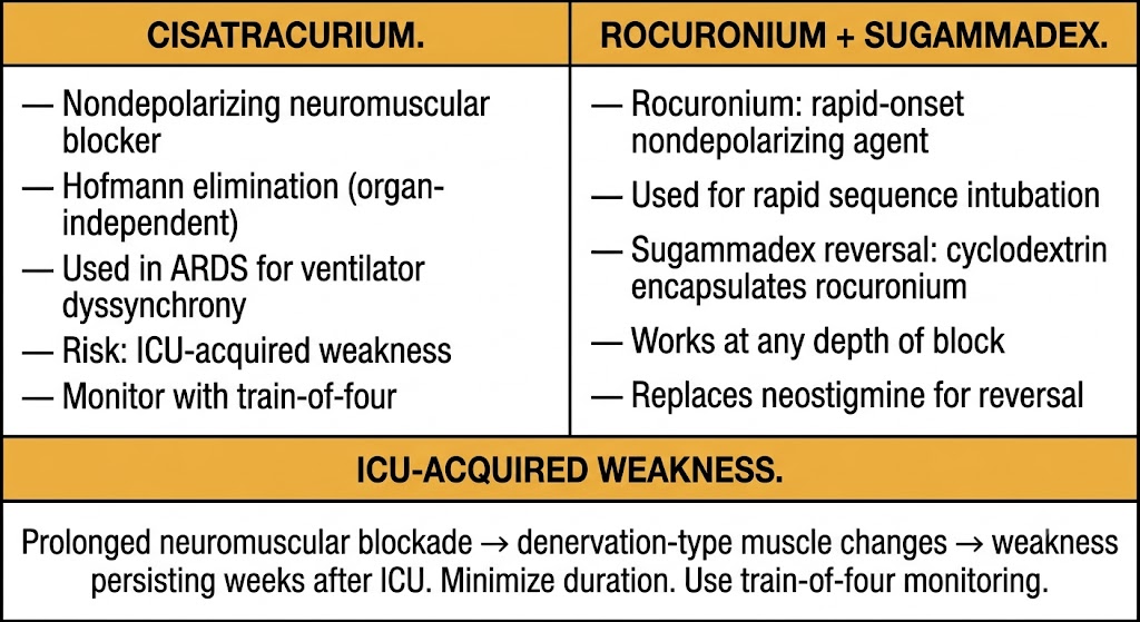
Cisatracurium is the neuromuscular blocking agent of choice in acute respiratory distress syndrome. It is an intermediate-duration nondepolarizing agent that undergoes Hofmann elimination — spontaneous non-enzymatic degradation at physiological temperature and pH, independent of hepatic or renal function. This property makes cisatracurium uniquely suitable for critically ill patients with multi-organ dysfunction, in whom liver and kidney failure would prolong the duration of action of renally or hepatically cleared agents. The ACURASYS trial suggested that 48 hours of early neuromuscular blockade with cisatracurium improved 90-day mortality in moderate-to-severe acute respiratory distress syndrome; however, the subsequent ROSE trial, which used deeper background sedation in the control group, did not replicate this mortality benefit. Current practice reserves neuromuscular blockade for patients with life-threatening hypoxemia or severe ventilator dyssynchrony refractory to optimized sedation.

ICU-Acquired Weakness and Rocuronium/Sugammadex

ICU-acquired weakness is the most clinically significant adverse consequence of prolonged neuromuscular blockade in critically ill patients. It manifests as generalized flaccid weakness that may persist for weeks to months after intensive care unit discharge and is associated with prolonged mechanical ventilation, delayed rehabilitation, and increased long-term mortality. The mechanism involves denervation-like changes in muscle, loss of thick (myosin) filaments, and mitochondrial dysfunction. Minimizing the duration of neuromuscular blockade and daily assessment of the need for continued blockade via train-of-four monitoring (targeting 1 to 2 twitches of 4 to minimize residual block while preventing overdose) are the principal preventive strategies.

Rocuronium is a rapid-onset nondepolarizing agent used in rapid sequence intubation and for shorter-term intensive care unit paralysis. Its primary clinical advantage is its reversibility with sugammadex — a modified gamma-cyclodextrin that encapsulates rocuronium (and vecuronium) molecules in a 1:1 complex, rapidly removing them from the neuromuscular junction and reversing paralysis within minutes regardless of depth of block. Sugammadex has largely replaced neostigmine/glycopyrrolate for reversal of rocuronium and vecuronium because it works at any depth of block, not just in the presence of spontaneous recovery, and has no cholinergic adverse effects.

Two-panel comparison of neuromuscular blocking agents: left panel shows cisatracurium with Hofmann elimination for organ-independent clearance, ARDS use for ventilator dyssynchrony, ICU-acquired weakness risk, and train-of-four monitoring; right panel shows rocuronium for rapid sequence intubation with sugammadex cyclodextrin reversal working at any depth of block replacing neostigmine. Shared box describes ICU-acquired weakness from prolonged neuromuscular blockade causing denervation-type muscle changes.
Cisatracurium (Hofmann elimination, ARDS) versus rocuronium with sugammadex reversal, and ICU-acquired weakness. Generated with Gemini AI for educational use.

Section 4

Inhaled Pulmonary Vasodilators in the ICU: Inhaled Nitric Oxide and Inhaled Epoprostenol

Selective pulmonary vasodilation through ventilation-perfusion matching, indications for rescue therapy in refractory hypoxemia, and the absence of survival benefit despite hemodynamic improvement

Inhaled pulmonary vasodilators produce selective pulmonary vasodilation by delivering drug only to ventilated lung units, avoiding systemic hypotension. They improve oxygenation in acute respiratory distress syndrome and pulmonary hypertension crises but have not been shown to improve mortality in acute respiratory distress syndrome — making them rescue therapies for refractory hypoxemia rather than standard treatment.

Inhaled Nitric Oxide

Nitric oxide is a potent vasodilator gas administered by inhalation at concentrations of 5 to 80 parts per million through the ventilator circuit. When inhaled, it diffuses into adjacent pulmonary arterial smooth muscle cells, activates soluble guanylate cyclase, raises cyclic GMP, and produces vasodilation — but only in ventilated alveolar units where the gas can reach the smooth muscle. Perfusion is selectively redirected from non-ventilated (flooded, collapsed) units toward ventilated units, improving the ventilation-perfusion ratio and arterial oxygenation. Nitric oxide is rapidly inactivated by hemoglobin in the pulmonary capillary blood, preventing systemic escape and limiting vasodilation to the pulmonary circulation. Rebound pulmonary hypertension occurs when inhaled nitric oxide is abruptly discontinued; weaning must be gradual. Methemoglobinemia can occur with high doses or prolonged use — nitric oxide oxidizes hemoglobin iron to the ferric state, producing methemoglobin that cannot carry oxygen. Nitrogen dioxide formation in the ventilator circuit is a toxic byproduct when nitric oxide contacts oxygen.

Inhaled Epoprostenol

Inhaled epoprostenol (prostacyclin) is an alternative to inhaled nitric oxide that produces pulmonary vasodilation through IP receptor activation and cyclic AMP elevation in pulmonary arterial smooth muscle. Unlike systemic epoprostenol, the inhaled route limits drug to ventilated lung regions, producing the same selective ventilation-perfusion matching improvement as inhaled nitric oxide. Inhaled epoprostenol is substantially less expensive than inhaled nitric oxide and does not require specialized delivery equipment, making it a practical alternative in many intensive care unit settings. Both agents produce similar degrees of oxygenation improvement in acute respiratory distress syndrome; neither has demonstrated mortality benefit in large randomized controlled trials. Both are used as bridge therapies in refractory hypoxemia while other interventions such as prone positioning or extracorporeal membrane oxygenation are being considered.


Section 5

Pharmacology of Ventilator Weaning

Corticosteroids to reduce post-extubation stridor and laryngeal edema, methylxanthines in neonatal apnea and premature weaning, and the pharmacological principles supporting liberation from mechanical ventilation

Ventilator weaning — the process of liberating a patient from mechanical ventilatory support — is primarily a clinical and physiological assessment, but several pharmacological interventions support the process in specific contexts.

Corticosteroids and Post-Extubation Stridor

Laryngeal edema from prolonged endotracheal intubation can cause post-extubation stridor and upper airway obstruction requiring reintubation. A cuff leak test — deflating the endotracheal tube cuff to assess whether air bypasses the tube, indicating adequate subglottic space — is used to identify patients at high risk for post-extubation stridor. Prophylactic methylprednisolone or dexamethasone, administered intravenously in the hours before planned extubation in patients who fail the cuff leak test, reduces the incidence of post-extubation stridor and reintubation by reducing laryngeal mucosal inflammation and edema. This is one of the few context-specific pharmacological interventions with clear benefit in the weaning phase.

Methylxanthines in Neonatal Apnea and Weaning

Caffeine, a methylxanthine chemically related to theophylline, is the drug of choice for apnea of prematurity in neonates. It stimulates the respiratory center through adenosine receptor antagonism, increasing the drive to breathe and reducing apneic episodes. The CAP trial demonstrated that caffeine therapy for apnea of prematurity not only reduces apnea but also accelerates successful extubation and reduces the rate of bronchopulmonary dysplasia and patent ductus arteriosus — outcomes with long-term significance. Caffeine has a far more favorable safety profile than theophylline in this setting due to its wider therapeutic window. In adult intensive care unit practice, methylxanthines are occasionally used to stimulate respiratory drive in patients with central hypoventilation or severe chronic obstructive pulmonary disease with hypercapnia who are difficult to wean, though the evidence base is weaker than in neonatal practice.

Ventilator Weaning Pharmacology Summary

Post-extubation stridor prevention: methylprednisolone or dexamethasone intravenously before extubation in patients who fail the cuff leak test. Reduces laryngeal edema and reintubation rate.

Neonatal apnea of prematurity: caffeine (methylxanthine) — adenosine receptor antagonism stimulates respiratory drive. CAP trial: reduces apnea, accelerates extubation, reduces bronchopulmonary dysplasia. Preferred over theophylline due to wider therapeutic window.

Sedation minimization: daily sedation interruption (spontaneous awakening trial) reduces total sedative exposure, ventilator days, and intensive care unit length of stay. The paired awakening and breathing trial protocol combines daily sedation interruption with spontaneous breathing trial on the same day for maximal benefit.


Suggested References
Author / OrganizationTitleSource
ARDS Definition Task Force, Ranieri VM, Rubenfeld GD, et alAcute respiratory distress syndrome: the Berlin DefinitionJAMA. 2012;307(23):2526–2533
Matthay MA, Zemans RL, Zimmerman GA, et alAcute respiratory distress syndromeNat Rev Dis Primers. 2019;5(1):18
Brower RG, Matthay MA, Morris A, et alVentilation with lower tidal volumes as compared with traditional tidal volumes for acute lung injury and the acute respiratory distress syndromeN Engl J Med. 2000;342(18):1301–1308
Villar J, Ferrando C, Martinez D, et alDexamethasone treatment for the acute respiratory distress syndrome: a multicentre, randomised controlled trialLancet Respir Med. 2020;8(3):267–276
Devlin JW, Skrobik Y, Gelinas C, et alClinical practice guidelines for the prevention and management of pain, agitation/sedation, delirium, immobility, and sleep disruption in adult patients in the ICUCrit Care Med. 2018;46(9):e825–e873
Jakob SM, Ruokonen E, Grounds RM, et alDexmedetomidine vs midazolam or propofol for sedation during prolonged mechanical ventilationJAMA. 2012;307(11):1151–1160
Papazian L, Forel JM, Gacouin A, et alNeuromuscular blockers in early acute respiratory distress syndromeN Engl J Med. 2010;363(12):1107–1116
National Heart, Lung, and Blood Institute PETAL Clinical Trials Network, Moss M, Huang DT, et alEarly neuromuscular blockade in the acute respiratory distress syndromeN Engl J Med. 2019;380(21):1997–2008
Adhikari NK, Burns KE, Friedrich JO, et alEffect of nitric oxide on oxygenation and mortality in acute lung injury: systematic review and meta-analysisBMJ. 2007;334(7597):779
Schmidt B, Roberts RS, Davis P, et alCaffeine therapy for apnea of prematurityN Engl J Med. 2006;354(20):2112–2121