Iodine-131 mechanism, rhTSH vs. hormone withdrawal, and low-iodine diet
Iodine-131 is the principal ablative agent for differentiated thyroid cancer remnants and for Graves’ disease. Its selective uptake by the sodium-iodide symporter concentrates the radioisotope in thyroid tissue, where beta particle emission destroys follicular cells over 6–12 weeks.
Iodine-131 is concentrated in thyroid follicular cells and differentiated thyroid cancer metastases by the sodium-iodide symporter. Beta particles deposit their energy within 1–2 mm of the absorbing cell, producing double-strand deoxyribonucleic acid breaks and progressive follicular destruction. After total thyroidectomy, residual normal thyroid tissue competes with metastatic disease for iodine-131 uptake; ablating this remnant eliminates competitive uptake and allows subsequent thyroglobulin measurements and whole-body scans to serve as interpretable markers of persistent or recurrent disease.
Thyroid-stimulating hormone above 30 mIU/L is required before radioactive iodine administration to maximize sodium-iodide symporter expression in remnant and metastatic tissue. This can be achieved by thyroid hormone withdrawal or by recombinant human thyroid-stimulating hormone injection. Thyroid hormone withdrawal requires stopping levothyroxine for 4 weeks (or liothyronine for 2 weeks if substituted) and produces sustained hypothyroidism with thyroid-stimulating hormone typically above 50 mIU/L but causes significant quality-of-life impairment.
Recombinant human thyroid-stimulating hormone (thyrotropin alfa) is administered as 0.9 mg intramuscularly on two consecutive days, with radioactive iodine given on the third day. Thyroid-stimulating hormone peaks above 100 mIU/L at 24 hours and returns to baseline by 72 hours. The recombinant protocol maintains euthyroidism throughout, preserves quality of life, reduces whole-body radiation exposure, and is approved for low-to-intermediate-risk differentiated thyroid cancer. Thyroid hormone withdrawal remains necessary for high-risk patients with known metastases requiring high-dose radioactive iodine with dosimetry.
A low-iodine diet (below 50 micrograms of iodine per day for 1–2 weeks before and 2 days after radioactive iodine) is required regardless of stimulation method, to deplete the competing stable iodine pool and maximize radioiodine uptake.
Recombinant human thyroid-stimulating hormone is equivalent to withdrawal for low-to-intermediate-risk remnant ablation and is preferred to avoid hypothyroidism. Withdrawal remains necessary for high-risk patients with known metastases where accurate dosimetry requires the patient to be in a euthyroid-depleted iodine state, reflecting their true pharmacokinetics. The choice is made in multidisciplinary consultation with nuclear medicine and endocrinology.
Risk-stratified targets, cardiovascular and skeletal consequences, and dynamic surveillance
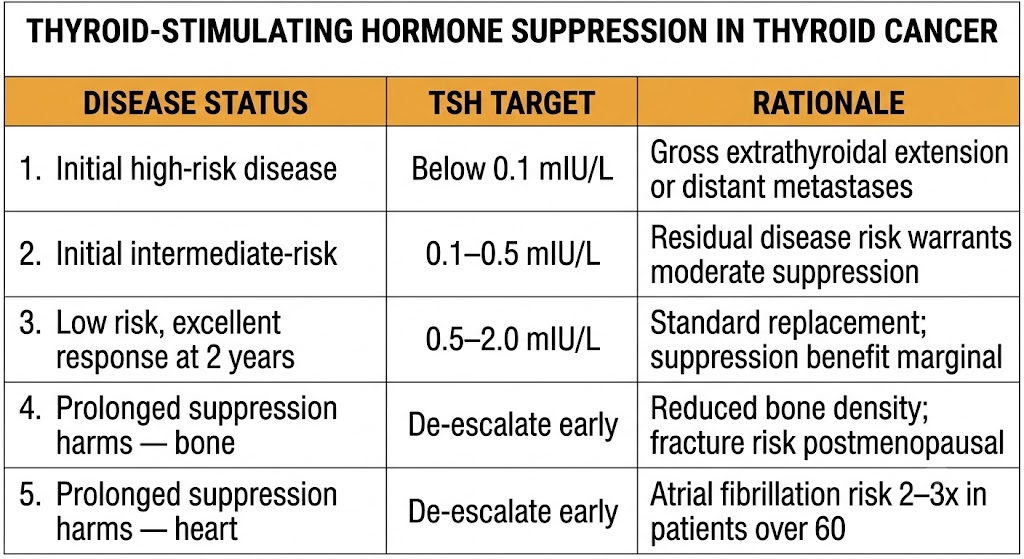
Supraphysiological levothyroxine doses suppress thyroid-stimulating hormone below the reference range in differentiated thyroid cancer, exploiting the trophic dependence of residual cancer cells on thyroid-stimulating hormone for proliferation. Targets are risk-stratified and reassessed dynamically with each follow-up.
High-risk disease (gross extrathyroidal extension, known distant metastases, incomplete resection) targets thyroid-stimulating hormone below 0.1 mIU/L in the initial post-treatment phase. Intermediate-risk disease targets 0.1–0.5 mIU/L. Low-risk disease after successful ablation and two years without evidence of recurrence is de-escalated to 0.5–2.0 mIU/L — the benefit of continued aggressive suppression is marginal at low recurrence risk and does not outweigh cumulative long-term harm from sustained subclinical thyrotoxicosis.
The skeleton bears the primary burden of sustained thyroid hormone excess. Osteoclast activity driven by thyroid hormone reduces bone mineral density, predominantly affecting cortical bone in postmenopausal women. Cumulative loss over years of suppression can reach the threshold for osteoporotic fracture risk. Bone mineral density screening and antiresorptive therapy should be considered in postmenopausal women on sustained suppression below 0.1 mIU/L.
In the cardiovascular system, suppression accelerates heart rate, increases left ventricular mass, and raises atrial fibrillation risk by two-to-threefold in older patients. Patients over 60 on suppressive therapy require scheduled atrial fibrillation monitoring. De-escalation to the lowest tolerable thyroid-stimulating hormone at the earliest clinical opportunity is the primary preventive strategy for both organ systems.
Patients are reclassified based on structural and biochemical response. Excellent response (negative imaging, undetectable thyroglobulin, negative anti-thyroglobulin antibodies) allows upward thyroid-stimulating hormone target revision — the majority of low-to-intermediate-risk patients achieve this within 2 years. Indeterminate, biochemically incomplete, or structurally incomplete response mandates maintained suppression with close surveillance. Serum thyroglobulin stimulated by recombinant human thyroid-stimulating hormone every 1–2 years is the primary biochemical surveillance tool; anti-thyroglobulin antibody positivity interferes with thyroglobulin assay and requires antibody trend monitoring as a surrogate.
Multi-kinase inhibitors, selective RET inhibitors, and BRAF-directed therapy
Radioiodine-refractory differentiated thyroid cancer, progressive medullary thyroid cancer, and anaplastic thyroid cancer are managed with targeted kinase inhibitors selected by tumor histology and molecular profile.
Radioiodine-refractory differentiated thyroid cancer develops when tumor cells dedifferentiate and lose sodium-iodide symporter expression, driven by activating mutations in the mitogen-activated protein kinase pathway — most commonly BRAF V600E and RAS mutations. Two multi-kinase inhibitors are approved: sorafenib (targets vascular endothelial growth factor receptor, platelet-derived growth factor receptor, and RAF kinases; improved median progression-free survival from 5.8 to 10.8 months in the DECISION trial) and lenvatinib (same target classes; achieved median progression-free survival of 18.3 months versus 3.6 months in the SELECT trial). Both are oral agents with significant toxicity including hypertension, hand-foot skin reaction, diarrhea, and hepatotoxicity.
Medullary thyroid cancer arises from calcitonin-secreting parafollicular C-cells and does not concentrate radioiodine. Heritable cases harbor germline RET proto-oncogene mutations; the specific codon mutation guides prophylactic thyroidectomy timing in multiple endocrine neoplasia type 2 families. Vandetanib and cabozantinib are approved multi-kinase inhibitors for progressive medullary thyroid cancer, both targeting RET kinase and vascular endothelial growth factor receptor. Selective RET inhibitors — selpercatinib and pralsetinib — are approved for RET-mutant medullary thyroid cancer and RET-fusion-positive differentiated thyroid cancer, with substantially better tolerability than multi-kinase inhibitors.
For BRAF V600E-mutant anaplastic thyroid cancer, dabrafenib plus trametinib (BRAF inhibitor plus mitogen-activated protein kinase kinase inhibitor) received approval in 2022 after demonstrating approximately 69% response rates in the ROAR basket trial — a meaningful result in a historically untreatable malignancy.
Type 1 vs. type 2 thyrotoxicosis: pathophysiology, differentiation, and treatment
Amiodarone causes thyroid dysfunction through multiple simultaneous mechanisms and produces a predictable pharmacological biochemical pattern that must be distinguished from true disease. When true thyrotoxicosis occurs, two pathophysiologically distinct types require different treatments.
Amiodarone contains 37% iodine by weight and releases approximately 6 mg of free inorganic iodide daily from a standard 200 mg tablet. Its elimination half-life of 40–55 days and extensive tissue accumulation mean thyroid effects persist for months after discontinuation. Amiodarone inhibits type 1 deiodinase, reducing thyroxine-to-triiodothyronine conversion and raising reverse triiodothyronine. In the first 3 months of treatment, this produces an expected pharmacological pattern: elevated free thyroxine, low triiodothyronine, high reverse triiodothyronine, and transiently elevated thyroid-stimulating hormone. This is a drug effect, not thyroid disease, and should not prompt treatment.
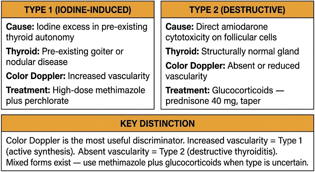
Type 1 amiodarone-induced thyrotoxicosis is iodine-induced hyperthyroidism occurring in patients with pre-existing thyroid autonomy — unrecognized Graves’ disease or toxic multinodular goiter. The iodine excess provides autonomous substrate driving unregulated thyroid hormone synthesis. Color Doppler ultrasound shows increased or normal vascularity. Treatment uses high-dose methimazole (40–60 mg per day), though efficacy is attenuated by high intrathyroidal iodine content. Potassium perchlorate can be added to deplete intrathyroidal iodine stores.
Type 2 amiodarone-induced thyrotoxicosis is a destructive thyroiditis caused by direct cytotoxic effects of amiodarone on thyroid follicular cells, releasing preformed hormone without new synthesis. It can occur in a structurally normal gland. Color Doppler ultrasound shows absent or markedly reduced vascularity. Treatment uses glucocorticoids (prednisone 40 mg per day, tapered over 3 months) to suppress the inflammatory destructive process. Thionamides are not effective in type 2 because there is no new synthesis to block.
Mixed forms with features of both type 1 and type 2 are common and difficult to distinguish. Combined therapy with methimazole plus glucocorticoids is used when the type is uncertain. Amiodarone discontinuation is often not feasible because it is being used for life-threatening arrhythmia — treatment of thyrotoxicosis must proceed without stopping the drug. When discontinuation is possible, the 40–55 day half-life means thyroid effects persist for months regardless.
Gestational thyrotoxicosis, Graves’ disease management, thionamide dosing goals, and postpartum thyroiditis
Pregnancy creates overlapping thyroid pharmacological challenges: normal gestational physiology mimics thyroid disease, drug effects extend to the fetus, and the consequences of both untreated hyperthyroidism and iatrogenic fetal hypothyroidism are severe.
Human chorionic gonadotropin shares structural homology with thyroid-stimulating hormone and weakly activates the thyroid-stimulating hormone receptor. It rises steeply in the first trimester, suppressing thyroid-stimulating hormone in up to 15% of normal pregnancies. Gestational transient thyrotoxicosis caused by this human chorionic gonadotropin-driven stimulation resolves spontaneously by 14–20 weeks as human chorionic gonadotropin declines and requires no antithyroid treatment. Distinguishing it from true Graves’ disease requires positive thyroid-stimulating hormone receptor antibodies, a diffuse goiter, and failure to resolve by 14–16 weeks.
The goal of antithyroid therapy in pregnancy is to maintain maternal free thyroxine in the upper third of the normal reference range using the lowest effective thionamide dose. Both thionamides cross the placenta and can suppress fetal thyroid hormone synthesis — overtreatment causing fetal hypothyroidism is as harmful as undertreatment causing fetal thyrotoxicosis. Thyroid function tests should be checked every 4 weeks with dose adjustment to target. Block-and-replace strategy is contraindicated in pregnancy because it requires higher cumulative thionamide doses that expose the fetus disproportionately.
As covered in Module 3, propylthiouracil is preferred over methimazole in the first trimester to avoid methimazole embryopathy during organogenesis, with transition back to methimazole at 16 weeks to minimize prolonged propylthiouracil hepatotoxicity risk. Thyroid-stimulating hormone receptor antibody measured at 28–32 weeks predicts neonatal Graves’ disease risk; levels above three times the upper reference limit prompt close neonatal monitoring.
Postpartum thyroiditis occurs in approximately 5–9% of women in the year following delivery and follows a characteristic triphasic course: a hyperthyroid phase (weeks 1–4 postpartum) caused by destructive autoimmune thyroiditis releasing preformed hormone, followed by a hypothyroid phase (months 4–8), followed by return to euthyroidism in most women. The hyperthyroid phase is managed symptomatically with beta-blockers; antithyroid drugs are not effective because there is no ongoing new thyroid hormone synthesis. The hypothyroid phase may require temporary levothyroxine. Anti-thyroid peroxidase antibody positivity before delivery is the strongest predictor of postpartum thyroiditis and identifies women at highest risk for permanent hypothyroidism (approximately 25–30% of affected women).
Transplacental thyroid-stimulating hormone receptor antibodies, the 3–7 day delay, and self-limited course
Neonatal Graves’ disease results from transplacental transfer of maternal thyroid-stimulating hormone receptor antibodies that continue to stimulate the neonatal thyroid after birth. Recognition requires understanding the characteristic delayed onset and its pharmacological explanation.
Neonatal Graves’ disease affects approximately 1–5% of neonates born to mothers with Graves’ disease and can occur even when the mother is euthyroid on antithyroid therapy, because thyroid-stimulating hormone receptor antibodies persist long after thyroid destruction. The clinical presentation typically begins 3–7 days after birth. When the mother has been on antithyroid drugs, the drug crosses the placenta and suppresses fetal thyroid function in utero; as the maternal drug clears over the first 3–7 days of life while thyroid-stimulating hormone receptor antibodies remain active, thyrotoxicosis emerges. A normal newborn screen thyroid-stimulating hormone does not exclude neonatal Graves’ disease with delayed onset.
Methimazole is the preferred antithyroid agent in neonates at 0.2–0.5 mg per kilogram per day divided every 8 hours, with dose titration by thyroid function tests every 1–2 weeks. Propylthiouracil is avoided given its hepatotoxicity risk. Propranolol controls tachycardia and adrenergic symptoms while antithyroid therapy establishes biochemical control. The course is self-limited: maternal thyroid-stimulating hormone receptor antibody titers decline over 3–6 months and thyrotoxicosis remits, allowing progressive withdrawal of antithyroid therapy.
Neonates born to mothers with thyroid-stimulating hormone receptor antibody levels above three times the upper reference limit at 28–32 weeks must be observed for at least 5–7 days with thyroid function testing at 48–72 hours and again at 7–10 days of life. Outpatient follow-up at 2 weeks is mandatory for all at-risk neonates. Early discharge before day 5 without a follow-up plan risks missing delayed-onset neonatal Graves’ disease at its most severe.
| Author / Organization | Title | Source |
|---|---|---|
| Haugen BR et al. | 2015 ATA management guidelines for thyroid nodules and differentiated thyroid cancer | Thyroid. 2016;26(1):1–133 |
| Schlumberger M et al. | Strategies of radioiodine ablation in patients with low-risk thyroid cancer (HiLo trial) | N Engl J Med. 2012;366(18):1663–1673 |
| Brose MS et al. | Sorafenib in radioactive iodine-refractory differentiated thyroid cancer (DECISION trial) | Lancet. 2014;384(9940):319–328 |
| Schlumberger M et al. | Lenvatinib versus placebo in radioiodine-refractory thyroid cancer (SELECT trial) | N Engl J Med. 2015;372(7):621–630 |
| Wirth LJ et al. | Efficacy of selpercatinib in RET-altered thyroid cancers | N Engl J Med. 2020;383(9):825–835 |
| Bogazzi F et al. | Approach to the patient with amiodarone-induced thyrotoxicosis | J Clin Endocrinol Metab. 2010;95(6):2529–2535 |
| Alexander EK et al. | 2017 ATA guidelines for thyroid disease during pregnancy and postpartum | Thyroid. 2017;27(3):315–389 |
| van der Kaay DC et al. | Management of neonates born to mothers with Graves’ disease | Pediatrics. 2016;137(4):e20151878 |