The autonomic nervous system (ANS) is the principal efferent pathway through which the central nervous system (CNS) regulates the function of visceral organs, glands, smooth muscle, and cardiac muscle. Unlike the somatic motor system, which operates largely under voluntary control and acts on skeletal muscle through a single motoneuron projecting directly from the spinal cord to the end organ, the ANS interpose a ganglion between the CNS and the target tissue, creating the two-neuron efferent arc that defines all autonomic outflow. This ganglionic synapse is one of the most pharmacologically exploited sites in clinical medicine.
The ANS is conventionally divided into three anatomically and functionally distinct components: the sympathetic (thoracolumbar) division, the parasympathetic (craniosacral) division, and the enteric nervous system (ENS). The sympathetic and parasympathetic divisions are both efferent pathways carrying commands from the CNS to peripheral organs, though they differ in their anatomical organization, neurotransmitter chemistry, and functional consequences at target tissues. The ENS is a semi-autonomous network of neurons embedded within the wall of the gastrointestinal (GI) tract that can regulate intestinal function independently of extrinsic innervation, earning it the designation of the "second brain." Each division will be examined in detail in subsequent sections; this section establishes the shared two-neuron architecture and the central regulatory context.1
The Two-Neuron Efferent Arc. All sympathetic and parasympathetic outflow is conducted via a two-neuron chain. The first neuron, the preganglionic neuron, has its cell body in the CNS (either in the brainstem or the spinal cord, depending on the division) and sends a myelinated axon to a ganglion located outside the CNS. At this ganglion, the preganglionic fiber synapses onto the second neuron, the postganglionic neuron, whose cell body resides within the ganglion and whose unmyelinated axon projects to the target organ. The neurotransmitter at the ganglionic synapse is acetylcholine (ACh) in both sympathetic and parasympathetic divisions, acting on nicotinic acetylcholine receptors (nAChRs) of the ganglionic (NN) subtype. This universal use of cholinergic ganglionic transmission means that ganglionic blocking drugs (which block NN receptors non-selectively) will disrupt both sympathetic and parasympathetic outflow simultaneously, a property that determines both their pharmacological effects and their clinical limitations.12
Hypothalamic Integration. The hypothalamus serves as the master integrating center for autonomic outflow, receiving input from higher cortical centers, the limbic system, the circumventricular organs, and peripheral visceral afferents, and translating this input into coordinated efferent commands to both sympathetic and parasympathetic preganglionic neurons. Cardiovascular regulation, thermoregulation, osmoregulation, feeding behavior, and the stress response are all organized at the hypothalamic level. The posterior hypothalamus drives sympathetic predominance; the anterior hypothalamus supports parasympathetic tone. This organization explains why hypothalamic lesions (stroke, tumor, trauma) produce complex autonomic syndromes that do not fit the pattern of a single peripheral nerve or ganglionic injury. For the pharmacologist, the hypothalamus is relevant because drugs that penetrate the CNS can modify autonomic output at this integrative level, a mechanism distinct from and additive to peripheral receptor effects.1
Visceral Afferents and the Reflex Arc. The ANS includes not only efferent pathways but also visceral afferent fibers that carry sensory information from visceral organs back to the CNS, completing the reflex arc. These visceral afferents travel alongside autonomic efferent fibers in peripheral nerves and enter the spinal cord via dorsal roots (or, for cranial visceral afferents, via cranial nerve nuclei in the brainstem). They are responsible for visceral pain, nausea, the baroreflex, the chemoreceptor reflex, and other homeostatic feedback loops. Several drug classes exert their clinical effects partly through visceral afferent pathways: opioids suppress visceral pain afferents; capsaicin depletes substance P from sensory terminals; the cardiac-sensitizing agent digoxin activates vagal afferents that contribute to its bradycardic effect. Understanding the afferent limb of autonomic reflexes is therefore essential for interpreting the full pharmacodynamic profile of drugs acting on the ANS.13
All sympathetic and parasympathetic efferent output passes through a ganglionic synapse where acetylcholine acts on nicotinic NN receptors. Ganglionic blockers (e.g., trimethaphan, mecamylamine) block both divisions simultaneously, producing orthostatic hypotension, paralytic ileus, urinary retention, and anhidrosis. Their indiscriminate blockade of all autonomic outflow renders them poorly tolerated for chronic use and restricts their application to hypertensive emergencies and controlled hypotension during surgery. The ganglion is also the site of action of nicotine at low doses. Every drug targeting the preganglionic synapse must be understood against this bilateral-blockade background.
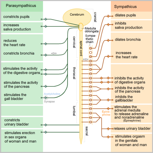
The sympathetic division coordinates the organism's response to stress, exercise, and emergency. Its anatomical architecture reflects this function: preganglionic neurons originate in a restricted spinal cord segment, diverge widely to reach paravertebral and prevertebral ganglia close to the spinal column, and from there project long postganglionic axons to widely distributed target organs. This organization allows a small number of preganglionic neurons to drive a large-scale, coordinated sympathetic response that simultaneously increases cardiac output, redistributes blood flow to skeletal muscle, dilates bronchi, and mobilizes fuel stores.
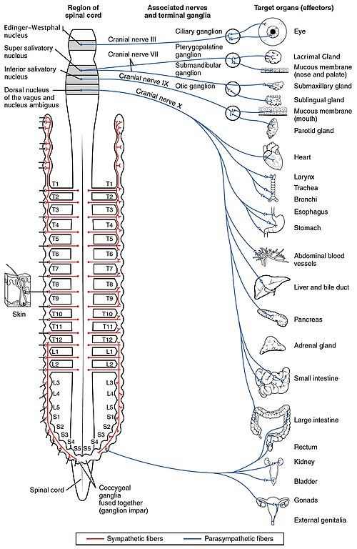
Thoracolumbar Preganglionic Origin. Sympathetic preganglionic neurons have their cell bodies in the intermediolateral cell column (also called the lateral horn) of the spinal cord gray matter, spanning from the first thoracic (T1) to the second or third lumbar (L2-L3) spinal segments. This is why the sympathetic division is termed "thoracolumbar." The myelinated preganglionic axons exit the spinal cord via the ventral roots, enter the spinal nerves briefly, and then leave via white rami communicantes to reach the sympathetic chain ganglia. The preganglionic fibers are relatively short compared to their parasympathetic counterparts, because the sympathetic ganglia are located close to the spinal column rather than at or near the target organ.24
Paravertebral (Sympathetic Chain) Ganglia. The paravertebral ganglia form two paired chains running bilaterally along the lateral surfaces of the vertebral column from the base of the skull to the coccyx. They are connected to each other by interganglionic rami, creating the sympathetic trunk or paravertebral chain. There are typically 22 to 23 ganglia per chain: 3 cervical, 10 to 12 thoracic, 4 lumbar, and 4 to 5 sacral, plus the ganglion impar at the coccyx where the two chains fuse. A preganglionic fiber entering the sympathetic chain via a white ramus communicans can take one of three routes: it may synapse with a postganglionic neuron at the same level, ascend or descend within the chain to synapse at a different level (which is how preganglionic fibers from thoracic levels T1 through T4 [T1-T4] reach the superior, middle, and inferior cervical ganglia to innervate the head, neck, and heart), or pass through the chain without synapsing to reach a prevertebral ganglion via a splanchnic nerve. Postganglionic fibers leaving the chain return to the spinal nerve via gray rami communicantes and travel with the somatic nerves to their peripheral targets.24
Prevertebral (Collateral) Ganglia. The prevertebral ganglia lie anterior to the vertebral column in the abdominal cavity, clustered around the origins of major abdominal arteries. The three principal prevertebral ganglia are the celiac ganglion (receiving input primarily from thoracic levels 5 through 12 [T5-T12] via the greater splanchnic nerve), the superior mesenteric ganglion (thoracic 10 through lumbar 1 [T10-L1] via the lesser splanchnic nerve), and the inferior mesenteric ganglion (lumbar 1 through 3 [L1-L3] via the lumbar splanchnic nerves). Preganglionic fibers reach these ganglia via the splanchnic nerves, which pass through the diaphragm without synapsing in the paravertebral chain. Postganglionic fibers from prevertebral ganglia innervate the abdominal and pelvic viscera, including the stomach, small intestine, large intestine, liver, pancreas, kidneys, adrenal glands, bladder, and reproductive organs. The long preganglionic fibers of the splanchnic nerves are surgically accessible, and splanchnic nerve block is used clinically for intractable visceral pain from pancreatic cancer and other abdominal malignancies.2
The Adrenal Medulla as a Modified Sympathetic Ganglion. The adrenal medulla is embryologically derived from neural crest cells that migrated to the adrenal gland and differentiated into chromaffin cells rather than classic postganglionic neurons. It is functionally homologous to a sympathetic ganglion: preganglionic sympathetic fibers travel via the greater splanchnic nerve to synapse directly on chromaffin cells using acetylcholine (ACh) at nicotinic ganglionic (NN) receptors, bypassing the usual postganglionic neuron entirely. Rather than releasing norepinephrine (NE) at a neuromuscular junction, chromaffin cells secrete a mixture of epinephrine (approximately 80%) and norepinephrine (approximately 20%) directly into the bloodstream, functioning as an endocrine organ. Epinephrine release from the adrenal medulla produces widespread sympathetic effects, with preferential activation of beta-2 adrenergic receptors (beta-2 ARs) in bronchial smooth muscle and peripheral vasculature because circulating epinephrine reaches these receptors in concentrations sufficient to engage beta-2 ARs, which have higher epinephrine affinity than the neurally released norepinephrine that dominates at alpha-1 adrenergic receptor (alpha-1 AR)-rich vascular beds. This distinction between neurally released NE (acting locally at the neuroeffector junction) and circulating epinephrine (acting via the bloodstream) is pharmacologically fundamental and explains why the adrenal medulla response has a different hemodynamic signature from direct sympathetic nerve activation.25
The cervical sympathetic chain (superior, middle, inferior/stellate ganglia) is clinically accessible: stellate ganglion block is used for complex regional pain syndrome, refractory ventricular arrhythmias, and hyperhidrosis. Horner syndrome (ptosis, miosis, anhidrosis, enophthalmos) results from interruption of the oculosympathetic pathway at any level from hypothalamus to superior cervical ganglion to iris dilator; localizing the lesion requires understanding the three-neuron oculosympathetic arc. The long preganglionic fibers of the splanchnic nerves make splanchnic nerve block feasible for visceral pain control. The adrenal medulla, innervated directly by preganglionic fibers, is the target in pheochromocytoma: catecholamine excess from chromaffin cell tumors produces hypertensive crises that must be managed with alpha-blockade before beta-blockade to avoid unopposed alpha-mediated vasoconstriction.
The parasympathetic division governs the organism's restorative, digestive, and energy-conserving functions under conditions of rest. Its anatomical design is the inverse of the sympathetic in a key respect: parasympathetic ganglia are located at or within the target organ itself, resulting in very long preganglionic axons and very short postganglionic axons. This means that parasympathetic postganglionic neurons are physically embedded in the target tissue, making them more accessible to locally applied drugs and less susceptible to ganglionic blocking agents that must reach distant ganglia.
Cranial Parasympathetic Outflow: Brainstem Nuclei. The cranial component of parasympathetic outflow arises from four paired preganglionic nuclei in the brainstem. The Edinger-Westphal nucleus (associated with cranial nerve III [CN III], the oculomotor nerve) sends preganglionic fibers to the ciliary ganglion located in the posterior orbit; postganglionic fibers from the ciliary ganglion innervate the ciliary muscle (controlling lens accommodation) and the iris sphincter (controlling pupillary constriction, or miosis). The superior salivatory nucleus (cranial nerve VII [CN VII], the facial nerve) sends preganglionic fibers via the chorda tympani nerve to the submandibular ganglion, which provides postganglionic innervation to the submandibular and sublingual salivary glands; the greater petrosal nerve branch of CN VII reaches the pterygopalatine ganglion to innervate the lacrimal gland and mucous membranes of the nose and palate. The inferior salivatory nucleus (cranial nerve IX [CN IX], the glossopharyngeal nerve) projects preganglionic fibers to the otic ganglion via the tympanic nerve and lesser petrosal nerve; the otic ganglion provides postganglionic innervation to the parotid gland. These cranial parasympathetic pathways control secretion from all major salivary and lacrimal glands, lens accommodation, and pupillary diameter, all of which are affected by muscarinic receptor antagonists (e.g., atropine produces mydriasis, cycloplegia, and xerostomia).26
The Vagus Nerve: Dominant Parasympathetic Pathway. Cranial nerve X, the vagus nerve, carries approximately 75% of all parasympathetic outflow and is by far the most clinically important parasympathetic pathway. Its preganglionic neurons originate in the dorsal nucleus of the vagus and the nucleus ambiguus in the medulla oblongata. The vagus descends bilaterally through the neck alongside the carotid artery and jugular vein within the carotid sheath, branches extensively in the thorax to innervate the heart (via the cardiac plexus) and lungs (via the pulmonary plexus), and enters the abdomen through the esophageal hiatus to supply the esophagus, stomach, small intestine, and proximal large intestine as far as the splenic flexure of the colon. Preganglionic vagal fibers synapse in ganglia located within or immediately adjacent to these organs; for cardiac innervation, ganglia are found in the epicardial fat pads near the sinoatrial (SA) node and atrioventricular (AV) node. The very short postganglionic fibers mean that systemic parasympathomimetic drugs acting at the neuroeffector junction (e.g., muscarinic agonists) produce their cardiac effects by activating muscarinic M2 receptors (M2 receptors) directly on pacemaker cells and nodal tissue, while the ganglionic synapse nearby is also accessible.26
Sacral Parasympathetic Outflow: Pelvic Splanchnic Nerves. The sacral component of parasympathetic outflow arises from preganglionic neurons in the intermediolateral cell column of spinal cord segments S2-S4 (and sometimes S1). These fibers exit via the ventral roots of the corresponding sacral nerves and form the pelvic splanchnic nerves (also called the nervi erigentes). The pelvic splanchnic nerves join the inferior hypogastric plexus (pelvic plexus) in the pelvis and distribute to ganglia located within or near the descending colon, sigmoid colon, rectum, bladder, and reproductive organs. Sacral parasympathetic outflow controls defecation (by stimulating colonic peristalsis and relaxing the internal anal sphincter), urination (by stimulating detrusor contraction and relaxing the internal urethral sphincter), and erection (by stimulating vasodilation in the corpora cavernosa via nitric oxide release from parasympathetic nerve terminals). This pelvic parasympathetic circuitry is directly relevant to the side effects of anticholinergic drugs, which cause urinary retention and constipation by blocking parasympathetic input to bladder and colon, and to the pharmacology of drugs used for overactive bladder, benign prostatic hyperplasia (BPH), and erectile dysfunction.26
Ganglionic Location and Drug Selectivity. The location of parasympathetic ganglia at or within target organs has a critical implication for drug selectivity that is often underappreciated. When a quaternary ammonium ganglionic blocker (which does not cross lipid membranes easily and does not penetrate the central nervous system [CNS]) is administered systemically, it must diffuse from the bloodstream to the ganglion. For the sympathetic division, the paravertebral and prevertebral ganglia are well perfused and readily accessible. For the parasympathetic division, ganglia are embedded in organ walls, but systemic concentrations of ganglionic blockers still reach them. However, the proximity of parasympathetic postganglionic terminals to the very cells they regulate means that drugs acting downstream of the ganglion at the muscarinic receptor level (e.g., atropine, ipratropium) produce more tissue-selective effects than drugs acting at the ganglionic nicotinic receptor level. This anatomical logic is one reason why muscarinic antagonists have largely replaced ganglionic blockers in clinical practice for conditions where selective parasympathetic blockade is desired.12
CN III / Ciliary ganglion: Muscarinic blockade (atropine, scopolamine, tropicamide) produces mydriasis and cycloplegia; muscarinic agonism (pilocarpine) produces miosis and accommodation used in glaucoma management. CN VII / Pterygopalatine + submandibular ganglia: Muscarinic blockade produces decreased lacrimation and xerostomia. CN X / Cardiac ganglia: Muscarinic M2 blockade (atropine) increases heart rate; M2 activation (neostigmine, pyridostigmine) slows conduction. Sacral / Pelvic ganglia: Muscarinic blockade (oxybutynin, solifenacin) relaxes detrusor for overactive bladder treatment; muscarinic activation (bethanechol) stimulates bladder contraction in urinary retention.
The enteric nervous system (ENS) is a vast, semi-autonomous neural network embedded within the wall of the gastrointestinal (GI) tract from the esophagus to the internal anal sphincter. Containing an estimated 200 to 600 million neurons, the ENS equals or exceeds the total neuron count of the spinal cord. It is capable of coordinating the full range of GI motor, secretory, and absorptive functions in the complete absence of input from the brain or spinal cord, a property demonstrated by the fact that the gut of an animal with severed vagal and splanchnic nerves continues to produce organized peristaltic contractions. The ENS is therefore not simply a relay station for central commands but a true integrative nervous system in its own right.
Myenteric Plexus (Auerbach's Plexus). The myenteric plexus is located between the longitudinal and circular layers of smooth muscle throughout the length of the GI tract. Its primary function is the regulation of GI motility. Excitatory motor neurons in the myenteric plexus release acetylcholine (ACh) and substance P onto circular and longitudinal smooth muscle, driving contraction. Inhibitory motor neurons release nitric oxide (NO) and vasoactive intestinal peptide (VIP), causing smooth muscle relaxation. The coordinated contraction of circular muscle behind the bolus and relaxation ahead of it, producing the peristaltic reflex, is organized entirely within the myenteric plexus without requiring central nervous system input. The descending inhibitory arm of this reflex, mediated by nitrergic and VIP-ergic neurons, is the target of disruption in achalasia, where loss of inhibitory myenteric neurons at the lower esophageal sphincter (LES) impairs relaxation. Botulinum toxin injection into the LES, by blocking ACh release from excitatory motor neurons and thereby shifting the balance toward the remaining inhibitory input, is a treatment for achalasia that exploits this ENS circuitry.78
Submucosal Plexus (Meissner's Plexus). The submucosal plexus lies between the circular muscle layer and the mucosa and is primarily responsible for regulating mucosal secretion and local blood flow. Neurons in the submucosal plexus detect luminal contents via sensory neurons that respond to mechanical deformation, chemical stimuli, and osmolality, and relay this information to secretomotor neurons that activate mucus, enzyme, and electrolyte secretion from mucosal epithelial cells. Cholinergic secretomotor neurons stimulate chloride secretion via muscarinic M3 receptors (M3 receptors) on enterocytes, and the resulting osmotic gradient drives water secretion into the lumen. The pharmacological relevance is direct: loperamide, used for diarrhea, acts on mu-opioid receptors in the submucosal plexus to reduce acetylcholine release from secretomotor neurons, decreasing fluid secretion and increasing smooth muscle tone. Clonidine, an alpha-2 adrenergic receptor (alpha-2 AR) agonist, reduces intestinal secretion via a similar pre-junctional mechanism.78
Extrinsic Autonomic Modulation of the ENS. Although the ENS is capable of autonomous function, it receives modulatory input from both sympathetic and parasympathetic divisions. Parasympathetic (vagal and pelvic) preganglionic fibers synapse on ENS neurons in both plexuses, generally facilitating GI motility and secretion. Sympathetic postganglionic fibers (from prevertebral ganglia) release norepinephrine (NE) onto both enteric neurons and directly onto GI smooth muscle and mucosal cells; NE inhibits ENS interneurons and motility neurons via alpha-2 AR and generally suppresses GI function. The "fight-or-flight" suppression of GI motility is mediated through this sympathetic override of ENS circuitry. Opioid receptors are abundantly expressed throughout both enteric plexuses: mu-opioid receptor activation reduces ACh release from enteric neurons, decreasing peristalsis and secretion, producing the constipation that is the most universal side effect of opioid analgesics. Opioid-induced bowel dysfunction (OBD) is thus an ENS-mediated phenomenon, and peripherally acting mu-opioid receptor antagonists (PAMORAs) such as methylnaltrexone and naloxegol, which do not cross the blood-brain barrier (BBB), are used specifically to reverse OBD without antagonizing central analgesia.789
Serotonin and the ENS. Approximately 95% of the body's total serotonin (5-hydroxytryptamine, 5-HT) is located in the GI tract, stored in enterochromaffin (EC) cells in the intestinal mucosa. Luminal mechanical stimulation triggers 5-HT release from EC cells, which activates 5-HT4 receptors on sensory neurons and 5-HT3 receptors on vagal afferents, initiating peristaltic and secretory reflexes. This central role of 5-HT in ENS function is the basis for several drug classes: metoclopramide and cisapride (now withdrawn due to cardiac arrhythmias) act as 5-HT4 receptor agonists to promote gastric emptying and small intestinal transit; prucalopride is a selective 5-HT4 agonist used for chronic constipation; ondansetron and the setron class block 5-HT3 receptors on vagal afferents to suppress chemotherapy-induced nausea and vomiting. The high concentration of 5-HT in the gut also means that drugs targeting serotonin reuptake transporters (SERTs), such as the selective serotonin reuptake inhibitors (SSRIs), produce GI side effects (nausea, diarrhea) by altering serotonergic signaling in the ENS.78
Opioids (mu-receptor): Reduce ACh release from enteric neurons → decreased peristalsis and secretion → constipation. PAMORAs (methylnaltrexone, naloxegol): Block peripheral mu-receptors selectively → reverse opioid-induced bowel dysfunction without crossing BBB. Loperamide: Mu-opioid agonist, peripherally restricted → antidiarrheal without CNS effect. Metoclopramide / prucalopride: 5-HT4 agonists → promote ENS-driven gastric emptying and colonic propulsion. Ondansetron: 5-HT3 antagonist on vagal afferents → antiemetic. Botulinum toxin: Blocks ACh release from excitatory myenteric neurons → used in achalasia and Hirschsprung's disease.
Most visceral organs receive input from both sympathetic and parasympathetic divisions of the autonomic nervous system (ANS), and the functional state of the organ at any given moment reflects the balance of these opposing influences. This dual innervation is not merely redundant; it allows precise, bidirectional modulation of organ function, with one division capable of counteracting or overriding the other in response to changing physiological demands. Understanding which division dominates under resting conditions, and how drugs shift this balance, is the foundation for predicting the clinical effects of autonomic pharmacological agents.
Resting Autonomic Tone by Organ. Under baseline conditions, different organs are dominated by different divisions of the ANS. The heart is under dominant parasympathetic (vagal) tone at rest: the intrinsic discharge rate of the sinoatrial (SA) node in a denervated heart is approximately 100 to 110 beats per minute (bpm), but resting heart rate in a healthy adult is 60 to 80 bpm, reflecting continuous vagal slowing. Administration of atropine (muscarinic M2 receptor [M2] blockade) at rest therefore increases heart rate toward the intrinsic SA node rate, demonstrating the degree of resting vagal dominance. Vascular smooth muscle tone, in contrast, is under dominant sympathetic influence: the resting arteriolar tone that maintains peripheral vascular resistance is sympathetically driven through tonic alpha-1 adrenergic receptor (alpha-1 AR) activation. Sympathectomy or alpha-1 blockade reduces peripheral resistance and lowers blood pressure. The gastrointestinal (GI) tract is generally under dominant parasympathetic influence promoting motility and secretion, whereas sympathetic input is inhibitory and is activated selectively during stress.1210
Organs With Predominantly Unilateral Innervation. Not all organs receive functionally significant input from both divisions. The sweat glands receive only sympathetic innervation (with the important exception that they use acetylcholine [ACh] as the neurotransmitter rather than norepinephrine (NE), via muscarinic M3 receptors (M3) on sweat gland epithelium, making them a sympathetic cholinergic exception). The adrenal medulla and piloerector muscles of the skin also receive only sympathetic innervation. Most blood vessels receive only sympathetic vasoconstrictor fibers, with vasodilation occurring through removal of sympathetic tone and, in some vascular beds (coronary, skeletal muscle), through active sympathetic beta-2 adrenergic receptor (beta-2 AR)-mediated vasodilation during exercise. Erection is an exception among vascular responses: penile arteriolar vasodilation is primarily a parasympathetic response mediated by nitric oxide (NO) released from pelvic parasympathetic nerve terminals. Understanding which organs lack dual innervation allows correct prediction of drug effects: an alpha-1 antagonist will produce marked vasodilation in peripheral resistance vessels (which have no opposing parasympathetic dilator) and reflex tachycardia (because baroreceptors detect the fall in blood pressure and withdraw vagal tone, increasing heart rate).210
Denervation Supersensitivity. When autonomic nerve terminals degenerate, target organs do not simply lose their responsiveness; they become hypersensitive to circulating catecholamines and to directly acting agonists. This phenomenon, termed denervation supersensitivity, results from two mechanisms: upregulation of receptor number and sensitivity at the postsynaptic membrane in the absence of tonic neurotransmitter stimulation, and loss of reuptake (via the norepinephrine transporter (NET)) and enzymatic degradation (via monoamine oxidase (MAO)) that normally terminate the action of released NE. The result is that a normally subthreshold concentration of NE or epinephrine produces an exaggerated response. Clinically, denervation supersensitivity is relevant in patients with sympathetic autonomic neuropathy (as in long-standing diabetes mellitus or Parkinson disease), where orthostatic hypotension is worsened by impaired vasoconstrictor responses but any remaining catecholamine exposure (including endogenous adrenal medulla secretion) may produce exaggerated pressor responses. It is also the basis for the hypersensitivity of transplanted hearts to direct-acting sympathomimetics.210
Functional Consequences of Autonomic Balance at Key Organs. The cardiovascular system illustrates the pharmacological consequences of dual innervation most clearly. Heart rate is increased by sympathetic activation (beta-1 adrenergic receptor [beta-1 AR]-mediated increase in SA node automaticity and atrioventricular [AV] node conduction velocity) and decreased by parasympathetic activation (muscarinic M2 receptor [M2 AR]-mediated slowing). A drug that blocks beta-1 ARs will lower heart rate and reduce myocardial contractility; a drug that blocks M2 ARs (atropine) will increase heart rate. In the bronchi, parasympathetic activation (muscarinic M3 receptor [M3 AR]) causes bronchoconstriction and mucus secretion, while sympathetic activation (beta-2 AR) causes bronchodilation; ipratropium (M3 AR antagonist) and albuterol (beta-2 agonist) exploit these reciprocal mechanisms for bronchodilation in obstructive airway disease. In the pupil, sympathetic alpha-1 AR activation contracts the radial dilator muscle (mydriasis), while parasympathetic M3 AR activation contracts the circular sphincter muscle (miosis); phenylephrine eye drops produce mydriasis through alpha-1 agonism, and pilocarpine produces miosis through M3 agonism. Every clinically useful autonomic drug exploits the dual innervation architecture to shift organ function in a desired direction.1210
Heart rate: Dominant parasympathetic (vagal) tone. Atropine increases HR; beta-blockers reduce HR less dramatically than expected from their receptor profile alone because vagal withdrawal partly compensates. Peripheral vasculature: Dominant sympathetic tone. Alpha-1 blockade (prazosin, doxazosin) reduces vascular resistance and causes orthostatic hypotension. GI motility: Dominant parasympathetic. Anticholinergics reduce motility; prokinetics enhance it. Bladder detrusor: Dominant parasympathetic. Anticholinergics reduce detrusor tone (urinary retention risk); bethanechol activates M3 to increase detrusor tone. Pupil: Balanced; sympathetic dilates (alpha-1), parasympathetic constricts (M3). Sweat glands: Sympathetic cholinergic only; anticholinergics produce anhidrosis, risking hyperthermia.
The anatomical organization of the autonomic nervous system (ANS) is not merely a structural curiosity; it directly determines where drugs can act, how selectively they can act, and what side-effect profiles they will produce. Moving from a purely mechanistic understanding of receptor pharmacology to a clinically actionable understanding of ANS drugs requires integrating receptor knowledge with ganglionic architecture, neurotransmitter geography, and the anatomical accessibility of different ANS components. This section synthesizes the preceding anatomical material into a framework for predicting drug behavior.
Preganglionic Selectivity: Ganglionic Blockers. Drugs acting at the ganglionic synapse (nicotinic NN receptor blockers) affect both sympathetic and parasympathetic divisions simultaneously, because both use ACh at nicotinic receptors at the ganglionic synapse. The net clinical effect of ganglionic blockade therefore reflects the removal of whichever division is dominant at each organ. In the cardiovascular system, where vagal tone dominates at the sinoatrial (SA) node and sympathetic tone dominates at blood vessels, ganglionic blockade produces tachycardia (vagal withdrawal from the SA node) combined with hypotension (sympathetic withdrawal from resistance vessels), a seemingly paradoxical combination. In the gastrointestinal (GI) tract, where parasympathetic tone dominates, ganglionic blockade produces ileus and constipation. In the bladder, ganglionic blockade produces urinary retention. This organ-by-organ analysis explains the constellation of side effects observed with ganglionic blockers and illustrates why these drugs cannot produce selective sympatholytic or parasympatholytic effects. Modern autonomic pharmacology has therefore moved almost entirely toward postganglionic receptor-selective agents.12
Postganglionic Selectivity: Receptor Subtype Targeting. Postganglionic autonomic pharmacology achieves selectivity primarily through receptor subtype specificity. In the sympathetic division, the major postganglionic neurotransmitter is norepinephrine (NE), which interacts with alpha-1 adrenergic receptors (alpha-1 AR) in vascular smooth muscle, alpha-2 adrenergic receptors (alpha-2 AR) on presynaptic terminals and platelets, beta-1 adrenergic receptors (beta-1 AR) in the heart and kidney, and beta-2 adrenergic receptors (beta-2 AR) in bronchial smooth muscle and peripheral vasculature. A selective beta-1 AR antagonist (e.g., metoprolol, atenolol) can reduce cardiac rate and contractility with minimal bronchospasm in patients with asthma or chronic obstructive pulmonary disease (COPD), because it avoids beta-2 AR blockade in airway smooth muscle. A selective alpha-1 AR antagonist (e.g., tamsulosin) can relax prostatic smooth muscle for benign prostatic hyperplasia (BPH) with less orthostatic hypotension than a non-selective alpha-blocker, because tamsulosin shows preferential affinity for alpha-1A AR in the prostate over alpha-1B AR in peripheral vasculature. This degree of receptor subtype selectivity is the direct pharmacological consequence of anatomical knowledge: knowing that the prostate is predominantly alpha-1A AR-expressing tissue and that vascular smooth muscle is predominantly alpha-1B AR-expressing tissue allows targeted drug design.211
Anatomical Accessibility and Route of Administration. The anatomical location of ganglia and terminal nerve fibers also determines the feasibility of regional administration. The stellate ganglion (the fusion of the inferior cervical and first thoracic sympathetic ganglia), located anterior to the neck of the first rib near the subclavian artery, is accessible under ultrasound guidance; stellate ganglion block with local anesthetics has been used for complex regional pain syndrome of the upper extremity, refractory ventricular arrhythmias, and menopausal hot flashes via temporary interruption of the upper thoracic and cervical sympathetic outflow. The celiac ganglion, accessible via endoscopic ultrasound or percutaneous computed tomography (CT)-guided injection, is the target for celiac plexus neurolysis in pancreatic cancer pain. Topical ocular delivery of muscarinic antagonists (tropicamide, cyclopentolate) or agonists (pilocarpine) exploits the proximity of the ciliary ganglion and its terminal branches to the anterior chamber, allowing local parasympathetic modulation of pupil and lens with minimal systemic absorption. Inhaled muscarinic antagonists (ipratropium, tiotropium) and inhaled beta-2 agonists (albuterol, salmeterol) exploit the high density of muscarinic M3 receptors (M3 ARs) and beta-2 ARs in airway smooth muscle and submucosal glands, delivering therapeutic concentrations locally while limiting systemic effects from ganglionic or distant receptor activation.212
The Pharmacological Significance of the Neuroeffector Junction. In contrast to the ganglionic synapse, where the synaptic cleft is a discrete, compact structure, the sympathetic neuroeffector junction in smooth muscle does not form a morphologically distinct synapse. Instead, sympathetic postganglionic fibers form varicosities along their length from which NE is released into a diffuse junctional cleft of variable width (20 to 100 nm), with the neurotransmitter reaching target adrenergic receptors by diffusion. This architecture has two pharmacological implications. First, drugs that interfere with NE synthesis, storage, or release (e.g., reserpine depletes vesicular NE; guanethidine prevents NE release from vesicles; alpha-methyldopa is a false substrate that produces a weaker false neurotransmitter) can reduce sympathetic neurotransmission at the neuroeffector junction without directly blocking adrenergic receptors. Second, the reuptake transporter norepinephrine transporter (NET) is the primary mechanism for terminating NE action at this junction, and drugs that block NET (tricyclic antidepressants, cocaine, atomoxetine) prolong and enhance sympathetic transmission, explaining their cardiovascular and central nervous system (CNS) effects. This neuroeffector junction architecture, and the drugs that target it, will be developed in detail in Module 2 of this series.25
Ganglionic blockers (NN antagonists): Affect both divisions; net effect determined by dominant tone at each organ. Cardiovascular result: tachycardia + hypotension. Clinical use now essentially limited to hypertensive emergencies (trimethaphan, rarely used) and research. Postganglionic sympatholytics: Act at the neuroeffector junction (reserpine, guanethidine) or at adrenergic receptors; division-selective effects possible depending on receptor specificity. Postganglionic parasympatholytics: Muscarinic receptor antagonists; organ selectivity achieved via route of administration (inhaled, topical, oral) and receptor subtype affinity. Indirect-acting agents (reuptake blockers, MAO inhibitors): Enhance or prolong endogenous neurotransmitter action; effects depend on which division is tonically active at the target organ.
The ANS efferent arc always involves two neurons: preganglionic (CNS to ganglion, ACh at NN receptor) and postganglionic (ganglion to target, ACh at muscarinic receptor for parasympathetic, NE at adrenergic receptor for sympathetic). Sympathetic ganglia: close to spinal cord (short preganglionic, long postganglionic); adrenal medulla is a modified ganglion releasing circulating catecholamines. Parasympathetic ganglia: at or within target organ (long preganglionic, short postganglionic); dominant vagal outflow via CN X. ENS: 200-600 million neurons, autonomous GI regulation, targeted by opioids, 5-HT agents, and cholinergic drugs. Most organs have dual innervation with predictable dominant tone; knowing which division dominates at each organ predicts the clinical consequence of selective stimulation or blockade.
Jänig W, McLachlan EM. Characteristics of function-specific pathways in the sympathetic nervous system. Trends Neurosci. 1992;15(12):475-481.
doi:10.1016/0166-2236(92)90092-MKatzung BG, ed. Basic and Clinical Pharmacology. 15th ed. New York: McGraw-Hill; 2021. Chapters 6-9 (Autonomic Nervous System Pharmacology).
Breit S, Kupferberg A, Rogler G, Hasler G. Vagus nerve as modulator of the brain-gut axis in psychiatric and inflammatory disorders. Front Psychiatry. 2018;9:44.
doi:10.3389/fpsyt.2018.00044Jansen ASP, Van Nguyen X, Karpitskiy V, Mettenleiter TC, Loewy AD. Central command neurons of the sympathetic nervous system: basis of the fight-or-flight response. Science. 1995;270(5236):644-646.
doi:10.1126/science.270.5236.644Goldstein DS, Eisenhofer G, Kopin IJ. Sources and significance of plasma levels of catechols and their metabolites in humans. J Pharmacol Exp Ther. 2003;305(3):800-811.
doi:10.1124/jpet.103.049270Wehrwein EA, Orer HS, Barman SM. Overview of the anatomy, physiology, and pharmacology of the autonomic nervous system. Compr Physiol. 2016;6(3):1239-1278.
doi:10.1002/cphy.c150037Furness JB. The enteric nervous system and neurogastroenterology. Nat Rev Gastroenterol Hepatol. 2012;9(5):286-294.
doi:10.1038/nrgastro.2012.32Bornstein JC, Costa M, Grider JR. Enteric motor and interneuronal circuits controlling motility. Neurogastroenterol Motil. 2004;16(Suppl 1):34-38.
doi:10.1111/j.1743-3150.2004.00472.xBecker G, Blum HE. Novel opioid antagonists for opioid-induced bowel dysfunction and postoperative ileus. Lancet. 2009;373(9670):1198-1206.
doi:10.1016/S0140-6736(09)60139-2Saper CB. The central autonomic nervous system: conscious visceral perception and autonomic pattern generation. Annu Rev Neurosci. 2002;25:433-469.
doi:10.1146/annurev.neuro.25.032502.111311Michel MC, Vrydag W. Alpha1-, alpha2- and beta-adrenoceptors in the urinary bladder, urethra and prostate. Br J Pharmacol. 2006;147(Suppl 2):S88-S119.
doi:10.1038/sj.bjp.0706619Cazzola M, Page CP, Calzetta L, Matera MG. Pharmacology and therapeutics of bronchodilators. Pharmacol Rev. 2012;64(3):450-504.
doi:10.1124/pr.111.004580