Anesthesia Pharmacology Chapter 10: Pharmacology of Antianginal Drugs
|
|
Chemical messenger- homeostatic activities
Cardiovascular tone
Platelet regulation
Immune regulation
CNS signaling
Gastrointestinal smooth muscle relaxation
Possible effector for volatile anesthetics
Precursor: amino acid L-arginine; synthetic enzyme: NO synthases
Target: (NO, a gas,diffuses from producing cells) , guanylate cyclase activation leading to increase cGMP concentration leading to vasodilatation
Half-life: < 5 seconds
NO binds to iron of heme proteins (inactivated by hemoglobin)
Metabolic transformation:
Interaction with hemoglobin yields nitrate
Interaction with oxygen yields nitrogen dioxide (NO2) -- pulmonary toxicant ("silo filler's disease")
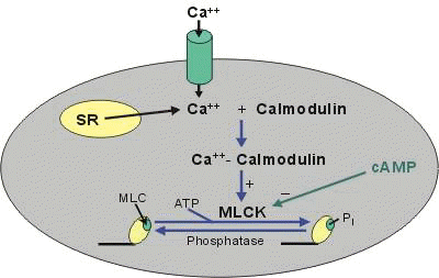
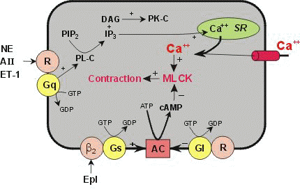
Mechanisms: Regulation of Myosin-light chain kinase activity and control of vascular smooth muscle tone: Modulation by nitric oxide and sympathomimetic amines


Increases in NO activate guanylyl cyclase causing increased formation of cGMP and vasodilation. The precise mechanisms by which cGMP relaxes vascular smooth muscle is unclear; however, cGMP can activate a cGMP-dependent protein kinase, activate K+ channels, decrease IP3, and inhibit calcium entry into the vascular smooth muscle -© 1999 Richard E. Klabunde, used with permission
Increases in NO activate guanylyl cyclase causing increased formation of cGMP and vasodilation.
The precise mechanisms by which cGMP relaxes vascular smooth muscle is unclear; however, cGMP can activate a cGMP-dependent protein kinase, activate K+ channels, decrease IP3, and inhibit calcium entry into the vascular smooth muscle-http://www.oucom.ohiou.edu/CVPhysiology/index.htm;above diagrams and descriptions: © 1999 Richard E. Klabunde, used with permission
Regulation of systemic vascular resistance/pulmonary vascular resistance-- secondary to:
Flow-induced shear stress: continual NO release
Pulsatile arterial flow: continual NO release
Endothelial NO production: determines cardiac output distribution, particularly
Pulmonary distribution
Cerebral distribution
Endothelial NO release: autoregulation {decreased oxygenation results in increased NO production}
NO opposes pulmonary hypertensive response to arterial hypoxemia
NO causes:
Negative inotropism
Negative chronotropism
Arteries generate more NO than veins (possible explanation why internal mammary artery bypass grafts associated with increased patency {compared to venous grafts})
NO: may contribute to bronchodilation
May be important in mediating ventilation- to-perfusion matching
Inhibit platelet aggregation and adhesion
Mechanism:
Activation of guanylate cyclase
Reduced intracellular calcium
NO effects -- synergistic with prostacyclin
Transmitter:
Brain
Spinal cord
Periphery
Peripheral Nervous System -- nonadrenergic & noncholinergic systems release NO as a neurotransmitter--possible innervation sites:
Peripheral nerves generating the myenteric plexus & GI tract smooth muscle relaxation
Innervation of the corpora cavernosa (responsible for penile erection)
NO:
Macrophage activation by cytokines {secondary to NO synthase induction}
Resultant high NO concentrations -- damage fungi, bacteria, protozoa
Inflammation modulation
Abnormal NO: Possible Pathophysiological consequences
Essential hypertension: reduced NO release
Septic shock hypotension: excess NO release
Defective NO production: possible prior role in atherosclerosis by inducing:
Platelet aggregation
Platelet-induced vasoconstriction
Leukocyte adhesion
Vasospastic reactions after subarachnoid hemorrhage (possibly secondary to reduced NO which inactivated by exposure to hemoglobin)
Possibly defective NO production:
Cause/contribute to pulmonary hypertension
Gastrointestinal:
Reduced NO activity in:
Pyloric stenosis and achalasia
NO- modulation of morphine-induced constipation
CNS: epilepsy pathogenesis
Suppression of NO synthesis by anesthetics could:
Decrease excitatory neurotransmission:
Reduce glutamate and cholinergic excitatory systems
Increase inhibitory neurotransmission
Increased GABA function
I-NO vent delivery system:
Adds NO to ventilator breathing system (inspired NO concentration = constant)
Therapy of pulmonary disease (NO rapidly inactivated by hemoglobin)
NO diffusion from alveoli to pulmonary vascular smooth muscle: rapid
Selective pulmonary vasodilator -- maybe useful in pulmonary hypertension {may follow cardiopulmonary bypass; endothelial dysfunction induced by bypass}
NO: treatment --persistent pulmonary hypertension in the newborn; reduces need for extracorporeal membrane oxygen therapy {mortality has not been demonstrated to decrease using this intervention}
Adult Respiratory Distress Syndrome
Findings: pulmonary hypertension & arterial hypoxemia
Management: IV pulmonary vasodilators including:
Nitroprusside, nitroglycerin, prostaglandin E1, prostacyclin, nefedipine result modest decrease in pulmonary artery pressure {large decreases in systemic BP/arterial oxygenation}
Inhaled NO: decrease pulmonary resistance & enhanced arterial oxygenation
Oxygenation improvement dependent on initial pulmonary vascular resistance prior to treatment
Improvement in arterial oxygenation: rationale --
Inhaled NO distributed based on ventilation resulting in associated vasodilation which improves blood flow to well- ventilated regions [improves ventilation/perfusion matching]
Associated with NO-mediated decreased pulmonary hypertension and improved arterial oxygenation which results in : increased time for pulmonary healing (improved survival: not yet proven)
Potential for NO-mediated pulmonary toxicity
Stoelting, R.K., "Peripheral Vasodilators", in Pharmacology and Physiology in Anesthetic Practice, Lippincott-Raven Publishers, 1999, pp. 313-315.
Definition: Angina are those symptoms of myocardial ischemia that occur when myocardial oxygen availability is insufficient to meet myocardial oxygen demand. Symptoms include:
Chest discomfort often described as heaviness, pressure, and squeezing. The sensation is localized typically in the sternal region.
Symptoms often last one to five minutes. Angina can radiate to the left shoulder, to both arms (ulnar surfaces of the forearm and hand), and can radiate to the neck, jaw, teeth, epigastrium and back.
In classical angina these symptoms often occur with exertion and are not due to sudden-onset vasospasm.
By contrast, in variant or Prinzmetal's angina which is caused by coronary vasospasm, reduction of coronary blood flow with angina may occur at rest.
Myocardial ischemia is usually caused by coronary vessel atherosclerosis. As the vessel lumen narrows blood flow is reduced.
Other causes that limit coronary blood flow include:
Arterial thrombi
Spasm
The extent of coronary vessel obstruction can vary as a function of vascular tone: ranging from spasm resulting in 90% obstruction to intermediate effects.
Coronary vasospasm that results in significant obstruction is the basis for Prinzmetal's variant angina.
If the plaque size results in less than about 50% reduction, sufficent coronary blood flow during exertion is still available and anginal symptoms are not present.
[Timmis, A.D. In Pocket Picture Guides: Cardiology, Gower Medical Publishing, London, 1985, p.21]
|
|
[Raehl, C.L., and Nolan, P.E. Ischemic Heart Disease: Anginal Syndromes in Applied Therapeutics: The Clinical Use of Drugs (Young, L.Y., and Koda-Kimble, M.A., eds), Applied Therapeutics, Inc., Vancouver, 1995, p 13-3.]
Coronary blood flow regulation:
Coronary blood flow is controlled by the myocardial oxygen demand and modulated by varying coronary vascular resistance considerably.
The myocardium extracts a fixed and high percentage of oxygen.
In the absence of atherosclerotic disease, intramyocardial resistance arterioles can significantly dilate.
By regulation of smooth muscle tone, intramyocardial arterioles (resistance vessels) a balance is maintained between coronary blood flow and myocardial oxygen requirement.
In healthy individuals, the large epicardial vessels are conductance vessels.
[Selwyn, A.P. and Braunwald, E Ischemic Heart Disease in Harrison's Principles of Internal Medicine (Isselbacher et al., eds) McGraw-Hill, Inc., New York, 1994, p 1077]
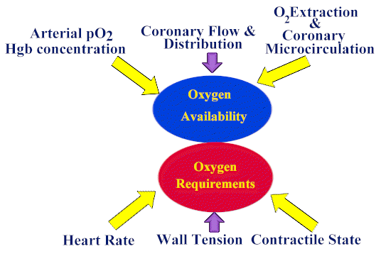
Factors Influencing Myocardial Oxygen Supply and Demand

This figure illustrates the factors that affect myocardial oxygen demand and supply.
Drugs that may prevent or terminate angina may:
Either increase oxygen supply (coronary vasodilators) and/or
Reduce oxygen demand (negative chrontropic drugs, vasodilators, and negative inotropic agents.)
[Raehl, C.L., and Nolan, P.E. Ischemic Heart Disease: Anginal Syndromes in Applied Therapeutics: The Clinical Use of Drugs (Young, L.Y., and Koda-Kimble, M.A., eds), Applied Therapeutics, Inc., Vancouver, 1995, p 13-3.]
Control of coronary blood flow involves:
Local metabolism
Nervous system regulation.
More important of these two mechanism is local myocardial metabolism, i.e. local arterial vasodilation is regulated by myocardial requirements.
Normally, increases in coronary blood flow, even in a denervated heart, occurs in response to increased myocardial contractility and rate.
Production of local vasodilator products may be responsible.
Candidates for these vasodilatory substances include:
Adenosine
Prostaglandins
Potassium ions
Bradykinin
Hydrogen ions
Carbon dioxide
Autonomic nervous system activity can affect coronary vasculature tone.
On the parasympathetic (vagal) side: there are so few fibers terminating on the coronary vasculature that the vagal dilating effects are minimal.
Coronary vascular bed has both alpha and beta-adrenoceptors.
Alpha receptor activation produces constriction, mainly in epicardial capacitance vessels when these receptors are mostly found.
Coronary vessel vasodilation is mediated by beta-adrenoceptors which are mainly localized in intramuscular arteries.
Sympathetic activation probably produces more constriction than dilation and in individuals with accentuated responses to alpha-receptor activation may be susceptible to vasospastic myocardial ischemia
Guyton, A.C. and Hall, J.E. in Textbook of Medical Physiology, W.B. Saunders & Co., Philadephia, 1984, p 258-259.
Angina: Treatment Objectives and Approaches: Treatment objectives include both acute management and prevention of anginal episodes:
Acute Management
These agents cause systemic venodilation and therefore reduce myocardial wall tension and oxygen requirements
Nitrates dilate epicardial coronary capacitance vessels thereby increasing blood flow in collateral vessels.
Although other nitrates may be used in management of angina, other preparations are less effective in acute management.
Chronic Management
Medical management is individualized for each patient, including patient education and counciling to assist in reduction of risks of coronary heart disease.
Risk management includes:
Optimal treatment or elimination of co-existing illness such as diabetes mellitus and hypertension.
Dietary and drug therapy of dyslipidemias
Smoking cessation
Adapation of activities, such as increasing the amount of time needed to accomplish a task which if performed more rapidly could induce angina.
Selwyn, A.P. and Braunwald, E Ischemic Heart Disease in Harrison's Principles of Internal Medicine (Isselbacher et al., eds) McGraw-Hill, Inc., New York, 1994, p 1081-2.
Pharmacological Interventions: Nitrates: The nitrates, by reducing myocardial wall tension and oxygen requirements, are very effective antianginal agents.
Sublingual nitroglycerin
Transdermal nitroglycerin
Isosorbide dinitrate, isosorbide-5-mononitrate
Other agents include amyl nitrite, erythrityl tetranitrate and pentaerythritol tetranitrate.
Beta-adrenoceptor Antagonists: Beta-receptor blockers reduce myocardial oxygen demand by reducing the increases in heart rate and contractility due to adrenergic activity. Therefore, during exercise normal positive chronotropic and inotropic responses are blunted. Propranolol, atenolol, and nadolol are examples of beta-receptor blockers used in management of chronic, classical angina
Calcium channel antagonists:
Nifedipine, verapamil, diltiazem are examples of calcium channel blockers that may be effective in chronic treatment of angina.
They are coronary vasodilators that reduce arterial pressure and myocardial contractility. Accordingly, they reduce myocardial oxygen requirements.
Some agents, such as verapamil and diltiazem are more likely to affect cardiac conduction and contractility, while others have more prominent effects on vascular smooth muscle. Variant angina (Prinzmetal's angina) is often effectively managed by calcium channel blockers.
Verapamil
Blocks cardiac calcium channels in slow response tissues, such as the sinus and AV nodes.
Useful in treating AV reentrant tachyarrhythmias and in management of high ventricular rates secondary to atrial flutter or fibrillation.
Useful also in treating angina since it reduces afterload and myocardial contractility.
Major adverse effects include: heart block or sinus bradycardia can also occur.
Diltiazem
Calcium channel blockers are effective in treating angina because because they:
Reduce peripheral resistance
Reduce dialate coronary vasculature
Decrease myocardial contractilty.
Arteriolar vascular tone depends on free intracellular Ca2+ concentration.
Calcium channel blockers reduce transmembrane movement of Ca2+
Reduce the amount reaching intracellular sites
and therefore reduce vascular smooth muscle tone.
Diltiazem has a direct negative chronotropic effect on the heart sufficient to block reflex-mediated tachycardia secondary to the decrease in peripheral resistance.
Adverse Effects
Diltiazem reduces myocardial contractility which may be undesirable in managing angina in patients with congestive heart failure.
SA nodal inhibition may lead to bradycardia or SA nodal arrest. This effect is more prominent if beta adrenergic antagonists are concurrently administered.
GI reflux.
Negative inotropic are augmented if beta-adrenergic receptor antagonists are concurrently administered.
Calcium channel blockers should not be administered if the patient has SA or AV nodal abnormalities or in patients with significant congestive heart failure.
Other Interventions: Non-pharmacological approaches involve mechanical revascularization, typically either percutaneous transluminal coronary angioplasty (angioplasty) or cornary artery bypass grafting.
[Selwyn, A.P. and Braunwald, E Ischemic Heart Disease in Harrison's Principles of Internal Medicine (Isselbacher et al., eds) McGraw-Hill, Inc., New York, 1994, p 1082]
Overview: Angina pectoris caused by temporary myocardial ischemia is responsive to treatment by organic nitrates.
Organic nitrates act primarily by vasodilation (especially venodilation) which reduces myocardial preload and therefore myocardial oxygen demand.
Nitrates also promote redistribution of blood flow to relative ischemic areas.
The organic nitrates and nitrites are denitrated to produce nitric oxide (NO) which activates guanylyl cyclase.
Activation of cyclase results in increased concentrations of cyclic guanosine 3',5'-monophosphate (cyclic GMP) which results in vasodilation.
NO activates guanylate cyclase, by binding to iron at the enzyme's heme moiety, causing cyclic GMP (cGMP) synthesis
cGMP mediated vasodilation occurs by
(1) decreasing calcium levels in the cell, thus decreasing calcium-calmodulin complex activation of myosin light chain kinase.
(2) Inhibition of myosin light chain kinase results in net dephosphorylation of myosin light chain and thus causes relaxation.
(3) cGMP also causes dephosphorylation of myosin light chain by activating other enzyme systems. [Ahlner et al., Pharmacol. Rev., 43 (3): 351-423, 1991] and D.K. Blumenthal, University of UtahNitric oxide synthetase produces endogenous nitrates by action on L-arginine
Nitric oxide synthetase produces endogenous nitrates by action on L-arginine.
Mechanism of Action of Nitrates in Treatment of Angina
Beneficial effects of low dose nitrates occurs primarily as a result of peripheral vasodilation.
Venodilation decreases end-diastolic left and right ventricular chamber size and pressures.
Arteriolar dilation, evidenced by flushing and dilation of menningeal arterial vessels, is responsible for headache associated with nitroglycern use.
At higher doses, additional venous pooling and arteriolar dilatation decrease blood pressure and cardiac output with attendant sympathetic nervous system activation.
Reflex-mediated tachycardia can be produced in this case. If cardiac output and blood pressure are sufficiently reduced, coronary blood flow will be compromised.
Tachycardia combined with a reduced coronary flow can in this case aggravate ischemia and induce an anginal attack.
Nitrate Effects on Regional and Total Coronary Blood Flow
In the presence of atherosclerotic disease with significant coronary occlusion proximal to small resistance arterioles, arterioles are maximally dilated by autoregulatory mechanisms.
Nitrates may still cause vasodilation of larger capacitance vessels but may exert antianginal activity by promoting coronary blood flow redistribution.
With coronary flow partially obstructed, there is a relative reduction in blood flow to the subendocardium.
Nitrates tend to redistribute coronary flow to the subendocardial region. Even if there is no significant increase in total coronary blood flow with nitrate administration, more blood may thus be directed to relatively ischemic areas.
Spasm of large epicardial vessels is responsible for Prinzmetal's variant angina. Effectiveness of sublingual nitroglycerin in treating this condition is due to direct vasodilation of the large vessel.
[Robertson, R. M. and Robertson, D., Drugs Used for the Treatment of Myocardial Ischemia In: Goodman and Gilman's The Pharmacological Basis of Therapeutics (Hardman et al., eds), McGraw-Hill, New York, 1996, p. 762.
Smooth Muscle Sites of Action
Nitrates (nitrovasodilators) act on nearly all smooth muscle, causing relaxation. Susceptible non-cardiac smooth muscle include:
Bronchiolar
Biliary Duct
Sphincter of Oddi
Smooth muscle of the gastrointestinal tract, including the esophagus
Uterine smooth muscle may be relaxed also, but the effects are less predictable compared to the above.
Esophageal spasm may produce symptoms similar to angina.
These symptoms are often relieved by nitroglycerin.
Accordingly, differential diagnosis between angina and "angina" secondary to biliary or esophageal spasm, based solely on response to nitrates may be problematic
Absorption and Metabolism
Organic nitrates are metabolized by reductive hydrolysis in the liver by glutathione-organic nitrate reductase.
Reaction products are more water-soluble denitrated compounds and inorganic nitrates.
Oral bioavailability and duration of action are determined by hepatic biotransformation.
Comparisons of nitrates relative to nitroglycerin:
Erythrityl tetranitrate is degraded 3 times faster.
Isosorbide dinitrates dinitrate is degraded at 1/6 the rate
Pentaerythritol nitrate is degraded at 1/10 the rate.
Isosorbide dintrate has long acting metabolites, isosorbide-2-mononitrate and isosorbide-5-mononitrate, with half-lives of 2 to 5 hours.
These metabolites are probably responsible for some of the therapeutic effectiveness of isosorbide-dinitrate.
Isosorbide-5-mononitrate may now be directly prescribed and, as expected, has a longer half-life than isosorbide dinitrate
Toxicities: Side effects are usually secondary to cardiovascular effects of nitrates, notable vasodilation. Adverse effects include:
Headache (meningeal vascular dilatation)- decreasing dosage may help.
Dizziness, weakness due to postural hypotension.
Some incidence of drug rash, especially with pentaerythritol tetranitrate.
[Robertson, R. M. and Robertson, D., Drugs Used for the Treatment of Myocardial Ischemia In: Goodman and Gilman's The Pharmacological Basis of Therapeutics (Hardman et al., eds), McGraw-Hill, New York, 1996, p. 764]
Calcium Channel Blockers: Vascular Effects
Calcium channel blockers cause vasodilation of the arterial vascular bed, thus:
Reducing afterload
Decreasing myocardial wall tension.
Afterload reduction: myocardial oxygen demand decreases.
Calcium channel antagonists have minimal effects on venous beds and thus have little effect on preload.
Calcium channel blockers cause vasodilation of the arterial vascular bed, thus:
Calcium channel blockers cause coronary arterial dilatation and have negative inotropic action.
Antianginal efficacy in exertional angina thus may result from a decrease in myocardial oxygen demand and an increase in coronary arterial blood flow
[Robertson, R. M. and Robertson, D., Drugs Used for the Treatment of Myocardial Ischemia In:Goodman and Gilman's The Pharmacological Basis of Therapeutics (Hardman et al., eds),McGraw-Hill, New York, 1996, p. 767-773]
Mechanism of Vascular Smooth Muscle Contraction
Three calcium-dependent mechanisms appear to be involved in vascular smooth muscle contraction:
Voltage-sensitive Ca2+ channels open following membrane depolarization and calcium ions, moving down its electrochemical potential, enter the cell.
Calcium channel blockers work at this step
Agonist-induced contractions occur without membrane depolarization but involves formation of inositol trisphosphate (IP3), a second messenger, from membrane phosphatidylinositol. Inositol trisphosphate causes release of Ca2+ from the sarcoplasmic reticulum.
Receptor-mediated Ca2+ channel activation as a function of receptor occupancy
The elevation of intracellular calcium results in:
Enhanced binding to calmodulin.
Calcium-calmodulin complex activates the enzyme myosin light-chain kinase.
Activated myosin light-chain kinase phosphorylates light-chain myosin.
Phosphorylated light-chain myosin favors actin and myosin interaction which results in smooth muscle contraction.
[Robertson, R. M. and Robertson, D., Drugs Used for the Treatment of Myocardial Ischemia In:Goodman and Gilman's The Pharmacological Basis of Therapeutics (Hardman et al., eds),McGraw-Hill, New York, 1996, p. 767-773]
In vascular smooth muscle, the major depolarizing current is due to Ca2+ .
In the heart, the "fast" response is mediated by Na+ whereas the slow response is Ca2+ mediated.
Note that in the specialized conduction system tissue, the SA and AV nodal tissue, depolarization is mainly due to calcium ion movement
In the cardiac myocyte, Ca2+ binds to troponin and reduces inhibitory effects of troponin on contraction, favoring muscle contraction.
However, calcium channel blockers reduce free intracellular calcium and therefore reduce contractility. Accordingly, calcium channel blockers are negative inotropic drugs.
Predominately Cardiac Effects:
Diltiazem (Cardiazem),Verapamil (Isoptin, Calan)
Moderate vasodilation
Moderate decreased contractility
Predominately Vascular Effects:
Amlodipine (Norvasc), Nicardipine (Cardene), Nifedipine (Procardia, Adalat)
Marked vasodilation
Minimal effects on contractility
Minimal or no effects on AV conduction
Absorption, Metabolism, Excretion
Calcium channel blockers are well absorbed after oral administration.
Bioavailability varies among these drugs due to varying degrees of first-pass hepatic metabolism.
Usually drug effects are seen with one hour; however, amlodipine (Norvasc), isradipine (DynaCirc) and felodipine (Plendil) are more slowly absorbed and are longer-acting.
Toxicity: In patients with liver cirrhosis half lives and bioavailability may be increased and dosages may have to be reduced.
Most common side effects caused by calcium channel blockers administration are secondary to excessive vasodilatation including dizziness, hypotension, flushing, and nausea.
Less commonly patients experience constipation, peripheral edema, coughing, pulmonary edema or wheezing. Relatively uncommon side effects include: rash, somnolence and liver function test changes.
Worsening of myocardial ischemia may be observed with dihydropyridines (e.g. amlodipine (Norvasc), felodipine (Plendil), nicardipine (Cardene), nitrendipine, nimodipine (Nimotop), nifedipine (Procardia, Adalat)) since they agents are more likely to produce excessive vasodilatation and reflex mediated tachycardia.
Mechanisms which may be responsible for worsening ischemia include:
Hypotension causing decreased coronary perfusion
Increasing coronary blood flow in non-ischemic regions at the expense of coronary flow in ischemic areas
Increase in myocardial oxygen demand due to tachycardia caused by reflex sympathetic stimulation.
Myocardial ischemia secondary to the use of calcium channel blockers is less frequently seen with verapamil and diltiazem since they are less likely to cause excessive arteriolar dilation.
If SA or AV nodal conduction disease is present, i.v. verapamil may cause bradycardia or transient asystole.
Intravenous verapamil (Isoptin, Calan) is contraindicated if administered with beta adrenoceptor antagonists because of increased likelihood of AV block and suppression of myocardial contractility
Both verapamil (Isoptin, Calan) or diltiazem (Cardiazem) should not be used in patients with ventricular dysfunction, SA or AV nodal conduction abnormalities, and systolic blood pressure below 90 mm Hg.
Therapeutic Uses of Calcium Channel Blockers
Variant Angina:
Calcium Channel Blockers are effective in treating variant angina (Prinzmetal's angina).
Variant angina is caused by coronary vasospasm that reduces coronary flow.
Calcium channel blockers exert their beneficial effects by direct coronary vasodilation (vasorelaxation), as opposed to peripheral hemodynamic effects.
Exertional Angina:
Calcium channel blockers are effective in managing exercise-induced angina probably by decreasing oxygen demand (decreased afterload and contractility) and/or increasing coronary blood flow.
Cardiac Arrhythmias: discussed elsewhere
Cerebral Vasospasm: Nimodipine is approved for use in those patients with CNS deficits after rupture of congential intracranial aneurysm.
Raynaud's Disease--provides symptomtic relief:
Nifedipine (Procardia, Adalat)
Diltiazem (Cardiazem)
Felodipine (Plendil)
Pharmacology of ß Adrenoceptor Antagonists: Antianginal Effects
Overview
Beta-adrenoceptor antagonists are beneficial in reducing:
Frequency
Severity of exertional angina attacks
Not effective in variant angina (may even worsen condition)
Effective antianginal drugs:
Propranolol (Inderal)
Metoprolol (Lopressor)
Timolol (Blocadren)
Atenolol (Tenormin)
Antianginal effects of beta-blockers are due to:
Decreased heart rate
Decreased contractility
Decreased blood pressure during exercise (reduced afterload)
Beta-adrenoceptor antagonists may be used in combination with nitrates and calcium channel blockers in select patients
Harmful effects
In patients with significantly reduced left ventricular function and limited myocardial reserve, beta-receptor blockade may precipitate heart failure by blocking essential sympathetic drive. Note, however, that beta-receptor antagonists are important drugs in management of congestive heart failure in many patients.
Clinical Pharmacology of Antianginal Agents
Nitrates: Beneficial Effects:
Angina pectoris caused by temporary myocardial ischemia is responsive to treatment by organic nitrates.
These agents act primarily by vasodilation (especially venodilation) which reduces myocardial preload and therefore myocardial oxygen demand.
Nitrates also promote redistribution of blood flow to relative ischemic areas.
Beneficial effects of low dose nitrates occurs primarily as a result of vasodilation.
Specifically, venodilation decreases end-diastolic left and right ventricular chamber size and pressures.
Some arteriolar dilation,evidenced by flushing and dilation of meningeal arterial vessels, is responsible for headache associated with nitroglycerin use.
At higher doses, additional venous pooling and arteriolar dilatation decrease blood pressure and cardiac output with attendant sympathetic nervous system activation.
Reflex-mediated tachycardia can be produced in this case. If cardiac output and blood pressure are sufficiently reduced, coronary blood flow will be compromised.
Beta-adrenoceptor antagonists are beneficial in reducing the frequency and severity of exertional angina attacks, but are not effective in variant angina and may even worsen that condition.
Propranolol (Inderal), timolol (Blocadren), metoprolol (Lopressor), atenolol (Tenormin) are effective antianginal drugs.
Antianginal effects of beta-blockers are due to:
Decreased heart rate
Decreased contractility
Decreased blood pressure during exercise (reduced afterload)
Calcium channel blockers cause vasodilation of the arterial vascular bed, thus reducing afterload and decreasing myocardial wall tension.
As a consequence, myocardial oxygen demand decreases.
Calcium channel antagonists have minimal effects on venous beds and thus have little effect on preload.
Calcium Channel Blockers are effective in treating variant angina (Prinzmetal's angina). Variant angina is caused by coronary vasospasm that reduces coronary flow. Calcium channel blockers exert their beneficial effects by direct coronary vasodilation (vasorelaxation), as opposed to peripheral hemodynamic effects.
Calcium channel blockers are effective in managing exercise-induced angina probably by decreasing oxygen demand (decreased afterload and contractility) and/or increasing coronary blood flow.
[Robertson, R. M. and Robertson, D., Drugs Used for the Treatment of Myocardial Ischemia In:Goodman and Gilman's The Pharmacological Basis of Therapeutics (Hardman et al., eds),McGraw-Hill, New York, 1996, p. 774-775]
Nitrates and Nitrites: amyl nitrite, erythrityl tetranitrate, isosorbide dinitrate (Isordil, Sorbitrate), nitroglycerin, pentaerythritol tetranitrate
Calcium Channel Blockers: amlodipine (Norvasc), bepridil (Vascor), diltiazem (Cardiazem), nimodipine (Nimotop) & felodipine (Plendil), isradipine (DynaCirc), nicardipine (Cardene), nifedipine (Procardia, Adalat), nisoldipine (Sular), verapamil (Isoptin, Calan)
ß-Adrenoceptor Antagonists: atenolol (Tenormin), metoprolol (Lopressor), nadolol (Corgard), propranolol (Inderal)
Angina pectoris caused by temporary myocardial ischemia is responsive to treatment by organic nitrates. These agents act primarily by vasodilation (especially venodilation) which reduces myocardial preload and therefore myocardial oxygen demand.
Isosorbide dinitrate is orally active and only slowly metabolized by the liver.
Comparisons of nitrates relative to nitroglycerin: isosorbide dinitrates dinitrate is degraded at 1/6 the rate
Isosorbide dintrate has long acting metabolites, isosorbide-2-mononitrate and isosorbide-5-mononitrate, with half-lives of 2 to 5 hours.
These metabolites are probably responsible for some of the therapeutic effectiveness of isosorbide-dinitrate.
Isosorbide-5-mononitrate may now be directly prescribed and, as expected, has a longer half-life than isosorbide dinitrate.
Nitrates also promote redistribution of blood flow to relative ischemic areas.
The organic nitrates and nitrites are denitrated to produce nitric oxide (NO) which activates guanylyl cyclase.
Activation of cyclase results in increased concentrations of cyclic guanosine 3',5'-monophosphate (cyclic GMP) which results in vasodilation by increasing the rate of dephosphorylation of myosin light chains.
Nitric oxide synthetase produces endogenous nitrates by action on L-arginine.
Some arteriolar dilation,evidenced by flushing and dilation of meningeal arterial vessels, is responsible for headache associated with nitroglycerin use.
Adverse Effects
At higher doses, additional venous pooling and arteriolar dilatation decrease blood pressure and cardiac output with attendant sympathetic nervous system activation.Reflex-mediated tachycardia can be produced in this case.
If cardiac output and blood pressure are sufficiently reduced, coronary blood flow will be compromised.
Sublingual nitroglycerin is used to relieve symptoms of angina or as a prophylactic before exertional activities that would otherwise cause angina.
Angina pectoris caused by temporary myocardial ischemia is responsive to treatment by organic nitrates.
These agents act primarily by vasodilation (especially venodilation) which reduces myocardial preload and therefore myocardial oxygen demand.
Nitrates also promote redistribution of blood flow to relative ischemic areas.The organic nitrates and nitrites are denitrated to produce nitric oxide (NO) which:
activates guanylyl cyclase.
Activation of cyclase results in increased concentrations of cyclic guanosine 3',5'-monophosphate (cyclic GMP) which results in vasodilation.
NO activates guanylate cyclase, by binding to iron at the enzyme's heme moiety, causing cyclic GMP (cGMP) synthesis
cGMP mediated vasodilation occurs by
(1) Decreasing calcium levels in the cell, thus decreasing calcium-calmodulin complex activation of myosin light chain kinase.
(2) Inhibition of myosin light chain kinase results in net dephosphorylation of myosin light chain and thus causes relaxation.
(3) cGMP also causes dephosphorylation of
myosin light chain by activating other enzyme systems. [Ahlner et al.,
Pharmacol. Rev., 43 (3): 351-423, 1991] and D.K.
Blumenthal, University of Utah
Nitric oxide synthetase produces endogenous nitrates by action on L-arginine.
Some arteriolar dilation,evidenced by flushing and dilation of meningeal arterial vessels, is responsible for headache associated with nitroglycerin use.
Adverse Effects
At higher doses, additional venous pooling and arteriolar dilatation decrease blood pressure and cardiac output with attendant sympathetic nervous system activation.
Reflex-mediated tachycardia can be produced in this case.
If cardiac output and blood pressure are sufficiently reduced, coronary blood flow will be compromised
Nimodipine (Nimotop) & Felodipine (Plendil)
Calcium channel blockers are effective in treating angina because they reduce peripheral resistance.
Nimodipine (Nimotop) and felodipine (Plendil) have relatively little effects on reducing myocardial contractility compared to verapamil (Isoptin, Calan) or diltiazem (Cardiazem). Arteriolar vascular tone depends on free intracellular Ca2+ concentration.
Calcium channel blockers reduce transmembrane movement of Ca2+ , reduce the amount reaching intracellular sites and therefore reduce vascular smooth muscle tone.
Adverse Effects
SA nodal inhibition may lead to bradycardia or SA nodal arrest. This effect is more prominent if beta adrenergic antagonists are concurrently administered.
GI reflux.
Negative inotropic are augmented if ß-adrenergic receptor antagonists are concurrently administered.
Calcium channel blockers should not be administered if the patient has SA or AV nodal abnormalities or in patients with significant congestive heart failure.
Calcium channel blockers are effective in treating angina because because they:
Reduce peripheral resistance
Reduce dialate coronary vasculature
and Decrease myocardial contractilty.
Arteriolar vascular tone depends on free intracellular Ca2+ concentration.
Calcium channel blockers reduce transmembrane movement of Ca2+
Reduce the amount reaching intracellular sites
Reduce vascular smooth muscle tone.
Diltiazem (Cardiazem) has a direct negative chronotropic effect on the heart sufficient to block reflex-mediated tachycardia secondary to the decrease in peripheral resistance.
Diltiazem (Cardiazem) also reduces myocardial contractility which may be undesirable in managing angina in patients with congestive heart failure.
Adverse Effects
SA nodal inhibition may lead to bradycardia or SA nodal arrest. This effect is more prominent if beta adrenergic antagonists are concurrently administered.
GI reflux.
Negative inotropic are augmented if beta-adrenergic receptor antagonists are concurrently administered.
Calcium channel blockers should not be administered if the patient has SA or AV nodal abnormalities or in patients with significant congestive heart failure.
Calcium channel blockers are effective in treating angina because reduce peripheral resistance is reduced.
Arteriolar vascular tone depends on free intracellular Ca2+ concentration.
Calcium channel blockers reduce transmembrane movement of Ca2+
Reduce the amount reaching intracellular sites and
Reduce vascular smooth muscle tone.
Nisoldipine, a dihydropyridine, is similar to nifedipine but five to ten times more potent as a vasodilator and little effect on myocardial contractility.
Adverse Effects
Dizziness, headache and peripheral edema.
Concurrent use of cimetadine, a fatty meal, or grapefruit juice may increase serum concentations and increase toxicity.
Verapamil: Greatest efficacy among calcium channel blockers for treatment of cardiac arrhythmias
Useful also in treating angina since it reduces afterload and myocardial contractility.
Pharmacokinetics:
Route of Administration: IV or oral
Extensive hepatic metabolism (following oral administration, bioavailability: 20%)
Cautious use in patients with hepatic dysfunction
Much larger oral dose required -- reflecting significant hepatic first pass metabolic effect
Following IV administration: 70% eliminated by the kidney (15% biliary excretion)
Dosage considerations:
Suppression of paroxysmal supraventricular tachycardia (5-10 mg IV over 1-3 minutes) -- followed by continues infusion of approximately 5 ug/kg/minute
Prior to verapamil administration: 1 g IV calcium gluconate: may decrease verapamil-induced hypotension without affecting verapamil's anti-rhythm it properties
Oral verapamil (80-120 mg every 6-8 hours):
Suppression of paroxysmal supraventricular tachycardia
Control of ventricular rate in atrial fibrillation or atrial flutter
Mechanism of Action/Cardiac Effects: Verapamil (and other calcium channel blockers)
Coronary artery and systemic arterial vasodilation (Moderate)
Diminishes calcium ion flux across slow-type Ca channels of smooth muscle and cardiac cells
Blocks both activated & inactivated calcium channels -- more effective on:
Tissues less completely polarized at rest (may fire more frequently)
Tissues the depend on calcium currents for activation: e.g. sinoatrial and atrioventricular nodes
AV nodal conduction (effective refractory period): prolonged
Significant AV nodal depression (reduced AV transmission)
Reduced Ca2+ flux: decreased diastolic depolarization rate (phase 4 depolarization)
Negative SA nodal chronotropic effect
Clinical Use:
Major Use:
Reentrant supraventricular tachycardia
Alternative drug: adenosine
Verapamil or adenosine preferable to previous therapy which included:
Propranolol, digoxin, edrophonium (anticholinesterase), vasoconstrictor agents (arrhythmia termination by reflex -- enhancing vagal tone), cardioversion
Occasionally useful: conversion of atrial flutter and fibrillation to normal sinus rhythm (NSR)
Rarely useful: ventricular arrhythmias
Side effects/Toxicities:
Cardiac:
Dose-related -- often avoidable
Common Error: administration of verapamil (IV) to patient with ventricular tachycardia which was misdiagnosed as supraventricular arrhythmia: hypotension & ventricular fibrillation may then occur
Exacerbation of left ventricular failure (CHF): due to negative inotropic properties of calcium channel blockers
A-V block (with large dose or in the presence of partial A-V block which may be treated with atropine, beta adrenergic receptor agonists, or calcium)
In patients with sinus node disease, precipitation of sinus arrest may occur
Extracardiac:
Constipation, nervousness, peripheral edema, lassitude