CHAPTER 8 · ANTIARRHYTHMIC DRUGS

Section 1

Mechanism of Antiarrhythmic Action

Four distinct pathways through which beta-blockers suppress arrhythmias

Beta-blockers competitively block beta-adrenergic receptors — primarily beta-1 receptors in the heart — reducing the effects of catecholamines (epinephrine and norepinephrine) on cardiac electrical activity. Their antiarrhythmic actions operate through four distinct mechanisms, each targeting a different aspect of arrhythmia generation or maintenance.
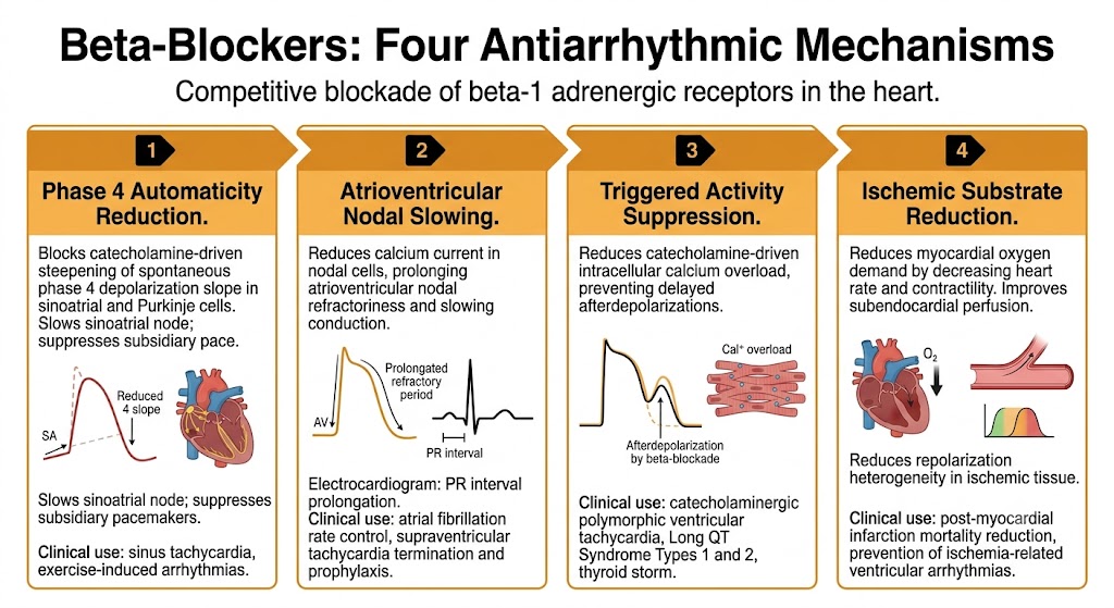
Four-panel diagram showing the antiarrhythmic mechanisms of beta-blockers: phase 4 automaticity reduction, atrioventricular nodal slowing with PR interval prolongation, triggered activity suppression via calcium overload prevention, and ischemic substrate reduction through decreased myocardial oxygen demand.
Source: AI-generated figure (Gemini). Educational use.

Reduction of Phase 4 Automaticity

Sympathetic stimulation steepens the slope of spontaneous phase 4 depolarization in sinoatrial nodal cells and Purkinje fibers, increasing the intrinsic firing rate. Beta-blockers reverse this effect by blocking catecholamine-driven acceleration of the pacemaker current, slowing the sinoatrial node and suppressing abnormal automaticity in subsidiary pacemakers. This is the mechanism responsible for rate control in sinus tachycardia and for suppressing exercise-induced ventricular arrhythmias driven by adrenergic excess.

Atrioventricular Nodal Slowing

Sympathetic tone accelerates conduction through the atrioventricular node by increasing calcium current in nodal cells. Beta-blockers attenuate this effect, prolonging atrioventricular nodal refractoriness and slowing conduction velocity through the node. On the electrocardiogram, this appears as PR interval prolongation. This mechanism underlies beta-blocker efficacy for ventricular rate control in atrial fibrillation and atrial flutter, and for terminating atrioventricular node-dependent re-entrant tachycardias where the atrioventricular node is a required limb of the circuit.

Suppression of Triggered Activity

Catecholamine excess causes intracellular calcium overload by increasing calcium entry and calcium release from intracellular stores. This calcium overload generates delayed afterdepolarizations — oscillatory membrane depolarizations during phase 4 that can trigger premature beats and sustained tachycardias. Beta-blockers interrupt this cycle by reducing adrenergic calcium loading, making them uniquely effective against delayed afterdepolarization-mediated arrhythmias: catecholaminergic polymorphic ventricular tachycardia, exercise-induced ventricular tachycardia, and arrhythmias complicating thyroid storm or pheochromocytoma.

Reduction of Ischemic Arrhythmia Substrate

Beta-blockers reduce myocardial oxygen demand by decreasing heart rate and contractility, and improve subendocardial perfusion by prolonging diastole. These anti-ischemic effects reduce the heterogeneity of repolarization that provides the substrate for re-entrant ventricular arrhythmias in the setting of coronary artery disease. This mechanism — combined with the suppression of catecholamine-driven triggered activity — accounts for the mortality benefit demonstrated in multiple large post-myocardial infarction trials.
Section 2

Key Beta-Blocker Agents

Selectivity, lipophilicity, and the properties that determine agent selection

Beta-blockers differ in their receptor selectivity, lipophilicity, and ancillary properties. These differences are not merely pharmacokinetic details — they determine which agent is safest and most effective in specific clinical situations.

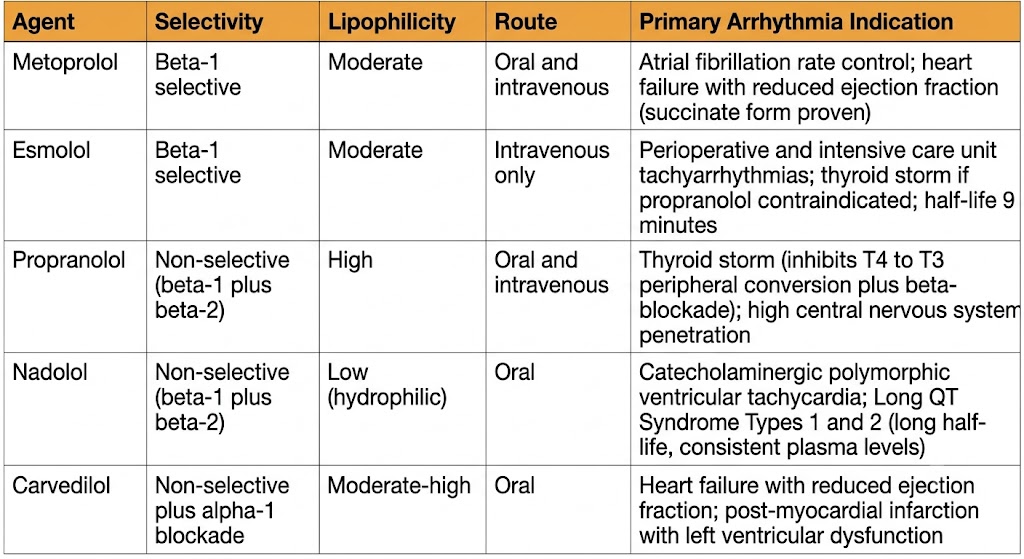
Reference table comparing five beta-blocker agents used in arrhythmia management — metoprolol, esmolol, propranolol, nadolol, and carvedilol — with columns for selectivity, lipophilicity, route, and primary arrhythmia indication.
Source: AI-generated figure (Gemini). Educational use.

Metoprolol

Metoprolol is beta-1 selective and the most commonly used beta-blocker for cardiac arrhythmia management. The immediate-release tartrate formulation is available for intravenous use, making it the standard agent for acute ventricular rate control in atrial fibrillation in hemodynamically stable patients. The extended-release succinate formulation provides once-daily dosing and is the guideline-preferred form for patients with heart failure with reduced ejection fraction, where it has demonstrated mortality benefit. Beta-1 selectivity makes metoprolol preferable to non-selective agents in patients with reactive airway disease, though caution is still warranted in severe asthma.

Esmolol

Esmolol is beta-1 selective and has an elimination half-life of approximately nine minutes, making it the only beta-blocker that can be rapidly titrated and quickly reversed. It is available only for intravenous use and is metabolized by red blood cell esterases independently of hepatic or renal function. This ultrashort duration makes esmolol the preferred agent when hemodynamic tolerance is uncertain — perioperative tachyarrhythmias, thyroid storm, intensive care unit rate control — and when rapid reversal of beta-blockade may be needed.

Propranolol

Propranolol is non-selective, blocking both beta-1 and beta-2 adrenergic receptors. It is highly lipophilic, penetrating the central nervous system readily, which accounts for its central nervous system adverse effects (fatigue, depression, vivid dreams) but also contributes to efficacy in arrhythmias with a central sympathetic drive component. Its defining clinical role among beta-blockers is in thyrotoxicosis-related arrhythmias: in addition to blocking adrenergic effects on the heart, propranolol inhibits the peripheral conversion of thyroxine to the more biologically active triiodothyronine through a mechanism involving beta-2 receptor blockade. This additional hormonal effect, unique to propranolol among the beta-blockers, makes it the preferred agent in thyroid storm.

Beta-2 blockade also means propranolol can cause clinically significant bronchospasm, making it contraindicated in asthma and severe reactive airway disease.

Nadolol

Nadolol is non-selective with a long half-life (approximately 14 to 24 hours) and is eliminated renally without significant hepatic metabolism. These properties — consistent, predictable plasma levels without the troughs that can permit breakthrough catecholamine-driven arrhythmias — make it the preferred agent for catecholaminergic polymorphic ventricular tachycardia and Long QT Syndrome Types 1 and 2. Its hydrophilicity results in low central nervous system penetration and a favorable central nervous system adverse effect profile. Dose reduction is required in chronic kidney disease.

Carvedilol

Carvedilol is non-selective with additional alpha-1 adrenergic receptor blocking activity, which produces peripheral vasodilation and reduces afterload. This ancillary vasodilatory property makes it particularly beneficial in heart failure with reduced ejection fraction, where it has demonstrated mortality benefit in large trials. Along with metoprolol succinate and bisoprolol, carvedilol is one of three beta-blockers with proven survival benefit in heart failure with reduced ejection fraction — a benefit that cannot be assumed for other agents in this drug class.

The Heart Failure with Reduced Ejection Fraction Rule

Only three beta-blockers have proven mortality benefit in heart failure with reduced ejection fraction: carvedilol, metoprolol succinate (extended-release), and bisoprolol. Other beta-blockers — including metoprolol tartrate (immediate-release), atenolol, and propranolol — do not carry this benefit and should not be substituted. When a patient with new heart failure with reduced ejection fraction is on a different beta-blocker, conversion to one of the three proven agents is a standard quality measure.

Critical initiation rule: beta-blockers must only be started in heart failure with reduced ejection fraction when the patient is clinically compensated and euvolemic. Starting during acute decompensation causes hemodynamic deterioration. Begin at the lowest dose and uptitrate gradually over weeks to months.


Section 3

Clinical Indications in Arrhythmia Management

Six arrhythmia contexts where beta-blockers are first-line or essential therapy

Beta-blockers are among the most broadly applicable antiarrhythmic agents. Their combination of nodal slowing, automaticity suppression, and triggered activity inhibition makes them first-line or essential therapy across a wide range of arrhythmia types.

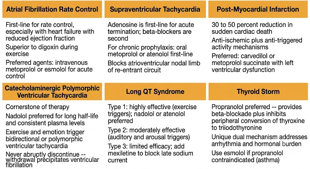
Six-panel summary diagram showing beta-blocker indications in arrhythmia management: atrial fibrillation rate control, supraventricular tachycardia prophylaxis, post-myocardial infarction mortality reduction, catecholaminergic polymorphic ventricular tachycardia, Long QT Syndrome by type, and thyroid storm with propranolol preferred for dual T4-to-T3 conversion blockade.
Source: AI-generated figure (Gemini). Educational use.

Ventricular Rate Control in Atrial Fibrillation and Flutter

Beta-blockers are first-line agents for ventricular rate control in atrial fibrillation, particularly in patients with sympathetically driven tachycardia (exercise, thyrotoxicosis, post-surgical atrial fibrillation) or in those with heart failure with reduced ejection fraction where non-dihydropyridine calcium channel blockers are contraindicated due to negative inotropy. Intravenous metoprolol or esmolol achieves rapid rate control in hemodynamically stable patients. Beta-blockers are superior to digoxin for rate control during exercise because digoxin's vagotonic mechanism loses effectiveness during sympathetic activation.

Supraventricular Tachycardias

For atrioventricular nodal reentrant tachycardia and atrioventricular reentrant tachycardia, beta-blockers can terminate acute episodes by blocking the atrioventricular nodal limb of the re-entrant circuit, though adenosine remains first-line for acute termination. For long-term prophylaxis in patients who decline catheter ablation or are not eligible for it, oral metoprolol or atenolol are first-line agents. Beta-blockers are preferred over calcium channel blockers for chronic suppression when there are contraindications to the latter or in patients who also have structural heart disease.

Post-Myocardial Infarction and Heart Failure with Reduced Ejection Fraction

Multiple large trials established beta-blockers as standard of care after myocardial infarction, with reductions in sudden cardiac death of 30 to 50 percent. The mechanism is multifactorial: suppression of catecholamine-driven triggered activity, anti-ischemic effects that reduce the re-entrant substrate, and prevention of the catecholamine-driven increases in calcium entry that predispose to ventricular fibrillation. Carvedilol and metoprolol succinate are guideline-preferred agents in post-myocardial infarction patients with left ventricular dysfunction.

Catecholaminergic Polymorphic Ventricular Tachycardia

Catecholaminergic polymorphic ventricular tachycardia is caused by mutations in proteins regulating calcium release from intracellular stores in cardiac muscle cells, resulting in pathologic calcium leak during adrenergic stimulation. Catecholamines trigger bidirectional or polymorphic ventricular tachycardia that characteristically occurs with exercise or emotional stress and terminates at rest. Beta-blockers are the cornerstone of treatment, with nadolol preferred because its long half-life and consistent pharmacokinetics minimize breakthrough arrhythmias at plasma concentration troughs. Beta-blockers must never be abruptly discontinued in catecholaminergic polymorphic ventricular tachycardia — withdrawal precipitates ventricular fibrillation.

Long QT Syndrome

In Long QT Syndrome Type 1, where arrhythmias are characteristically triggered by sympathetic activation during swimming or sudden exertion, beta-blockers are highly effective and reduce event rates substantially. Nadolol or atenolol are preferred for their consistent pharmacokinetics. In Long QT Syndrome Type 2, where arrhythmias are triggered by auditory stimuli and arousal, beta-blockers are moderately effective. In Long QT Syndrome Type 3, which involves a gain-of-function sodium channel mutation, beta-blockers have limited efficacy and mexiletine is typically added to block the abnormal late sodium current that drives QT prolongation in this subtype.

Thyroid Storm

Thyroid storm produces extreme adrenergic activation from massive thyroid hormone release, causing sinus tachycardia, atrial fibrillation, and occasionally ventricular tachycardia driven by catecholamine hypersensitivity. Propranolol is the preferred agent in thyroid storm for two reasons: it provides non-selective beta-blockade to control the adrenergic manifestations, and it additionally inhibits peripheral conversion of thyroxine to the more potent triiodothyronine through a mechanism involving beta-2 receptor blockade — an effect not shared by selective agents. This dual action — sympatholytic plus hormonal — makes propranolol the only beta-blocker that addresses both the arrhythmia and the thyroid hormone burden simultaneously. If propranolol is contraindicated (reactive airway disease), esmolol provides titratable intravenous rate control without the hormonal conversion benefit.


Section 4

Adverse Effects, Contraindications, and Withdrawal

Predicting and managing the class-specific risks of beta-adrenergic blockade

The adverse effects of beta-blockers follow directly from their mechanism — each predictable consequence of blocking beta-adrenergic receptors in cardiac and non-cardiac tissue. Understanding which property (selectivity, lipophilicity) determines each adverse effect allows rational agent selection to minimize risk in individual patients.

Adverse Effects

Bradycardia and atrioventricular block are the most clinically significant cardiac adverse effects, arising from excessive suppression of sinoatrial automaticity and atrioventricular nodal conduction. Risk is greatest at initiation, with dose increases, and in patients with pre-existing conduction system disease. All beta-blockers carry this risk equally.

Bronchospasm is mediated by beta-2 receptor blockade impairing bronchodilation in the airways. Non-selective agents (propranolol, nadolol, carvedilol) carry the greatest risk. Beta-1 selective agents (metoprolol, atenolol) are substantially safer in patients with reactive airway disease, though no beta-blocker is completely free of bronchospastic risk at high doses. Non-selective agents are contraindicated in severe asthma.

Central nervous system effects — fatigue, depression, sleep disturbance, and vivid dreams — are most prominent with lipophilic agents (propranolol, metoprolol) that readily cross the blood-brain barrier. Hydrophilic agents (nadolol, atenolol) have lower central nervous system penetration and are preferred when central nervous system adverse effects are a concern.

Metabolic effects include masking of hypoglycemic symptoms (particularly tachycardia) in insulin-dependent diabetics and blunting of the compensatory tachycardia that normally signals hypoglycemia. Non-selective agents carry greater risk. Beta-1 selective agents are preferable in insulin-dependent diabetics when beta-blockade is necessary.

Peripheral vasoconstriction from beta-2 blockade can exacerbate Raynaud's phenomenon and peripheral vascular disease. Beta-1 selective or vasodilatory agents (carvedilol) are preferred in these patients.

Key Contraindications

Absolute contraindications include decompensated heart failure with acute pulmonary edema (initiate only after stabilization), cardiogenic shock, high-degree atrioventricular block (second or third degree) without a functioning pacemaker, sick sinus syndrome without pacemaker backup, and severe asthma with active bronchospasm. Cocaine-induced tachycardia is a specific contraindication: beta-blockade in this setting causes unopposed alpha-adrenergic stimulation, worsening coronary vasospasm and potentially precipitating myocardial infarction.

Beta-Blocker Withdrawal Syndrome

Chronic beta-blockade causes upregulation of beta-adrenergic receptor density as a compensatory response to sustained blockade. When beta-blockers are abruptly discontinued, this supersensitive receptor population is suddenly exposed to endogenous catecholamines, producing rebound tachycardia, hypertension, angina, and — in patients with underlying coronary artery disease — potentially ventricular fibrillation. The risk is highest in the first 24 to 48 hours after abrupt discontinuation.

Beta-blockers should never be abruptly discontinued in patients with coronary artery disease, prior myocardial infarction, catecholaminergic polymorphic ventricular tachycardia, or Long QT Syndrome. Taper over one to two weeks when discontinuation is planned. In patients on chronic beta-blocker therapy who require surgery, the beta-blocker must be continued perioperatively — abrupt withdrawal on hospital admission is a preventable cause of perioperative arrhythmia.

Never Abruptly Stop Beta-Blockers in High-Risk Patients

Upregulation of beta-adrenergic receptors during chronic therapy means abrupt discontinuation exposes a hypersensitive receptor population to endogenous catecholamines. In patients with coronary artery disease, prior myocardial infarction, catecholaminergic polymorphic ventricular tachycardia, or Long QT Syndrome, this rebound can precipitate ventricular fibrillation. Always taper. Always continue perioperatively. If discontinuation is unavoidable, intravenous esmolol can bridge the gap until the oral agent is restarted.


Suggested References
Author / Organization Title Source
Bers DM Cardiac excitation-contraction coupling Nature. 2002;415(6868):198–205
Frishman WH Beta-adrenergic blockers: a 50-year historical perspective Am J Ther. 2008;15(6):565–576
Priori SG, Napolitano C, Tiso N, et al Mutations in the cardiac ryanodine receptor gene (hRyR2) underlie catecholaminergic polymorphic ventricular tachycardia Circulation. 2001;103(2):196–200
Yusuf S, Peto R, Lewis J, Collins R, Sleight P Beta blockade during and after myocardial infarction: an overview of the randomized trials Prog Cardiovasc Dis. 1985;27(5):335–371
Priori SG, Wilde AA, Horie M, et al HRS/EHRA/APHRS expert consensus statement on the diagnosis and management of patients with inherited primary arrhythmia syndromes Heart Rhythm. 2013;10(12):1932–1963
Joglar JA, Chung MK, Armbruster AL, et al 2023 ACC/AHA/ACCP/HRS guideline for the diagnosis and management of atrial fibrillation J Am Coll Cardiol. 2024;83(1):109–279
Priori SG, Blomstrom-Lundqvist C, Mazzanti A, et al 2015 ESC Guidelines for the management of patients with ventricular arrhythmias and the prevention of sudden cardiac death Eur Heart J. 2015;36(41):2793–2867
Psaty BM, Koepsell TD, Wagner EH, LoGerfo JP, Inui TS The relative risk of incident coronary heart disease associated with recently stopping the use of beta-blockers JAMA. 1990;263(12):1653–1657
Packer M, Coats AJ, Fowler MB, et al Effect of carvedilol on survival in severe chronic heart failure N Engl J Med. 2001;344(22):1651–1658