CHAPTER 35  ·  ANTIBACTERIAL AGENTS
Section 1

Mechanism of Action and Spectrum

Ribosomal binding, concentration-dependent killing, and the gram-negative-predominant spectrum

Aminoglycosides kill gram-negative bacteria by an irreversible mechanism involving ribosomal binding and inner membrane disruption. Their concentration-dependent bactericidal activity and prolonged post-antibiotic effect distinguish them from beta-lactams and support once-daily dosing strategies that exploit peak drug concentration as the pharmacodynamic driver of efficacy.

Uptake and Mechanism of Action

Aminoglycosides bind the 30S ribosomal subunit, causing misreading of messenger RNA and production of non-functional proteins that disrupt the bacterial inner membrane, producing rapid, irreversible bactericidal activity. Entry into bacterial cells requires aerobic metabolism, which is why obligate anaerobes are intrinsically resistant to aminoglycosides regardless of in vitro susceptibility results.

Spectrum of Activity

Aminoglycosides are primarily active against aerobic gram-negative bacilli: most Enterobacteriaceae (Escherichia coli, Klebsiella pneumoniae, Enterobacter, Serratia, Proteus), Pseudomonas aeruginosa, and Acinetobacter baumannii. They have no useful activity against obligate anaerobes, Streptococcus pneumoniae, or most streptococci when used as monotherapy. Low-level synergistic activity against enterococci and streptococci is exploited in combination with cell wall-active agents for endocarditis.

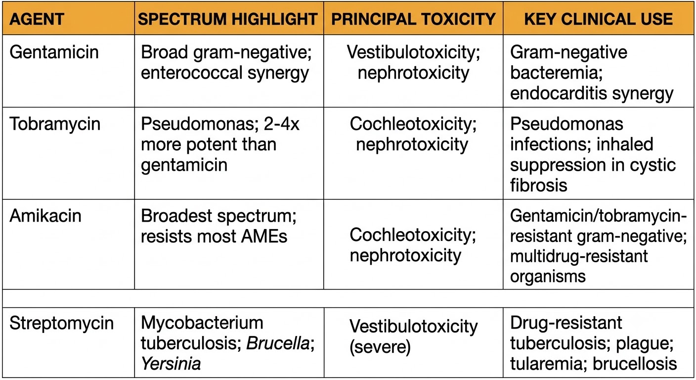
Among the agents, tobramycin is approximately two- to four-fold more potent than gentamicin against Pseudomonas aeruginosa, making it the preferred agent for Pseudomonas pulmonary infections. Amikacin has the broadest spectrum because it resists most aminoglycoside-modifying enzymes, making it the agent of choice when gentamicin or tobramycin resistance is suspected.

Pharmacodynamics

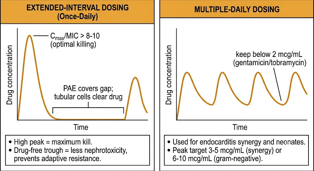
Aminoglycosides are concentration-dependent killers: the key pharmacodynamic driver is the ratio of peak drug concentration to the minimum inhibitory concentration (Cmax/MIC). Maximum bactericidal activity is achieved when this ratio exceeds 8–10. The post-antibiotic effect against gram-negative bacteria is substantial (2–8 hours), meaning significant bacterial growth suppression persists well after drug concentrations fall below the minimum inhibitory concentration. Both properties support extended-interval (once-daily) dosing: a high peak optimizes killing, and a drug-free interval exploits the post-antibiotic effect while limiting tubular cell accumulation and adaptive resistance.
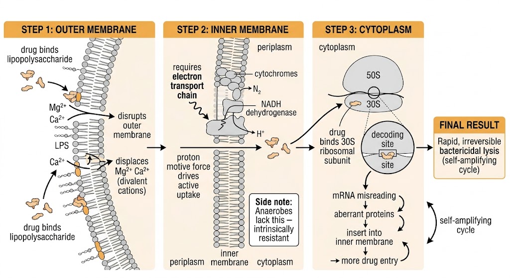
Three-step mechanism diagram showing aminoglycoside entry into gram-negative bacteria: outer membrane binding displacing Mg2+ and Ca2+, proton motive force-driven active uptake across the inner membrane, and 30S ribosomal subunit binding causing mRNA misreading and self-amplifying bactericidal lysis.
Aminoglycoside mechanism: two-stage uptake, ribosomal binding, and self-amplifying bactericidal killing. Generated figure.

Section 2

Pharmacokinetics and Dosing Strategies

Distribution, renal elimination, extended-interval dosing, and therapeutic drug monitoring

The pharmacokinetics of aminoglycosides are shaped by their polycationic nature: negligible oral bioavailability, distribution primarily in extracellular fluid, almost exclusive renal elimination, and a propensity to accumulate in renal cortical tissue and inner ear endolymph—the two compartments responsible for their principal toxicities.

Distribution and Elimination

Aminoglycosides are not absorbed orally and must be administered intravenously or intramuscularly. The volume of distribution is approximately 0.25–0.3 L/kg in euvolemic adults, reflecting distribution predominantly in extracellular fluid. This increases substantially in patients with third-space fluid accumulation (sepsis, ascites, edema) and requires weight-based dosing adjustments using ideal body weight corrected for obesity. Aminoglycosides are eliminated essentially unchanged by glomerular filtration, with a plasma half-life of approximately 2–3 hours in patients with normal renal function. Dose adjustment is required in renal impairment.

Extended-Interval (Once-Daily) Dosing

Extended-interval dosing administers the entire daily aminoglycoside dose as a single infusion, generating a high peak concentration that optimizes the Cmax/MIC ratio while allowing a drug-free trough period that limits proximal tubular accumulation and adaptive resistance development. This strategy has been demonstrated to be equivalent or superior to multiple-daily dosing for most gram-negative infections, with a consistent trend toward reduced nephrotoxicity.

The Hartford nomogram is widely used to individualize extended-interval dosing intervals. A single serum level drawn 6–14 hours after the infusion is plotted against sampling time; the result determines whether the next dose should be given at 24, 36, or 48 hours based on the patient's clearance. The nomogram is not validated for neonates, pregnancy, significant burns, or ascites.

Therapeutic Drug Monitoring

Therapeutic drug monitoring (TDM) is mandatory for aminoglycosides because of their narrow therapeutic index and the direct relationship between drug exposure and toxicity. For extended-interval dosing, a single level drawn 6–14 hours post-infusion is plotted on the Hartford nomogram. For multiple-daily dosing (used in endocarditis synergy regimens and neonates), both peak levels and trough levels are monitored: target troughs for gentamicin and tobramycin are below 2 mcg/mL (ideally below 1 mcg/mL) to minimize nephrotoxicity; target peaks are 6–10 mcg/mL for gram-negative infections and 3–5 mcg/mL for synergy regimens.

Two-panel diagram contrasting extended-interval once-daily dosing (high Cmax/MIC peak with drug-free trough) versus multiple-daily dosing (lower repeated peaks with sustained troughs), with pharmacodynamic targets and clinical indications for each strategy.
Aminoglycoside dosing strategies: extended-interval versus multiple-daily dosing pharmacodynamic profiles. Generated figure.

Section 3

Nephrotoxicity and Ototoxicity

Mechanisms, risk factors, and prevention of the two major dose-limiting toxicities

The two principal dose-limiting toxicities of aminoglycosides—nephrotoxicity and ototoxicity—both result from drug accumulation in specific cellular compartments with limited elimination capacity. Both can be irreversible when recognized late, and many risk factors are modifiable.

Nephrotoxicity

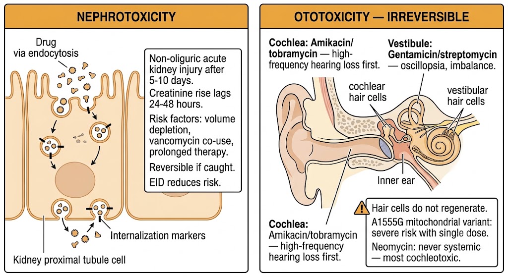
Aminoglycoside nephrotoxicity results from preferential accumulation in proximal tubular epithelial cells of the renal cortex. The drug is filtered at the glomerulus and accumulates in proximal tubular cells to concentrations far exceeding plasma levels, causing tubular cell death. The clinical manifestation is a non-oliguric acute kidney injury that typically develops after 5–10 days of therapy, with serum creatinine rises that may lag behind actual tubular injury by 24–48 hours. Incidence with multiple-daily dosing ranges from 10–25%; extended-interval dosing reduces nephrotoxicity risk because the drug-free trough period allows clearance of proximal tubular drug before the next accumulation cycle.

Independent risk factors include pre-existing renal impairment, volume depletion, hypokalemia, hypomagnesemia, prolonged therapy duration, older age, liver disease, and concomitant nephrotoxin exposure. The combination of vancomycin and an aminoglycoside carries substantially higher nephrotoxicity risk than either agent alone and requires daily renal function monitoring. Prevention strategies include maintaining adequate hydration, correcting electrolyte deficits, avoiding additional nephrotoxins, using extended-interval dosing, and limiting course duration.

Ototoxicity: Cochlear and Vestibular

Aminoglycoside ototoxicity results from irreversible destruction of sensory hair cells in the cochlea (causing sensorineural hearing loss) and vestibular apparatus (causing vertigo and chronic disequilibrium). Unlike nephrotoxicity, cochlear hair cell destruction is permanent because mammalian hair cells do not regenerate. Cochlear toxicity manifests initially as high-frequency hearing loss above the speech range, detectable only with audiometry; speech-frequency loss develops with more severe injury. Vestibular toxicity manifests as oscillopsia (inability to stabilize visual images during head movement), imbalance, and chronic disequilibrium that may persist years after drug discontinuation.

The relative toxicity profile differs among agents: streptomycin and gentamicin preferentially cause vestibular toxicity; amikacin and tobramycin preferentially cause cochlear toxicity; neomycin is the most cochleotoxic aminoglycoside and must never be given systemically. A rare pharmacogenomic variant in mitochondrial ribosomal RNA dramatically increases cochlear susceptibility and can cause severe hearing loss after a single conventional dose in carriers.

Vancomycin-Aminoglycoside Combination: High Nephrotoxicity Risk

Combining vancomycin with an aminoglycoside carries substantially higher nephrotoxicity risk than either agent alone, with acute kidney injury rates of 20–35% or higher reported in observational studies. When this combination is used for specific indications (enterococcal endocarditis, selected gram-negative bacteremia), daily renal function monitoring, aggressive hydration, and avoidance of additional nephrotoxins are mandatory. The rationale for continuing the combination must be reassessed daily against the toxicity risk.

Two-panel toxicity diagram: left panel shows aminoglycoside accumulation in kidney proximal tubular cells causing non-oliguric acute kidney injury with risk factors; right panel shows irreversible cochlear and vestibular hair cell destruction with agent-specific toxicity profiles and A1555G genetic risk.
Aminoglycoside nephrotoxicity and ototoxicity: mechanisms, risk factors, and agent-specific profiles. Generated figure.

Section 4

Clinical Use, Synergy, and Resistance

Current indications, endocarditis synergy, and aminoglycoside-modifying enzyme resistance

The clinical role of aminoglycosides has narrowed as less toxic alternatives have emerged for many gram-negative infections. They retain important niches: serious Pseudomonas aeruginosa infections, enterococcal endocarditis synergy regimens, multidrug-resistant gram-negative bacteremia, and inhaled tobramycin for cystic fibrosis lung disease.

Gram-Negative Infections and Pseudomonas aeruginosa

Current evidence does not support the routine addition of an aminoglycoside to a beta-lactam for gram-negative bacteremia caused by susceptible organisms in non-immunocompromised patients, as combination therapy does not improve mortality compared to beta-lactam monotherapy while substantially increasing nephrotoxicity. However, aminoglycosides retain a role in initial empiric coverage of septic shock when multidrug-resistant gram-negative organisms are suspected, with the aminoglycoside typically discontinued once susceptibility data and clinical stability are established.

Tobramycin is preferred over gentamicin for Pseudomonas aeruginosa infections due to superior intrinsic potency. Inhaled tobramycin (tobramycin inhalation solution or tobramycin inhalation powder) is approved for chronic suppression of Pseudomonas aeruginosa in patients with cystic fibrosis, achieving high airway concentrations with minimal systemic exposure and reducing exacerbation frequency without the toxicity burden of parenteral dosing.

Endocarditis Synergy Regimens

Low-dose aminoglycosides exploit synergy with cell wall-active agents to enhance bactericidal killing against enterococci and streptococci. For enterococcal endocarditis caused by gentamicin-susceptible Enterococcus faecalis, the combination of ampicillin plus ceftriaxone has demonstrated equivalent efficacy to ampicillin plus gentamicin with substantially less nephrotoxicity and is now preferred at many centers. Gentamicin synergy remains guideline-supported for streptococcal endocarditis where short-course combination therapy (two weeks) has established benefit. High-level aminoglycoside resistance (defined as gentamicin minimum inhibitory concentration at or above 500 mcg/mL) eliminates synergistic activity and must be tested before relying on this approach.

Resistance Mechanisms

The dominant resistance mechanism is enzymatic inactivation by aminoglycoside-modifying enzymes (AMEs) that chemically modify the drug, abolishing ribosomal binding. AMEs are encoded on mobile genetic elements (plasmids, transposons, integrons), enabling horizontal transfer among Enterobacteriaceae, Pseudomonas aeruginosa, and Acinetobacter baumannii. Amikacin resists most AMEs, making it the agent of choice when other aminoglycosides have failed.

A second resistance mechanism involves ribosomal RNA methyltransferases that methylate the aminoglycoside binding site on the ribosome, conferring high-level resistance to all aminoglycosides including amikacin. These enzymes are increasingly co-located with carbapenemase genes (including New Delhi metallo-beta-lactamase) on mobile plasmids, creating organisms resistant to essentially all conventional antibiotics. Detection requires genotypic testing.

Reference table comparing gentamicin, tobramycin, amikacin, and streptomycin by spectrum highlight, principal toxicity, and key clinical use.
Aminoglycoside agents: spectrum, toxicity, and clinical niche comparison. Generated figure.

Suggested References
Author / OrganizationTitleSource
Mingeot-Leclercq MP, Glupczynski Y, Tulkens PMAminoglycosides: activity and resistanceAntimicrob Agents Chemother. 1999;43(4):727-737
Moazed D, Noller HFInteraction of antibiotics with functional sites in 16S ribosomal RNANature. 1987;327(6121):389-394
Paul M, Lador A, Grozinsky-Glasberg S, Leibovici LBeta lactam antibiotic monotherapy versus beta lactam-aminoglycoside antibiotic combination therapy for sepsisCochrane Database Syst Rev. 2014;1:CD003344
Craig WAPharmacokinetic/pharmacodynamic parameters: rationale for antibacterial dosing of mice and menClin Infect Dis. 1998;26(1):1-12
Nicolau DP, Freeman CD, Belliveau PP, Nightingale CH, Ross JW, Quintiliani RExperience with a once-daily aminoglycoside program administered to 2,184 adult patientsAntimicrob Agents Chemother. 1995;39(3):650-655
Streetman DS, Nafziger AN, Destache CJ, Bertino JS JrIndividualized pharmacokinetic monitoring results in less aminoglycoside-associated nephrotoxicity and fewer associated costsPharmacotherapy. 2001;21(4):443-451
Rybak MJ, Lomaestro BM, Rotschafer JC, et al.Therapeutic monitoring of vancomycin in adult patients: a consensus review of the American Society of Health-System Pharmacists, the Infectious Diseases Society of America, and the Society of Infectious Diseases PharmacistsAm J Health Syst Pharm. 2009;66(1):82-98
Mingeot-Leclercq MP, Tulkens PMAminoglycosides: nephrotoxicityAntimicrob Agents Chemother. 1999;43(5):1003-1012
Corcoran TE, Smaldone GC, Bahrani-Mougeot FK, et al.Pharmacokinetics and safety of inhaled tobramycin in critically ill patientsJ Aerosol Med. 2006;19(1):68-78
Fischel-Ghodsian NGenetic factors in aminoglycoside toxicityPharmacogenomics. 2005;6(1):27-36
Ramsey BW, Pepe MS, Quan JM, et al.Intermittent administration of inhaled tobramycin in patients with cystic fibrosisN Engl J Med. 1999;340(1):23-30
Fernandez-Hidalgo N, Almirante B, Gavalda J, et al.Ampicillin plus ceftriaxone is as effective as ampicillin plus gentamicin for treating Enterococcus faecalis infective endocarditisClin Infect Dis. 2013;56(9):1261-1268