CHAPTER 37  ·  ANTIFUNGAL AGENTS
Section 1

Candidiasis

Mucosal, invasive, and bloodstream disease — matching drug to host and organism

Candida species are the most common cause of invasive fungal infections in hospitalized patients and the fourth most common cause of healthcare-associated bloodstream infections in the United States. The clinical spectrum ranges from superficial mucosal disease, which is common and readily treated, to invasive candidiasis and candidemia, which carry mortality rates of 30 to 40 percent and require rapid, appropriate antifungal therapy guided by species identification and susceptibility data.

Mucosal Candidiasis

Oropharyngeal candidiasis (thrush) presents as white plaques on erythematous mucosa in the mouth, pharynx, or tongue that can be scraped off, distinguishing them from leukoplakia. It occurs predominantly in patients receiving inhaled corticosteroids (who should rinse the mouth after each use), those with human immunodeficiency virus (HIV) infection with low CD4 counts, patients receiving chemotherapy or antibiotics, denture wearers, and neonates. Fluconazole 200 mg on day one followed by 100 mg daily for seven to 14 days is the drug of choice; topical nystatin suspension is an alternative for mild disease. Esophageal candidiasis presents with odynophagia and dysphagia and requires systemic therapy — fluconazole is significantly more effective than nystatin swish-and-swallow for this indication and is first-line. Vulvovaginal candidiasis, caused predominantly by Candida albicans, is treated with either a single oral fluconazole dose (150 mg) or topical azole preparations; recurrent vulvovaginal candidiasis (four or more episodes per year) warrants maintenance fluconazole suppression.

Invasive Candidiasis and Candidemia

Candidemia — Candida isolated from blood culture — requires immediate systemic antifungal therapy and a thorough search for deep-seated foci including Candida endophthalmitis (present in five to ten percent of candidemia cases and requiring ophthalmologic evaluation), hepatosplenic candidiasis in patients recovering from neutropenia, Candida endocarditis in patients with prosthetic valves or prolonged candidemia, and osteomyelitis. Risk factors for invasive candidiasis include prolonged broad-spectrum antibiotic use, central venous catheters, total parenteral nutrition (TPN), abdominal surgery, renal replacement therapy, and immunosuppression.

An echinocandin — caspofungin, micafungin, or anidulafungin — is the preferred initial therapy for candidemia in most patients, given superior fungicidal activity and retained efficacy against fluconazole-resistant Candida glabrata. The central venous catheter should be removed as soon as possible in patients with candidemia, as catheter removal improves outcomes and reduces time to blood culture clearance. Step-down to oral fluconazole is appropriate once the patient is clinically stable, blood cultures are negative, and the species is confirmed fluconazole-susceptible. Treatment duration for uncomplicated candidemia is 14 days from the first negative blood culture. Candida auris, the multidrug-resistant emerging pathogen, is typically echinocandin-susceptible; echinocandin therapy is recommended while susceptibility data are pending.

Candidemia Treatment Algorithm

Step 1: Start echinocandin (caspofungin, micafungin, or anidulafungin) empirically. Step 2: Remove central venous catheter. Step 3: Ophthalmology consult for dilated funduscopic examination. Step 4: Repeat blood cultures daily until negative. Step 5: At day five to seven, if clinically stable, blood cultures negative, and species confirmed fluconazole-susceptible, step down to oral fluconazole. Step 6: Treat for 14 days from first negative blood culture. Never use fluconazole empirically as initial therapy in severely ill patients or those with prior azole exposure.


Section 2

Invasive Aspergillosis

Risk stratification, diagnosis, primary treatment, and salvage strategies

Invasive aspergillosis (IA) is a life-threatening mold infection caused predominantly by Aspergillus fumigatus and occurring almost exclusively in immunocompromised hosts. Mortality rates range from 30 to over 80 percent depending on the host immune status, site of infection, and time to diagnosis and treatment initiation. Early empirical treatment before culture confirmation is often necessary and life-saving.

Risk Factors and Presentation

The populations at highest risk for invasive aspergillosis are patients with prolonged profound neutropenia (below 500 neutrophils per microliter for more than ten days), allogeneic hematopoietic stem cell transplant (HSCT) recipients, solid organ transplant recipients (especially lung transplant), patients receiving high-dose corticosteroids, and patients with severe combined immunodeficiency or advanced HIV infection with CD4 counts below 50 cells per microliter. Invasive pulmonary aspergillosis (IPA) is the most common form, presenting with fever, cough, pleuritic chest pain, and hemoptysis in a neutropenic or transplant patient who is not responding to broad-spectrum antibacterial therapy. The halo sign — a nodule surrounded by ground-glass opacity representing hemorrhagic infarction — on high-resolution computed tomography (CT) of the chest is a characteristic early finding of IPA. Sinusitis, tracheobronchitis, and central nervous system (CNS) disease are less common presentations.

Diagnosis

Diagnosis relies on a combination of clinical, radiologic, and mycological criteria as defined by the European Organization for Research and Treatment of Cancer (EORTC) and the Mycoses Study Group (MSG) consensus criteria. Serum galactomannan — a cell wall polysaccharide antigen shed by Aspergillus hyphae — is a validated biomarker for invasive aspergillosis in neutropenic patients and HSCT recipients, with sensitivity of approximately 70 to 80 percent and high specificity in these populations. Beta-d-glucan is a pan-fungal cell wall marker detectable in aspergillosis but less specific than galactomannan. Bronchoalveolar lavage (BAL) galactomannan and culture improve diagnostic yield in patients with pulmonary infiltrates. Culture of Aspergillus from a normally sterile site (blood, tissue biopsy) is definitive but blood cultures are almost always negative; tissue biopsy demonstrating septate hyphae with acute-angle branching is the histologic standard. Because Aspergillus molds can colonize respiratory tracts, a positive respiratory culture alone is not diagnostic in the absence of supporting clinical and radiologic evidence.

Primary Treatment

Voriconazole is the established first-line agent for invasive aspergillosis, based on the landmark 2002 randomized trial by Herbrecht et al. demonstrating superior survival and response rates compared to amphotericin B deoxycholate. Isavuconazole is an alternative first-line agent following the SECURE trial demonstrating non-inferiority to voriconazole with better tolerability. Liposomal amphotericin B (L-AmB) is an alternative when azoles are contraindicated or not tolerated. The combination of voriconazole plus anidulafungin has shown potential benefit over voriconazole monotherapy in a subgroup analysis of patients with positive galactomannan, though current Infectious Diseases Society of America (IDSA) guidelines consider combination therapy an option rather than a standard recommendation. Treatment duration is typically six to 12 weeks, continued until radiologic resolution and immune reconstitution. Therapeutic drug monitoring (TDM) of voriconazole (target trough 1.0 to 5.5 mg/L) or isavuconazole is recommended.
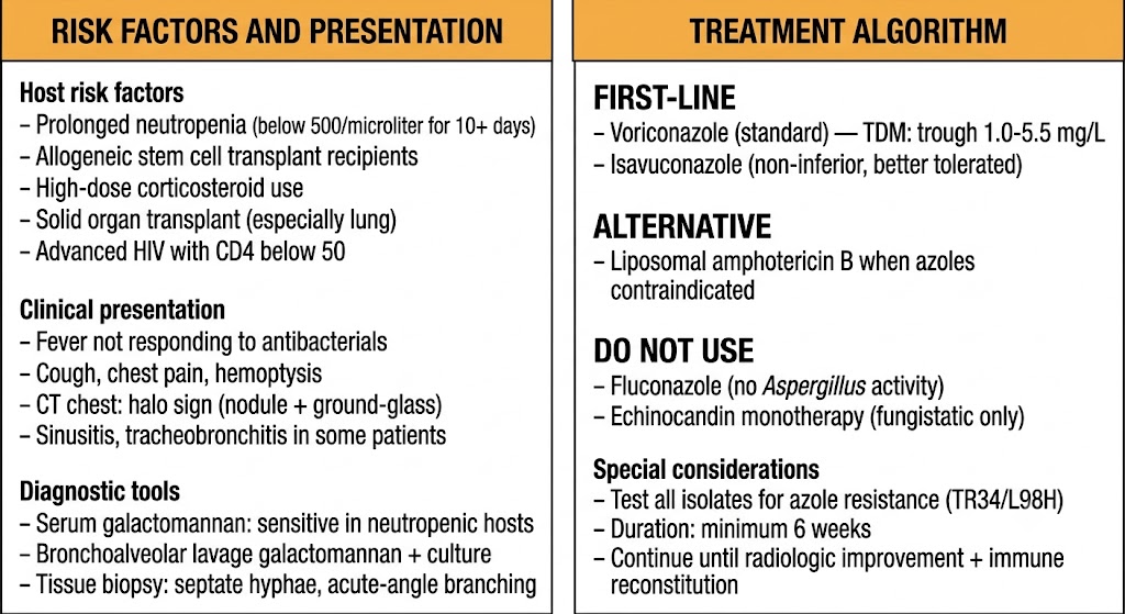
Two-panel diagram for invasive aspergillosis. Left panel lists host risk factors including prolonged neutropenia, allogeneic stem cell transplant, high-dose corticosteroids, solid organ transplant, and advanced HIV, along with clinical presentation features and diagnostic tools including serum galactomannan, bronchoalveolar lavage, and tissue biopsy showing septate hyphae. Right panel shows treatment algorithm with voriconazole and isavuconazole as first-line agents, liposomal amphotericin B as alternative, and a do-not-use list excluding fluconazole and echinocandin monotherapy, plus special considerations including azole resistance testing.
Invasive aspergillosis: risk factors, clinical presentation, diagnostic tools, and treatment algorithm. Gemini-generated figure.
Aspergillosis Treatment Rules

First-line: voriconazole or isavuconazole (non-inferior, better tolerated). Alternative: liposomal amphotericin B when azoles contraindicated. Do not use: fluconazole (no Aspergillus activity), itraconazole (not first-line for invasive disease), echinocandin monotherapy (fungistatic only). Monitor voriconazole trough (target 1.0 to 5.5 mg/L). Duration: minimum six weeks; continue until radiologic improvement and immune reconstitution. Test all isolates for azole resistance (TR34/L98H) — pan-azole-resistant isolates require liposomal amphotericin B or combination therapy.

Section 3

Cryptococcosis

Cryptococcal meningitis in HIV-positive patients and immunocompetent hosts, induction-consolidation-maintenance strategy

Cryptococcus neoformans causes life-threatening meningitis predominantly in patients with advanced HIV infection (CD4 count below 100 cells per microliter) and in other severely immunosuppressed hosts including transplant recipients and patients on high-dose corticosteroids. Cryptococcus gattii causes disease in immunocompetent hosts, particularly in endemic regions of the Pacific Northwest and tropical areas. Untreated cryptococcal meningitis is uniformly fatal; even with optimal therapy, mortality in HIV-associated cryptococcal meningitis ranges from 20 to 30 percent in resource-rich settings and significantly higher in resource-limited settings.

Clinical Presentation

Cryptococcal meningitis typically presents subacutely over days to weeks with headache, fever, malaise, and altered mental status. The subacute course reflects the predominantly fungal rather than inflammatory nature of the infection — the classic signs of bacterial meningitis (meningismus, photophobia, Kernig and Brudzinski signs) are often absent or mild. Cerebrospinal fluid (CSF) examination characteristically shows mild pleocytosis (typically below 50 white blood cells per microliter), mildly elevated protein, and low glucose, with large encapsulated yeast visible on India ink staining in approximately 75 percent of cases. CSF cryptococcal antigen — a lateral flow assay for the Cryptococcus capsular polysaccharide — is positive in essentially all cases of cryptococcal meningitis and is the most sensitive and specific diagnostic test. Elevated CSF opening pressure (above 25 cm H2O) is present in the majority of patients and correlates with mortality; management of elevated intracranial pressure through therapeutic lumbar punctures is a critical and often life-saving component of treatment independent of antifungal therapy.

Treatment: Induction, Consolidation, and Maintenance

Treatment of cryptococcal meningitis is organized into three sequential phases. Induction therapy aims to rapidly reduce the fungal burden in the CSF and brain and is the most critical determinant of early mortality. The 2022 World Health Organization (WHO) guidelines recommend a one-week regimen of liposomal amphotericin B (10 mg/kg as a single dose, or 3 to 4 mg/kg daily for one week) plus flucytosine plus fluconazole for settings with access to all three drugs. Where liposomal amphotericin B is unavailable, amphotericin B deoxycholate (1 mg/kg per day) plus flucytosine for one week followed by fluconazole consolidation is an acceptable alternative, superior to fluconazole monotherapy and to amphotericin B without flucytosine. The IDSA 2010 guidelines for non-HIV settings recommend amphotericin B plus flucytosine for at least two weeks of induction.

Consolidation therapy follows induction and aims to maintain fungal suppression while immune function recovers: fluconazole 400 mg daily for eight weeks is standard. Maintenance therapy (secondary prophylaxis) follows consolidation: fluconazole 200 mg daily until the CD4 count exceeds 200 cells per microliter on antiretroviral therapy (ART) for at least six months. Cryptococcal meningitis is an AIDS-defining illness, and initiating ART is essential but must be timed carefully: starting ART too early (within the first two weeks of antifungal induction) dramatically worsens outcomes due to immune reconstitution inflammatory syndrome (IRIS), and current guidelines recommend deferring ART initiation for two to four weeks after antifungal induction.

Cryptococcal Meningitis Treatment Sequence

Induction (1 week, WHO 2022): liposomal amphotericin B plus flucytosine plus fluconazole. Where L-AmB unavailable: amphotericin B deoxycholate plus flucytosine for one week. Consolidation (8 weeks): fluconazole 400 mg daily. Maintenance: fluconazole 200 mg daily until CD4 above 200 cells per microliter for six months on antiretroviral therapy. Manage elevated intracranial pressure with serial lumbar punctures. Defer ART initiation two to four weeks to reduce immune reconstitution inflammatory syndrome risk.


Section 4

Mucormycosis

Rhinoorbitalcerebral and pulmonary forms, angioinvasion, surgical management, and antifungal selection

Mucormycosis is caused by molds of the order Mucorales — primarily Rhizopus arrhizus, Mucor species, and Lichtheimia corymbifera — and is characterized by aggressive angioinvasion with rapid progression, high mortality, and the requirement for combined surgical and antifungal management. It occurs almost exclusively in immunocompromised or metabolically compromised hosts.

Risk Factors and Pathogenesis

The most important risk factors for mucormycosis are uncontrolled diabetes mellitus with diabetic ketoacidosis (DKA), prolonged neutropenia, high-dose corticosteroid use, deferoxamine therapy (which provides iron to Mucorales organisms), hematologic malignancy, solid organ transplantation, and trauma or burns with soil contamination. The Mucorales are characterized by their ability to grow in high-glucose, acidic environments and to utilize free iron — conditions created by diabetic ketoacidosis. Angioinvasion by fungal hyphae causes thrombosis and tissue necrosis, which limits antifungal penetration and is the reason why surgical debridement is essential and cannot be replaced by antifungal therapy alone. The classic presentation is rhinoorbitalcerebral mucormycosis (ROCM) in diabetic patients: nasal stuffiness progressing to palatal or nasal eschar, periorbital swelling, proptosis, ophthalmoplegia, and ultimately intracranial extension. Pulmonary mucormycosis presents similarly to invasive aspergillosis and may be indistinguishable clinically; the key distinction is voriconazole has no activity against Mucorales while liposomal amphotericin B, posaconazole, and isavuconazole do.

Treatment

Management of mucormycosis requires three simultaneous interventions: antifungal therapy, surgical debridement, and reversal of the underlying predisposing condition. Liposomal amphotericin B at five mg/kg per day is the preferred primary antifungal, initiated as early as possible — even before histologic confirmation when clinical suspicion is high. Surgical debridement of infected tissue is essential; delay in surgery worsens outcomes, and repeated debridements are often necessary. Control of diabetic ketoacidosis, reduction of corticosteroid doses, and discontinuation of deferoxamine when possible are critical adjuncts. Step-down to oral posaconazole or isavuconazole after clinical stabilization with liposomal amphotericin B has become standard practice in many centers, allowing continuation of therapy in an outpatient setting and reducing the toxicity burden of prolonged intravenous (IV) amphotericin B. The duration of therapy is not standardized and is guided by clinical and radiologic response and degree of immune reconstitution.

A critical diagnostic distinction: Mucorales are not covered by voriconazole, and patients receiving voriconazole prophylaxis have developed breakthrough mucormycosis. When mucormycosis is suspected in a patient on voriconazole, the antifungal must be switched to liposomal amphotericin B immediately, without waiting for mycologic confirmation.

Mucormycosis Management Rules

Antifungal: liposomal amphotericin B 5 mg/kg per day — start immediately on clinical suspicion. Surgery: urgent debridement is essential and not optional. Reverse predisposing factors: control diabetic ketoacidosis, reduce corticosteroids, stop deferoxamine. Step-down: posaconazole or isavuconazole oral after stabilization. Critical distinction: voriconazole has NO activity against Mucorales — switch immediately if mucormycosis suspected in a patient on voriconazole. Do not wait for culture confirmation to switch antifungal therapy.


Section 5

Endemic Mycoses

Histoplasmosis, coccidioidomycosis, and blastomycosis — geographic distribution, clinical spectrum, and treatment by severity

The endemic mycoses are caused by dimorphic fungi — organisms that exist as molds in the environment and convert to yeast forms (or spherules in the case of Coccidioides) at body temperature. They are geographically restricted, cause infection through inhalation of spores, and produce a spectrum of disease ranging from asymptomatic self-limited pulmonary infection to life-threatening disseminated disease, particularly in immunocompromised hosts.

Histoplasmosis

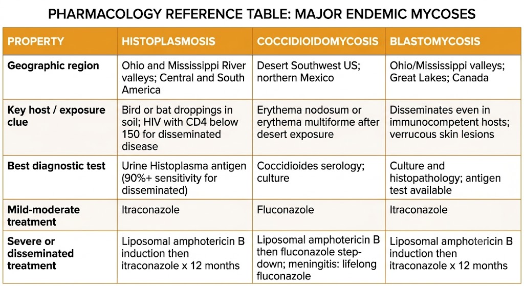
Histoplasma capsulatum is endemic to the Ohio and Mississippi River valleys of the United States, and to parts of Central and South America. Infection occurs through inhalation of microconidia from soil enriched with bird or bat droppings. The vast majority of infections in immunocompetent hosts are asymptomatic or cause a self-limited flu-like illness that resolves without treatment. Clinically significant disease includes acute pulmonary histoplasmosis (presenting with fever, headache, myalgia, and non-productive cough after heavy exposure), chronic pulmonary histoplasmosis (resembling tuberculosis, occurring in patients with underlying lung disease), and progressive disseminated histoplasmosis (PDH) in immunocompromised patients — particularly those with HIV infection and CD4 counts below 150 cells per microliter — presenting with fever, weight loss, hepatosplenomegaly, mucocutaneous lesions, and cytopenias. Urine Histoplasma antigen is the most sensitive diagnostic test for disseminated and moderate-to-severe pulmonary disease (sensitivity exceeding 90 percent for PDH).

Treatment is severity-based. Mild to moderate pulmonary histoplasmosis: itraconazole 200 mg three times daily for three days (loading), then twice daily for six to twelve weeks. Moderately severe to severe histoplasmosis and PDH in immunocompromised patients: liposomal amphotericin B induction for one to two weeks, followed by itraconazole step-down for at least twelve months. Secondary prophylaxis with itraconazole 200 mg daily is recommended for HIV-positive patients with a history of disseminated disease until the CD4 count exceeds 150 cells per microliter on antiretroviral therapy for at least six months.

Coccidioidomycosis

Coccidioides immitis (California) and Coccidioides posadasii (other endemic areas) cause coccidioidomycosis in the desert Southwest United States, northern Mexico, and parts of Central and South America. Infection occurs through inhalation of arthroconidia. Approximately 60 percent of infections are asymptomatic; symptomatic primary pulmonary coccidioidomycosis (Valley Fever) presents with fever, cough, chest pain, fatigue, and characteristic skin findings including erythema nodosum and erythema multiforme. The majority of immunocompetent patients recover without antifungal therapy. Dissemination — occurring in less than one percent of immunocompetent patients but in significantly higher proportions of immunocompromised patients, pregnant women, and individuals of African or Filipino ancestry — can involve bone, joints, skin, and the central nervous system (CNS). Coccidioidal meningitis is the most feared complication and requires lifelong antifungal suppression because it cannot be cured with currently available agents.

Treatment: fluconazole is the drug of choice for most non-meningeal and meningeal coccidioidomycosis because of its excellent CNS penetration. Itraconazole is preferred for bone and joint disease. Liposomal amphotericin B is used for severe disseminated disease requiring initial induction before step-down to azole therapy. Coccidioidal meningitis requires lifelong fluconazole suppression at 400 mg daily; intrathecal amphotericin B, historically used for meningitis refractory to azole therapy, is rarely employed today.

Blastomycosis

Blastomyces dermatitidis is endemic to the Ohio and Mississippi River valleys, Great Lakes region, and parts of Canada. It causes pulmonary infection through inhalation and disseminates to skin, bone, and genitourinary tract more readily than other endemic fungi even in immunocompetent hosts. Cutaneous blastomycosis — verrucous or ulcerative skin lesions — is the most common manifestation of disseminated disease. Treatment follows a severity-based approach: itraconazole for mild to moderate pulmonary and non-life-threatening disseminated disease; liposomal amphotericin B induction for moderately severe to severe pulmonary disease, central nervous system (CNS) involvement, or immunocompromised hosts, followed by itraconazole step-down for twelve months.

Reference table comparing histoplasmosis, coccidioidomycosis, and blastomycosis across five properties: geographic region, key host and exposure clue, best diagnostic test, mild-to-moderate treatment, and severe or disseminated treatment. Histoplasmosis and blastomycosis both use itraconazole for mild disease and liposomal amphotericin B induction then itraconazole for severe disease. Coccidioidomycosis uses fluconazole for most disease including meningitis, where it must be continued lifelong.
Endemic mycoses comparison: histoplasmosis, coccidioidomycosis, and blastomycosis across geographic distribution, host clues, diagnosis, and treatment by severity. Gemini-generated figure.
Module 7 Summary — Drug of Choice by Syndrome

Candidiasis (invasive): echinocandin first-line; step down to fluconazole when stable and susceptible. Invasive aspergillosis: voriconazole or isavuconazole first-line; liposomal amphotericin B alternative. Cryptococcal meningitis induction: liposomal amphotericin B plus flucytosine plus fluconazole (one week, WHO 2022). Consolidation: fluconazole 400 mg daily for eight weeks. Maintenance: fluconazole 200 mg daily. Mucormycosis: liposomal amphotericin B plus urgent surgical debridement; step down to posaconazole or isavuconazole oral; voriconazole has no activity. Histoplasmosis (mild-moderate): itraconazole. Histoplasmosis (severe or disseminated): liposomal amphotericin B then itraconazole. Coccidioidomycosis: fluconazole (including meningitis, lifelong). Blastomycosis (mild-moderate): itraconazole. Blastomycosis (severe or CNS): liposomal amphotericin B then itraconazole.

Suggested References
Author / Organization Title Source
Pappas PG, Kauffman CA, Andes DR, et al. Clinical practice guideline for the management of candidiasis: 2016 update by the Infectious Diseases Society of America Clin Infect Dis. 2016;62(4):e1-e50
Kullberg BJ, Arendrup MC Invasive fungal disease in the patient with cancer N Engl J Med. 2015;373(15):1445-1456
Patterson TF, Thompson GR, Denning DW, et al. Practice guidelines for the diagnosis and management of aspergillosis: 2016 update by the Infectious Diseases Society of America Clin Infect Dis. 2016;63(4):e1-e60
Herbrecht R, Denning DW, Patterson TF, et al. Voriconazole versus amphotericin B for primary therapy of invasive aspergillosis N Engl J Med. 2002;347(6):408-415
Perfect JR, Dismukes WE, Dromer F, et al. Clinical practice guidelines for the management of cryptococcal disease: 2010 update by the Infectious Diseases Society of America Clin Infect Dis. 2010;50(3):291-322
World Health Organization Guidelines for Diagnosing, Preventing and Managing Cryptococcal Disease Among Adults, Adolescents and Children Living with HIV Geneva: WHO; 2022
Cornely OA, Alastruey-Izquierdo A, Arenz D, et al. Global guideline for the diagnosis and management of mucormycosis: an initiative of the European Confederation of Medical Mycology in cooperation with the Mycoses Study Group Education and Research Consortium Lancet Infect Dis. 2019;19(12):e405-e421
Marr KA, Schlamm HT, Herbrecht R, et al. Combination antifungal therapy for invasive aspergillosis: a randomized trial Ann Intern Med. 2015;162(2):81-89
Wheat LJ, Freifeld AG, Kleiman MB, et al. Clinical practice guidelines for the management of patients with histoplasmosis: 2007 update by the Infectious Diseases Society of America Clin Infect Dis. 2007;45(7):807-825
Galgiani JN, Ampel NM, Blair JE, et al. 2016 Infectious Diseases Society of America (IDSA) clinical practice guideline for the treatment of coccidioidomycosis Clin Infect Dis. 2016;63(6):e112-e146