CHAPTER 41  ·  ANTI-INFLAMMATORY DRUGS
Section 1

Glucocorticoid Receptor Mechanisms and Anti-Inflammatory Actions

Receptor activation, nuclear translocation, transactivation and transrepression, and the multi-target suppression of inflammation

Abbreviations used in this module: glucocorticoid receptor (GR), glucocorticoid (GC), nuclear factor kappa B (NF-κB), activator protein-1 (AP-1), phospholipase A2 (PLA2), cyclooxygenase (COX), cyclooxygenase-2 (COX-2), lipoxygenase (LOX), nonsteroidal anti-inflammatory drug (NSAID), hypothalamic-pituitary-adrenal (HPA), cytochrome P450 3A4 (CYP3A4), inhaled corticosteroid (ICS), long-acting beta-2 agonist (LABA), chronic obstructive pulmonary disease (COPD), adrenocorticotropic hormone (ACTH), proton pump inhibitor (PPI).

Corticosteroids exert their anti-inflammatory effects through a single intracellular receptor — the GR — that functions as a ligand-activated transcription factor. Once activated, GR enters the nucleus and simultaneously turns on anti-inflammatory genes and turns off pro-inflammatory genes. This transcriptional duality explains both the therapeutic breadth and the extensive adverse effect profile of corticosteroid therapy: the GR is expressed in virtually every nucleated cell in the body.

Receptor Activation and Nuclear Translocation

In the absence of ligand, the GR resides in the cytoplasm held in an inactive complex with heat shock proteins (including heat shock protein 90) that keep it sequestered and prevent inadvertent gene activation. GCs are lipophilic and freely cross the cell membrane. When a GC binds the GR, the heat shock protein complex dissociates, a nuclear localization signal is exposed, and the GC-GR complex is actively transported into the nucleus within minutes. This rapid nuclear entry is why the GR activation step itself is fast, even though the downstream transcriptional changes unfold over hours.

Transactivation — Turning On Anti-Inflammatory Genes

In the nucleus, the GC-GR complex can dimerize and bind to specific DNA sequences called glucocorticoid response elements in the promoter regions of target genes. This direct DNA binding (transactivation) upregulates genes whose protein products have anti-inflammatory roles. The most pharmacologically important transactivation target is annexin A1 (formerly called lipocortin-1), an endogenous inhibitor of PLA2. By inducing annexin A1, corticosteroids suppress the rate-limiting step in eicosanoid synthesis — the liberation of arachidonic acid from membrane phospholipids — blocking both the COX pathway and the LOX pathway simultaneously. This upstream PLA2 block is the critical mechanistic distinction from NSAIDs, which act downstream of PLA2 and block only the COX pathway.

Other transactivation targets include anti-inflammatory signaling proteins that dampen the inflammatory cascade at multiple points. However, transactivation also drives many of the metabolic side effects of corticosteroids: upregulation of gluconeogenic enzymes, promotion of lipolysis and adipose redistribution, and catabolism of muscle protein are all GRE-driven transactivation events.
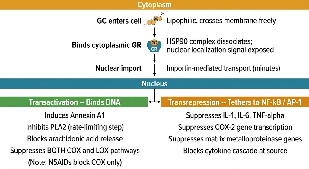
Flow diagram of glucocorticoid receptor activation: GC enters cell and binds cytoplasmic GR causing HSP90 dissociation and nuclear localization signal exposure; importin-mediated nuclear import occurs within minutes; in the nucleus, transactivation via DNA binding induces annexin A1 to inhibit PLA2 and block both COX and LOX pathways; transrepression via tethering to NF-kB and AP-1 suppresses IL-1, IL-6, TNF-alpha, COX-2 gene transcription, and matrix metalloproteinase genes.
Glucocorticoid receptor activation pathway: cytoplasmic GR-ligand binding, nuclear translocation, and the two nuclear mechanisms — transactivation (PLA2 inhibition via annexin A1) and transrepression (NF-κB/AP-1 silencing). Figure generated with Gemini AI for educational use.
Transrepression — Silencing Pro-Inflammatory Genes

The second nuclear mechanism — transrepression — does not require the GR to bind DNA directly. Instead, the activated GR tethers to pro-inflammatory transcription factors, principally NF-κB and AP-1, through protein-protein interactions. This tethering prevents NF-κB and AP-1 from binding to their target gene promoters, blocking transcription of a broad array of pro-inflammatory mediators: interleukin-1 beta, interleukin-2, interleukin-6, interleukin-8, tumor necrosis factor-alpha, COX-2, and matrix metalloproteinases. The suppression of COX-2 gene transcription by this mechanism adds a second layer of eicosanoid inhibition on top of the upstream PLA2 block from annexin A1 induction.

This combination — upstream PLA2 inhibition via annexin A1 and downstream COX-2 gene suppression via NF-κB transrepression — explains why corticosteroids are more potent anti-inflammatory agents than any NSAID at equivalent doses. NSAIDs can only block the COX enzyme; corticosteroids reduce the amount of arachidonic acid entering the pathway and simultaneously suppress the inducible COX-2 gene that produces the inflammatory enzyme in the first place.

Effects on Leukocytes and Vascular Permeability

Beyond eicosanoid suppression and cytokine gene transrepression, corticosteroids produce coordinated changes in leukocyte behavior that reduce inflammatory cell infiltration into tissues. Neutrophils are rapidly demarginated from vascular endothelium (because corticosteroids reduce adhesion molecule expression), causing a characteristic neutrophilia in the blood within hours of a corticosteroid dose. However, neutrophil migration into inflamed tissues is simultaneously reduced through suppression of chemokine production. The net effect is more circulating neutrophils but less tissue neutrophil infiltration.

Eosinophils are exquisitely sensitive to corticosteroids: circulating eosinophil counts fall within hours because corticosteroids promote eosinophil apoptosis and reduce the interleukin-5 and granulocyte-macrophage colony-stimulating factor survival signals that normally sustain them. This rapid eosinophil reduction is the basis for corticosteroids' effectiveness in eosinophilic asthma, allergic rhinitis, and eosinophilic esophagitis. Lymphocytes are redistributed from the bloodstream into lymphoid organs, producing lymphopenia; at high doses, lymphocyte apoptosis also occurs, contributing to immunosuppression. Vascular permeability is reduced through suppression of histamine release and downregulation of vascular endothelial growth factor, attenuating the edema component of inflammation.

Corticosteroids vs NSAIDs — Why Steroids Are Broader

NSAIDs: block COX enzyme only; leave LOX pathway and cytokine production intact; no effect on leukocyte trafficking. Corticosteroids: induce annexin A1 → inhibit PLA2 → block both COX and LOX branches; transrepress NF-κB and AP-1 → suppress cytokine genes and COX-2 gene; promote eosinophil apoptosis; reduce vascular permeability. Corticosteroids suppress inflammation at more points in the cascade simultaneously, producing greater potency at the cost of broader adverse effects including immunosuppression, metabolic changes, and HPA axis suppression that NSAIDs do not produce.

Section 2

Pharmacokinetics, Potency, and Drug Interactions

Prednisone prodrug activation, CYP3A4 metabolism and key inhibitor interactions, comparative glucocorticoid and mineralocorticoid potency, and dose equivalence

Corticosteroids share several pharmacokinetic features but differ meaningfully in potency, mineralocorticoid activity, half-life, and metabolic pathway. The comparative potency table — anchored to hydrocortisone as the reference compound — is essential clinical knowledge for dose conversions when switching agents and for predicting which adverse effects are most likely with a given drug.

Oral Absorption and Prodrug Activation

Most oral corticosteroids are well absorbed from the gastrointestinal tract. Prednisone is the most widely prescribed oral corticosteroid and is a pharmacologically inactive prodrug. It requires hepatic conversion by 11-beta-hydroxysteroid dehydrogenase type 1 to its active form, prednisolone. In patients with severe hepatic disease, this conversion may be unreliable, and prednisolone (the active form) is preferred because it bypasses the activation step. The oral-to-intravenous conversion is approximately 1:1 for methylprednisolone and prednisolone, simplifying route transitions.

CYP3A4 Metabolism and Drug Interactions

All systemic corticosteroids are substrates of CYP3A4, the primary hepatic and intestinal wall metabolizing enzyme for this drug class. CYP3A4 inhibitors reduce corticosteroid clearance and raise plasma levels, potentially causing Cushing syndrome features at doses not normally associated with toxicity. The most clinically dangerous inhibitor interaction involves ritonavir — a component of many antiretroviral regimens — which profoundly inhibits CYP3A4 and can raise fluticasone (inhaled) or triamcinolone (injected) systemic exposure to levels producing iatrogenic Cushing syndrome and secondary adrenal insufficiency, even when the corticosteroid dose itself is non-systemic. Azole antifungals (ketoconazole, itraconazole, voriconazole) are potent CYP3A4 inhibitors frequently co-prescribed with corticosteroids in immunosuppressed patients; corticosteroid dose adjustment is appropriate when azoles are initiated. Diltiazem, clarithromycin, and erythromycin are additional moderate inhibitors.

CYP3A4 inducers accelerate corticosteroid metabolism. Rifampin is the most potent inducer clinically, reducing prednisolone plasma exposure by 50 to 75%, potentially causing therapeutic failure and in dependent patients precipitating adrenal crisis when rifampin is started without corticosteroid dose adjustment. This interaction is encountered in patients with tuberculosis who are also receiving corticosteroids for another indication.

Comparative Potency and Mineralocorticoid Activity

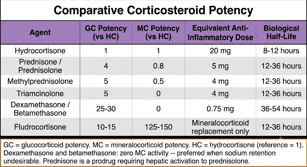
Corticosteroid potency is expressed relative to hydrocortisone, which serves as the reference compound with a glucocorticoid potency of 1 and a mineralocorticoid potency of 1. Mineralocorticoid activity is clinically important: activation of the mineralocorticoid receptor in the renal collecting duct promotes sodium retention and potassium excretion, causing hypertension, hypokalemia, and edema. The glucocorticoid-to-mineralocorticoid activity ratio differs substantially across agents, which determines clinical agent selection.

Dexamethasone and betamethasone have negligible mineralocorticoid activity (potency approximately 0), making them preferred when sodium retention is undesirable — as in cerebral edema management, fetal lung maturation, and treatment of congenital adrenal hyperplasia. Hydrocortisone, with equal glucocorticoid and mineralocorticoid potency, is the preferred agent for physiological replacement in adrenal insufficiency because it replaces both hormones simultaneously at physiological doses. Fludrocortisone has extremely high mineralocorticoid potency and negligible anti-inflammatory activity at clinically used doses; it is used exclusively for mineralocorticoid replacement.

Anti-inflammatory dose equivalence is the practical conversion tool: hydrocortisone 20 mg equals prednisone 5 mg equals methylprednisolone 4 mg equals dexamethasone 0.75 mg in anti-inflammatory potency. These equivalences allow safe dose conversion when switching agents for clinical reasons such as reducing mineralocorticoid side effects or changing to a longer-acting agent.

Comparative corticosteroid potency table with five columns: agent name, glucocorticoid potency relative to hydrocortisone, mineralocorticoid potency relative to hydrocortisone, equivalent anti-inflammatory dose, and biological half-life. Rows include hydrocortisone (1, 1, 20 mg, 8-12 h), prednisone/prednisolone (4, 0.8, 5 mg, 12-36 h), methylprednisolone (5, 0.5, 4 mg, 12-36 h), triamcinolone (5, 0, 4 mg, 12-36 h), dexamethasone/betamethasone (25-30, 0, 0.75 mg, 36-54 h), and fludrocortisone (10-15, 125-150, mineralocorticoid replacement only, 12-36 h).
Comparative corticosteroid potency: glucocorticoid (GC) and mineralocorticoid (MC) potency relative to hydrocortisone, with equivalent anti-inflammatory doses and biological half-lives. Figure generated with Gemini AI for educational use.
Dose Conversion Anchors

Hydrocortisone 20 mg = Prednisone or prednisolone 5 mg = Methylprednisolone or triamcinolone 4 mg = Dexamethasone or betamethasone 0.75 mg. Dexamethasone and betamethasone have zero mineralocorticoid activity — use when sodium retention is undesirable. Prednisone is the standard oral agent for most chronic indications; methylprednisolone sodium succinate is preferred intravenously for acute situations. In severe hepatic disease, use prednisolone (active form) rather than prednisone (prodrug).


Section 3

Systemic Clinical Indications and Inhaled Corticosteroids

Emergency and acute applications, chronic autoimmune indications, adrenal insufficiency replacement, and the pharmacokinetic basis of ICS therapeutic advantage

Systemic corticosteroids have the broadest clinical application of any drug class in medicine. They are used across the spectrum from acute emergencies to lifelong hormone replacement. Inhaled corticosteroids represent one of the most successful applications of targeted drug delivery in pharmacology — achieving local airway anti-inflammatory potency while minimizing systemic exposure through pharmacokinetic design.

Emergency and Acute Applications

In anaphylaxis, corticosteroids are given as adjunctive therapy after epinephrine — hydrocortisone or methylprednisolone intravenously — with the rationale of preventing or attenuating a biphasic anaphylactic reaction occurring hours after the initial event. Epinephrine remains the first and only essential drug in anaphylaxis; corticosteroids are supportive.

In acute severe asthma, systemic corticosteroids administered within the first hour of emergency presentation reduce hospital admission rates. Both intravenous methylprednisolone and oral prednisolone are effective and approximately equivalent in outcome when the oral route is feasible. The anti-inflammatory mechanism takes hours to manifest, so corticosteroids complement bronchodilators rather than replace them in the acute setting.

In giant cell arteritis, high-dose prednisone must be started immediately when the clinical picture is consistent with the diagnosis, without waiting for temporal artery biopsy results. The ophthalmic and posterior ciliary arteries supplying the retina can occlude from granulomatous inflammation, causing rapid and permanent vision loss. The biopsy should be performed within one to two weeks of starting corticosteroids — histological findings persist for this period despite steroid treatment.

In cerebral edema from brain tumors or metastases, dexamethasone is preferred because its negligible mineralocorticoid activity avoids sodium retention and because its long biological half-life enables twice-daily or four-times-daily dosing. Dexamethasone is ineffective in cytotoxic edema from ischemic stroke and should not be used in that setting.

Chronic Autoimmune and Inflammatory Indications

Corticosteroids are used as induction therapy for many serious autoimmune and inflammatory conditions, typically followed by steroid-sparing immunosuppressive agents as maintenance. In systemic lupus erythematosus with nephritis or major organ involvement, high-dose prednisone combined with cyclophosphamide or mycophenolate is standard induction. In polymyalgia rheumatica, low-dose prednisone produces dramatic symptomatic relief within days — a response so characteristic that it serves as a diagnostic criterion. In autoimmune hemolytic anemia and immune thrombocytopenic purpura, prednisone at 1 mg per kg per day is first-line. In organ transplantation, corticosteroids remain a core component of immunosuppression protocols and first-line treatment for acute cellular rejection.

Adrenal Insufficiency Replacement

In primary adrenal insufficiency (Addison disease), both glucocorticoid and mineralocorticoid replacement are required. Hydrocortisone given in divided doses — approximately two-thirds in the morning and one-third in the early afternoon — mimics the normal diurnal cortisol pattern and avoids HPA axis suppression better than once-daily dosing. Fludrocortisone provides mineralocorticoid replacement. In secondary adrenal insufficiency from pituitary disease or exogenous corticosteroid withdrawal, mineralocorticoid replacement is not required because aldosterone secretion via the renin-angiotensin-aldosterone system is intact. All patients with adrenal insufficiency require sick day rules education and an emergency injectable hydrocortisone kit.

Inhaled Corticosteroids — Pharmacokinetic Basis of the Therapeutic Advantage

ICS achieve high local airway concentrations while minimizing systemic exposure through two complementary pharmacokinetic features. First, inhaled particles deposit drug directly on inflamed airway mucosa, producing high local concentrations with a small administered dose. Second, the fraction of drug that reaches systemic circulation — from both pulmonary absorption and swallowed oropharyngeal drug — undergoes rapid first-pass hepatic metabolism by CYP3A4, limiting systemic bioavailability. Fluticasone propionate and fluticasone furoate have essentially zero oral bioavailability due to near-complete hepatic first-pass extraction; the oropharyngeal fraction that is swallowed contributes negligibly to systemic exposure. This pharmacokinetic design gives ICS a therapeutic window that is far wider than equivalent systemic doses.

ICS are the cornerstone of chronic asthma management. They reduce airway inflammation, decrease exacerbation frequency, and improve symptom control across all levels of asthma severity requiring maintenance therapy. At higher asthma severity steps, ICS are combined with a LABA — a combination that is more effective than doubling the ICS dose, because LABAs and corticosteroids are pharmacologically synergistic: LABAs enhance GR nuclear translocation and corticosteroid responsiveness, while corticosteroids prevent the LABA-induced beta-2 receptor downregulation that would otherwise limit bronchodilator efficacy. LABAs must never be used as asthma monotherapy without an ICS — clinical trial evidence demonstrates increased asthma mortality with LABA alone.

In COPD, the role of ICS is more restricted. Triple inhaled therapy combining an ICS with a LABA and a long-acting muscarinic antagonist reduces exacerbation rates and mortality in patients with frequent exacerbations and blood eosinophil counts above 300 cells per microliter. Blood eosinophil count is a practical biomarker that predicts ICS responsiveness in COPD; ICS confer little benefit and increase pneumonia risk in COPD patients with low eosinophil counts.

Local adverse effects of ICS — oropharyngeal candidiasis and dysphonia — are reduced by using a spacer device, rinsing the mouth after each dose, and choosing prodrug ICS that are inactive at the oropharyngeal mucosa (such as ciclesonide).

Giant Cell Arteritis — Do Not Wait for Biopsy

When the clinical picture is consistent with giant cell arteritis — age above 50, new headache, jaw claudication, tender temporal artery, elevated erythrocyte sedimentation rate or C-reactive protein — start high-dose prednisone (40 to 60 mg per day) immediately. The ophthalmic artery can occlude from ongoing granulomatous inflammation, causing permanent bilateral blindness. Temporal artery biopsy should be performed within one to two weeks — histological findings persist for this period. Never delay corticosteroid treatment pending biopsy results.


Section 4

HPA Axis Suppression, Stress Dosing, and Tapering

Mechanism and predictors of HPA axis suppression, secondary adrenal insufficiency, stress dosing principles, and tapering strategy

HPA axis suppression is the single most common and potentially most dangerous consequence of systemic corticosteroid therapy. Understanding its mechanism, predicting its likelihood, and managing withdrawal safely are mandatory clinical competencies for any prescriber who uses corticosteroids beyond short courses.

Mechanism of HPA Axis Suppression

Cortisol normally regulates its own production through negative feedback at the hypothalamus and pituitary. Exogenous GCs bind GRs in hypothalamic and pituitary cells, suppressing the transcription of corticotropin-releasing hormone and ACTH respectively. As exogenous corticosteroid therapy continues, chronically low ACTH levels cause progressive adrenal cortical atrophy — the adrenal gland literally decreases in size and functional capacity because its primary trophic stimulus (ACTH) has been removed.

When exogenous corticosteroids are withdrawn, the atrophied adrenal gland cannot immediately resume normal cortisol production. The hypothalamus and pituitary also require time to restore their normal pulsatile secretion patterns after prolonged suppression. During this recovery period, the patient is in a state of secondary adrenal insufficiency — they cannot generate the surge in cortisol output (from a basal rate of approximately 8 to 10 mg per day to 75 to 150 mg per day) that physiological stress requires.

Predictors of HPA Suppression

Any patient who has received more than the equivalent of prednisone 20 mg per day for more than three weeks is likely to have clinically significant HPA suppression. Lower doses and longer durations may also cause suppression. Morning dosing causes less suppression than evening or divided dosing because exogenous morning drug overlaps with the natural cortisol peak rather than suppressing the trough that normally triggers corticotropin-releasing hormone and ACTH release. Dexamethasone's long biological half-life (36 to 54 hours) makes it particularly prone to HPA suppression even at modest doses and is a reason it is not preferred for long-term anti-inflammatory maintenance therapy despite its high potency. Alternate-day dosing — giving the total dose every 48 hours rather than daily — substantially reduces HPA suppression while preserving anti-inflammatory efficacy in many indications.

Secondary Adrenal Insufficiency — Clinical Features

Secondary adrenal insufficiency from exogenous corticosteroid use differs from primary adrenal insufficiency (Addison disease) in two clinically important ways. First, mineralocorticoid production is preserved because aldosterone is regulated by the renin-angiotensin-aldosterone system, not ACTH — so salt craving, hyperkalemia, and marked hypotension are less prominent features. Second, hyperpigmentation does not occur because ACTH is low rather than high. Secondary adrenal insufficiency presents with fatigue, nausea, anorexia, arthralgia, myalgia, and orthostatic hypotension. The dangerous clinical event is an adrenal crisis triggered by physiological stress — surgery, severe infection, or major trauma — in a patient whose adrenal axis cannot mount the required cortisol response. Patients on chronic corticosteroids should carry a steroid emergency card and a prefilled hydrocortisone injection kit (100 mg intramuscularly) and must be taught sick day rules.

Stress Dosing and Tapering

Stress dosing provides supplemental corticosteroid during physiological stress in patients with actual or presumed HPA suppression. For minor illness with fever, doubling or tripling the usual oral corticosteroid dose is generally appropriate. For major surgery or intensive care admission, hydrocortisone 50 to 100 mg intravenously every 6 to 8 hours provides adequate stress coverage, with tapering to the basal dose over 24 to 48 hours postoperatively as the patient recovers. Patients who cannot take oral medications due to vomiting or surgery require parenteral hydrocortisone regardless of the clinical situation's severity.

Tapering strategy must balance two risks: relapse of the underlying condition if tapered too quickly, and prolonged HPA suppression if the dose is maintained unnecessarily. Short corticosteroid courses — less than three weeks at any dose — can generally be stopped abruptly without a formal taper, because HPA suppression is unlikely. Longer courses require gradual reduction. A practical approach reduces the dose in steps of 5 to 10 mg per week until the physiological replacement range (approximately 5 to 7.5 mg per day of prednisone) is reached, then reduces more slowly — by 1 mg every one to two weeks — while monitoring for symptoms of adrenal insufficiency. A morning serum cortisol below 3 micrograms per deciliter indicates incomplete recovery and warrants a temporary pause in the taper.

Sick Day Rules — Every Patient on Chronic Corticosteroids

Any corticosteroid course exceeding 20 mg per day prednisone for more than three weeks: counsel patient on the following. Febrile illness or significant stress: double or triple the oral dose until recovered. Vomiting or inability to take oral medications: inject 100 mg hydrocortisone intramuscularly immediately and seek emergency care. Major surgery: inform the anesthetic team; expect supplemental stress-dose hydrocortisone perioperatively. Never stop corticosteroids abruptly after more than three weeks of use. Carry the steroid emergency card and injectable hydrocortisone at all times during any taper period.


Suggested References
Author / Organization Title Source
Oakley RH, Cidlowski JA The biology of the glucocorticoid receptor: new signaling mechanisms in health and disease J Allergy Clin Immunol. 2013;132(5):1033-1044
Coutinho AE, Chapman KE The anti-inflammatory and immunosuppressive effects of glucocorticoids, recent developments and mechanistic insights Mol Cell Endocrinol. 2011;335(1):2-13
Barnes PJ Corticosteroid effects on cell signalling Eur Respir J. 2006;27(2):413-426
Rhen T, Cidlowski JA Antiinflammatory action of glucocorticoids — new mechanisms for old drugs N Engl J Med. 2005;353(16):1711-1723
Franchimont D Overview of the actions of glucocorticoids on the immune response Ann N Y Acad Sci. 2004;1024:124-137
Liu D, Ahmet A, Ward L, et al A practical guide to the monitoring and management of the complications of systemic corticosteroid therapy Allergy Asthma Clin Immunol. 2013;9(1):30
Nicolaides NC, Pavlaki AN, Chrousos GP Glucocorticoid therapy and adrenal suppression Endotext. MDText.com; 2018
Czock D, Keller F, Rasche FM, Haussler U Pharmacokinetics and pharmacodynamics of systemically administered glucocorticoids Clin Pharmacokinet. 2005;44(1):61-98
Spies CM, Strehl C, van der Goes MC, Bijlsma JW, Buttgereit F Glucocorticoids Best Pract Res Clin Rheumatol. 2011;25(6):891-900
Derendorf H, Hochhaus G, Meibohm B, Mollmann H, Barth J Pharmacokinetics and pharmacodynamics of inhaled corticosteroids J Allergy Clin Immunol. 1998;101(4 Pt 2):S440-S446
Global Initiative for Asthma (GINA) Global Strategy for Asthma Management and Prevention Updated 2023. ginasthma.org/gina-reports/
Dinsen S, Baslund B, Klose M, et al Why glucocorticoid withdrawal may sometimes be as dangerous as the treatment itself Eur J Intern Med. 2013;24(8):714-720
Rushworth RL, Torpy DJ, Falhammar H Adrenal crises: perspectives and research directions Endocrine. 2017;55(2):336-345