CHAPTER 14  ·  GENERAL ANESTHESIA

Section 1

Introduction

The central nervous system and cardiovascular system are simultaneously the targets and the dose-limiting toxicity sites of inhalational anesthetics

Inhalational anesthetics produce their desired effects — unconsciousness and immobility — through actions on the central nervous system. But the same central nervous system effects extend beyond consciousness suppression to alter cerebral blood flow, cerebral metabolic rate, intracranial pressure, and seizure threshold. Simultaneously, the cardiovascular system is profoundly affected: blood pressure, heart rate, myocardial contractility, and vascular resistance are all modified in ways that are both agent-specific and dose-dependent. Understanding these effects is the foundation for safe agent selection, dose management, and anticipation of adverse events — especially in patients with neurological or cardiac disease.


Section 2

Central Nervous System Effects: General Principles

How inhalational agents suppress neuronal activity and the key tension between metabolic protection and intracranial pressure elevation

All inhalational anesthetics suppress central nervous system neuronal activity, producing unconsciousness through potentiation of gamma-aminobutyric acid type A receptor-mediated inhibition and, for nitrous oxide, N-methyl-D-aspartate receptor antagonism. This suppression produces characteristic dose-dependent electroencephalographic changes: progressive slowing, increasing amplitude, burst suppression, and ultimately an isoelectric tracing at supratherapeutic concentrations. Isoflurane most readily produces burst suppression at 1.5 to 2 minimum alveolar concentration — a property exploited to provide maximal metabolic protection during brief periods of deliberate vascular occlusion in neurosurgery.

Two central nervous system effects exist in clinical tension with each other. All volatile agents reduce the cerebral metabolic rate for oxygen — a desirable property that reduces the brain's vulnerability to ischemia. At the same time, all volatile agents cause cerebral vasodilation and increase cerebral blood flow, which raises intracranial blood volume and can elevate intracranial pressure in patients with reduced intracranial compliance. Managing this tension — maximizing metabolic protection while controlling intracranial pressure — is the defining challenge of neuroanesthesia.


Section 3

Seizure Activity

Enflurane is the only volatile anesthetic with clinically significant epileptogenic potential

The relationship between inhalational anesthetics and seizure activity is agent-specific. Enflurane is the clear outlier: at concentrations above approximately 2 minimum alveolar concentration, particularly in the presence of hypocapnia (which lowers seizure threshold), enflurane produces electroencephalographic spike-and-wave complexes and can induce generalized tonic-clonic seizures even in patients without pre-existing epilepsy. This epileptogenic property is concentration-dependent and was a primary reason for enflurane's displacement by isoflurane and sevoflurane.

Isoflurane, sevoflurane, and desflurane do not have meaningful epileptogenic potential at clinical doses and are safe in patients with seizure disorders. Isoflurane at high doses produces burst suppression — an antiepileptiform pattern. Isolated reports of electroencephalographic spike activity with sevoflurane during high-concentration induction exist, particularly in children, but overt clinical seizures attributable to sevoflurane are exceedingly rare. Halothane has no epileptogenic potential. Nitrous oxide may provide mild anticonvulsant effect through its N-methyl-D-aspartate receptor antagonism.

Seizure Risk Summary

Contraindicated in seizure disorders: enflurane — the only epileptogenic volatile anesthetic.

Safe in seizure disorders: isoflurane, sevoflurane, desflurane, halothane, nitrous oxide.

Aggravating factor: hypocapnia lowers seizure threshold and potentiates enflurane's epileptogenic effect.


Section 4

Cerebral Blood Flow and Autoregulation

Volatile agents dilate cerebral vessels; this effect can be attenuated by controlled hyperventilation

Cerebral Vasodilation

All volatile halogenated anesthetics cause dose-dependent cerebral vasodilation, increasing cerebral blood flow at concentrations above approximately 0.5 minimum alveolar concentration. This effect is a direct action on cerebrovascular smooth muscle, independent of changes in cerebral metabolic rate. The magnitude differs among agents: halothane produces the greatest cerebral blood flow increase at equivalent doses, while isoflurane and sevoflurane produce more modest increases. Desflurane is intermediate and similar to isoflurane.

Critically, this vasodilation can be substantially counteracted by controlled hyperventilation. Reducing the arterial carbon dioxide partial pressure through deliberate hyperventilation causes cerebral vasoconstriction via carbon dioxide-mediated changes in perivascular pH, largely offsetting the anesthetic-induced vasodilation. This is the pharmacological rationale for mild hyperventilation in neurosurgical patients — it blunts the anesthetic-induced rise in cerebral blood flow and intracranial pressure. The carbon dioxide vasomotor reactivity of cerebral vessels is preserved during inhalational anesthesia, making this a reliable and rapid intervention.
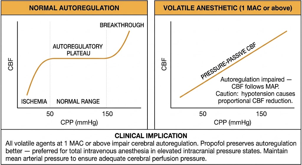
Two-panel diagram of cerebral autoregulation. Left panel shows the normal autoregulation curve with a flat plateau between 50 and 150 mmHg cerebral perfusion pressure, with ischemia below 50 mmHg and breakthrough above 150 mmHg. Right panel shows the pressure-passive linear relationship under volatile anesthetic at 1 MAC or above, where cerebral blood flow follows mean arterial pressure directly. A shared clinical implication box states that propofol preserves autoregulation better and is preferred for total intravenous anesthesia in elevated intracranial pressure states.
Cerebral autoregulation: normal curve versus pressure-passive flow under volatile anesthetic at 1 minimum alveolar concentration or above. Figure generated by Gemini AI.
Cerebral Autoregulation

Cerebral autoregulation is the ability of the cerebral vasculature to maintain relatively constant blood flow across a range of cerebral perfusion pressures (approximately 50 to 150 mmHg in healthy adults) by adjusting cerebrovascular resistance. Below the lower limit, vasodilation is maximal and blood flow falls passively with further pressure decreases, risking ischemia. Above the upper limit, forced vasodilation occurs with risk of hypertensive encephalopathy.

Volatile anesthetic agents impair cerebral autoregulation in a dose-dependent fashion. At concentrations of 1 minimum alveolar concentration or above, all volatile agents substantially reduce autoregulatory capacity, making cerebral blood flow more directly dependent on mean arterial pressure. Under deep volatile anesthesia, hypotension causes proportional reductions in cerebral blood flow, while hypertension causes proportional increases and potentially elevated intracranial pressure. This has direct implications for hemodynamic management in neurosurgical patients, where maintaining mean arterial pressure within a range that supports adequate cerebral perfusion pressure is a primary goal. Propofol, at clinical doses, preserves cerebral autoregulation better than volatile agents — one rationale for preferring total intravenous anesthesia in patients with compromised intracranial compliance.

Section 5

Intracranial Pressure

The Monro-Kellie doctrine explains why volatile agent-induced cerebral vasodilation raises intracranial pressure in vulnerable patients

The Monro-Kellie Doctrine

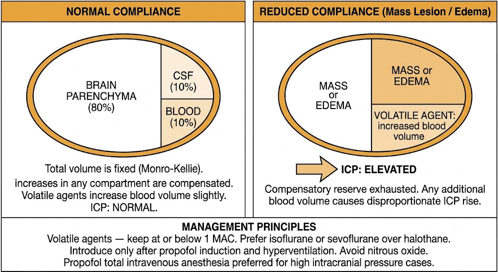
The cranial vault is a rigid compartment of fixed total volume. Its contents — brain parenchyma, cerebrospinal fluid, and intracranial blood — must sum to a constant. Any increase in one component must be compensated by a decrease in another, or intracranial pressure rises. In a patient with normal intracranial compliance (normal brain, no mass lesion), the modest increase in intracranial blood volume caused by volatile agent-induced vasodilation produces only a transient, easily compensated rise in intracranial pressure. In a patient with reduced intracranial compliance — mass lesion, traumatic brain injury, cerebral edema, or hydrocephalus — the same increase in blood volume produces a disproportionate and potentially dangerous intracranial pressure elevation.

Two-panel diagram of the Monro-Kellie doctrine. Left panel shows normal intracranial compliance with brain parenchyma at 80 percent, cerebrospinal fluid at 10 percent, and blood at 10 percent in a fixed oval cranial vault, with intracranial pressure normal. Right panel shows reduced compliance where mass or edema plus volatile agent-induced increased blood volume exhausts compensatory reserve, causing disproportionate intracranial pressure elevation. A shared management principles box outlines volatile agent precautions and preference for propofol total intravenous anesthesia in high intracranial pressure cases.
The Monro-Kellie doctrine: normal intracranial compliance versus reduced compliance with volatile agent-induced blood volume increase. Figure generated by Gemini AI.
Clinical Principles for Volatile Agent Use in Elevated Intracranial Pressure

When volatile agents are used in patients with elevated intracranial pressure or reduced intracranial compliance, several principles apply. Concentrations should be kept at or below 1 minimum alveolar concentration. Agents with the least cerebral vasodilatory effect — isoflurane or sevoflurane — are preferred over halothane. Volatile agents should be introduced only after induction with propofol (which reduces cerebral blood flow and cerebral metabolic rate without vasodilation) and after establishing hyperventilation. Nitrous oxide should generally be avoided in neurosurgical patients because it increases cerebral blood flow, increases cerebral metabolic rate, and can expand any intracranial gas collection.

Total intravenous anesthesia with propofol is often preferred for high-intracranial-pressure cases because propofol reduces both cerebral blood flow and cerebral metabolic rate without the vasodilatory liability of volatile agents, and it better preserves cerebral autoregulation.

Drugs Used to Reduce Intracranial Pressure

Several drug classes are used to reduce intracranial pressure by targeting different compartments of the Monro-Kellie equation. These are high-yield for understanding the pharmacological management of neurosurgical patients.

Intracranial Pressure Reduction

Mannitol

  • Osmotic diuretic — draws free water from brain parenchyma into plasma along an osmotic gradient, reducing brain water content and volume
  • Onset: 15 to 30 minutes; duration approximately 90 to 120 minutes
  • Requires an intact blood-brain barrier for full efficacy
  • Also transiently reduces blood viscosity, improving cerebral microcirculation
  • Monitor serum osmolality — risk of acute kidney injury at osmolality above 320 mOsm/kg

Intracranial Pressure Reduction

Hyperventilation and Dexamethasone

  • Hyperventilation: reduces arterial carbon dioxide partial pressure → cerebral vasoconstriction → reduces intracranial blood volume and intracranial pressure; fastest available intervention; effect wanes over 4 to 6 hours as cerebrospinal fluid bicarbonate adapts
  • Dexamethasone: glucocorticoid; reduces vasogenic cerebral edema associated with brain tumors and abscesses by decreasing blood-brain barrier permeability; onset hours to days; not effective for cytotoxic edema (stroke, traumatic brain injury)

Section 6

Cardiovascular Effects of Inhalational Agents

All volatile agents reduce blood pressure, but through different mechanisms with different clinical implications

All inhalational anesthetics reduce mean arterial pressure in a dose-dependent fashion, but the mechanism differs importantly among agents and determines which patients tolerate each agent better.

Mechanisms of Blood Pressure Reduction

Halothane reduces blood pressure primarily through direct myocardial depression — it reduces cardiac output by depressing contractility and suppressing sinoatrial node automaticity, causing bradycardia. Systemic vascular resistance is relatively maintained. Patients with impaired myocardial function tolerate this mechanism poorly.

Isoflurane, sevoflurane, and desflurane (at stable concentrations) reduce blood pressure primarily through peripheral vasodilation, lowering systemic vascular resistance. Cardiac output is relatively preserved because reduced afterload maintains stroke volume, and baroreceptor reflexes increase heart rate to compensate for vasodilation. This mechanism is better tolerated in patients with impaired myocardial function compared to halothane, because cardiac output is maintained despite falling blood pressure.

Heart Rate Effects

Halothane causes bradycardia through direct depression of sinoatrial node automaticity and sensitization to vagal tone — this is distinct from all modern agents. Isoflurane, sevoflurane, and nitrous oxide maintain or mildly increase heart rate through baroreceptor-mediated sympathetic reflexes in response to vasodilation. Desflurane at stable concentrations has a similar profile to isoflurane, but when the inspired concentration is rapidly increased, a marked transient sympathetic discharge produces tachycardia and hypertension — a property unique to desflurane and clinically important in patients with coronary artery disease.

Hypoxic Pulmonary Vasoconstriction Inhibition

Hypoxic pulmonary vasoconstriction is the physiological mechanism by which pulmonary blood flow is diverted away from poorly ventilated lung segments, maintaining ventilation-perfusion matching and oxygenation. All volatile halogenated agents inhibit hypoxic pulmonary vasoconstriction in a dose-dependent manner, worsening intrapulmonary shunting and contributing to intraoperative hypoxemia — this is particularly relevant during one-lung ventilation for thoracic surgery. Propofol does not inhibit hypoxic pulmonary vasoconstriction, making total intravenous anesthesia with propofol the preferred technique when oxygenation is marginal during one-lung ventilation.

Cardiovascular Profile

Halothane

  • Blood pressure reduction: myocardial depression
  • Heart rate: bradycardia (sinoatrial node depression)
  • Cardiac output: reduced (reduced contractility)
  • Catecholamine sensitization: YES — ventricular arrhythmias
  • Worst tolerated in: impaired myocardial function

Cardiovascular Profile

Isoflurane / Sevoflurane

  • Blood pressure reduction: peripheral vasodilation
  • Heart rate: maintained or mildly increased (reflex tachycardia)
  • Cardiac output: relatively preserved
  • Catecholamine sensitization: NO
  • Better tolerated than halothane in impaired myocardial function

Cardiovascular Profile

Desflurane

  • Stable concentrations: similar to isoflurane
  • Rapid concentration increase: sympathetic surge → tachycardia, hypertension
  • Avoid rapid increases in coronary artery disease or hypertensive heart disease
  • Catecholamine sensitization: NO

Suggested References
Author / Organization Title Source
Miller RD, ed. Miller's Anesthesia, 8th ed. Chapters 27, 57 Philadelphia: Elsevier; 2015
Stoelting RK, Hillier SC Pharmacology and Physiology in Anesthetic Practice, 4th ed. Philadelphia: Lippincott Williams & Wilkins; 2006
Franks NP Molecular targets underlying general anaesthesia Br J Pharmacol. 2006;147(Suppl 1):S72–S81
Voss LJ, et al. The howling cortex: seizures and general anesthetic drugs Anesth Analg. 2008;107(5):1689–1703
Cottrell JE, Patel P, eds. Cottrell and Patel's Neuroanesthesia, 6th ed. Chapters 3, 4, 9 Philadelphia: Elsevier; 2017
Naguib M, et al. Conceptual and technical insights into the basis of neuromuscular monitoring Anaesthesia. 2017;72(Suppl 1):16–37
Weiskopf RB, et al. Rapid increase in desflurane concentration is associated with greater transient cardiovascular stimulation than rapid increases in isoflurane concentration in humans Anesthesiology. 1994;80(5):1035–1045
Johnston RR, Eger EI 2nd, Wilson C A comparative interaction of epinephrine with enflurane, isoflurane, and halothane in man Anesth Analg. 1976;55(5):709–712
Flood P, Rathmell JP, Shafer SL, eds. Stoelting's Pharmacology and Physiology in Anesthetic Practice, 5th ed. Philadelphia: Wolters Kluwer; 2015