CHAPTER 36  ·  ANTIVIRAL PHARMACOLOGY
1. Immunocompromised Host · 2. CMV Beyond Retinitis · 3. EBV and PTLD · 4. Adenovirus and BK Virus · 5. JC Virus and PML · 6. HHV-6 and IRIS ↑ Top
Module Contents
Section 1
The Immunocompromised Host: Risk Stratification and Surveillance
Immune deficits, viral reactivation risk, and the framework for antiviral prophylaxis decisions

Opportunistic viral infections emerge when quantitative or qualitative defects in cellular immunity — most commonly T-lymphocyte depletion or dysfunction — allow latent herpesviruses and other DNA viruses to reactivate or permit primary infections to escape immune control. The degree and duration of immunosuppression are the primary determinants of infection risk, and the antiviral pharmacological approach must be calibrated accordingly.

The immunocompromised populations at highest risk for opportunistic viral infections can be stratified by the dominant immune defect. Hematopoietic stem cell transplant (HSCT) recipients experience the most profound and prolonged immunosuppression of any clinical population, with cellular immune reconstitution requiring 6–12 months after allogeneic transplantation and longer in patients with graft-versus-host disease (GVHD) requiring intensified immunosuppression. The combination of conditioning-induced lymphodepletion, graft-versus-host disease prophylaxis, and treatment of GVHD creates a window of extreme vulnerability spanning the engraftment period through at least the first year post-transplant. Solid organ transplant (SOT) recipients receive calcineurin inhibitor-based immunosuppression (cyclosporine or tacrolimus) that selectively suppresses T-lymphocyte activation, producing a risk profile shaped more by the intensity and duration of immunosuppression than by total lymphocyte count. HIV-positive patients with cluster of differentiation 4 (CD4) counts below 200 cells/mm³ — the threshold defining acquired immunodeficiency syndrome (AIDS) — are at risk for all opportunistic viral infections; those with CD4 below 50 cells/mm³ are at risk for the most severe manifestations including CMV retinitis, disseminated CMV, and progressive multifocal leukoencephalopathy (PML).

Surveillance for viral reactivation uses quantitative PCR-based viral load assays for CMV, EBV, adenovirus, and BK polyomavirus (BKPyV). CMV surveillance by PCR in peripheral blood is the cornerstone of pre-emptive therapy strategies in SOT and HSCT recipients, allowing treatment initiation before clinical disease develops. EBV viral load monitoring in HSCT recipients — particularly in the setting of T-cell depletion — provides an early signal for post-transplant lymphoproliferative disorder (PTLD), enabling pre-emptive rituximab before frank lymphoma develops. BKPyV surveillance by urine and plasma PCR after kidney transplantation detects nephropathy at the stage of viremia rather than established fibrosis. The pharmacological response to surveillance findings in each case depends on the specific virus, the magnitude of viral load, the clinical context, and available antiviral options — which vary widely across these pathogens.

Antiviral Availability by Opportunistic Virus

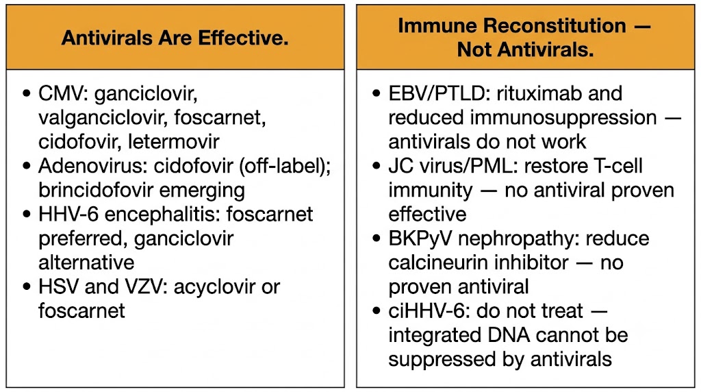
CMV: licensed antivirals available (ganciclovir, valganciclovir, foscarnet, cidofovir, letermovir, maribavir). Adenovirus and BKPyV: cidofovir is the primary antiviral (off-label for BKPyV); brincidofovir has emerging data. EBV/PTLD: no antiviral with proven clinical efficacy; management is immunological (rituximab, reduction of immunosuppression). JC virus/PML: no effective licensed antiviral; management centers on immune reconstitution. HHV-6: foscarnet and ganciclovir have activity but clinical benefit in transplant recipients is uncertain.

Section 2
CMV Beyond Retinitis: Colitis, Pneumonitis, and CNS Disease
End-organ manifestations, diagnostic pitfalls, and antiviral management by syndrome

CMV end-organ disease encompasses a spectrum of manifestations that differ in diagnostic approach, severity, and antiviral management. Understanding the syndrome-specific nuances — particularly the poor sensitivity of blood PCR for gastrointestinal CMV disease — is essential for avoiding misdiagnosis and delayed treatment.

CMV colitis is the most common gastrointestinal CMV manifestation in immunocompromised patients, presenting with profuse watery or bloody diarrhea, tenesmus, and abdominal cramping. A critical diagnostic pitfall is that blood CMV PCR is frequently undetectable or low-level in isolated CMV colitis — the virus replicates locally within colonic mucosa without generating systemic viremia sufficient to be detected in peripheral blood in many patients. Diagnosis therefore requires colonoscopy with biopsy demonstrating characteristic CMV cytopathic effect (enlarged cells with intranuclear and intracytoplasmic inclusions) confirmed by immunohistochemistry or in situ hybridization for CMV DNA. Treatment is intravenous ganciclovir induction (5 mg/kg every 12 hours) for 3–6 weeks followed by oral valganciclovir consolidation; response is monitored clinically and endoscopically, not by blood PCR. CMV esophagitis presents with odynophagia and may be indistinguishable from Candida esophagitis clinically; endoscopy with biopsy is required for diagnosis.
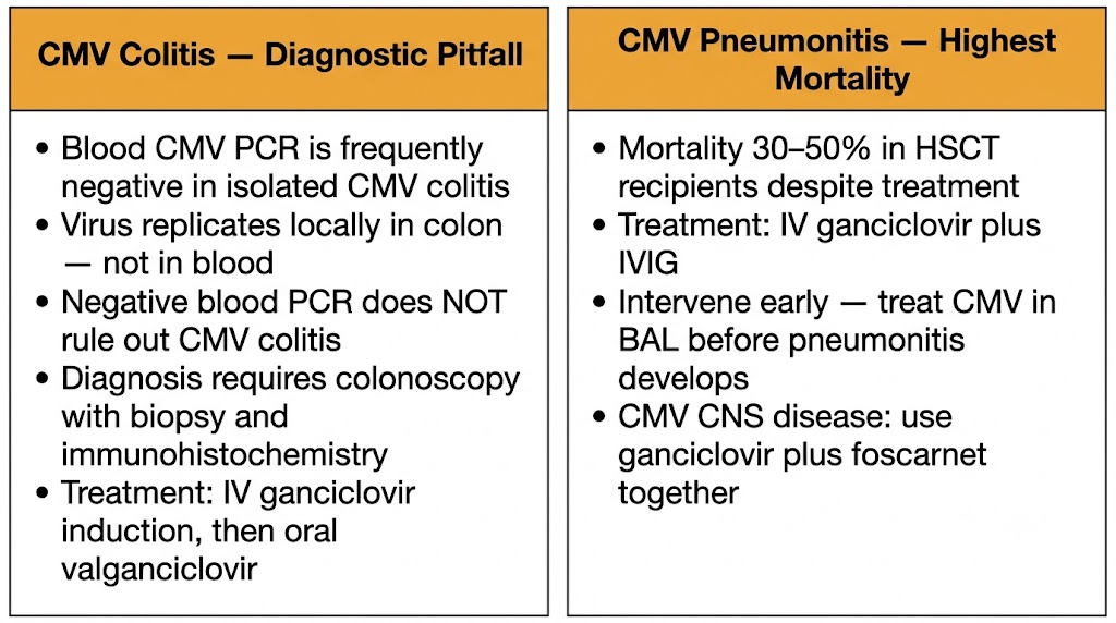
CMV colitis diagnostic pitfall and CMV pneumonitis management

CMV pneumonitis in HSCT recipients is the most life-threatening CMV end-organ manifestation, carrying mortality rates of 30–50% despite antiviral therapy in established disease. It typically presents 1–4 months post-transplant with progressive hypoxia, non-productive cough, and bilateral interstitial infiltrates. The combination of intravenous ganciclovir with intravenous immunoglobulin (IVIG) is standard of care based on retrospective cohort data demonstrating survival benefit over ganciclovir alone, though randomized controlled trial evidence is lacking. Progression from upper respiratory tract CMV infection to lower respiratory tract disease in HSCT recipients — which carries far higher mortality than upper respiratory tract disease — is the target of pre-emptive treatment with valganciclovir at the stage of CMV detection in bronchoalveolar lavage (BAL) samples.

CMV CNS disease encompasses encephalitis, ventriculoencephalitis, and polyradiculomyelopathy, occurring predominantly in patients with CD4 counts below 50 cells/mm³ in HIV disease and in HSCT recipients with severe T-cell depletion. CMV ventriculoencephalitis in advanced HIV presents with cognitive deterioration, confusion, and cranial nerve palsies, with MRI showing periventricular enhancement. CMV polyradiculomyelopathy presents with ascending flaccid paraparesis, bladder dysfunction, and sacral paresthesias — a distinctive syndrome that can be confused with other causes of myelopathy. CSF PCR for CMV DNA is the diagnostic test of choice, with sensitivity above 80% in CNS CMV disease. Treatment of CMV CNS disease uses combination ganciclovir plus foscarnet, exploiting mechanistic complementarity to achieve higher antiviral potency than either agent alone; this combination is used despite greater toxicity burden because of the severity of the syndrome and the importance of maximal viral suppression.
Section 3
EBV and Post-Transplant Lymphoproliferative Disorder
Pathogenesis, EBV viral load monitoring, rituximab, and the absence of effective antivirals

Post-transplant lymphoproliferative disorder (PTLD) is a spectrum of EBV-driven lymphoid proliferations ranging from polyclonal B-cell hyperplasia to frank diffuse large B-cell lymphoma (DLBCL), arising in the context of T-lymphocyte immunosuppression that removes the normal immune surveillance constraining EBV-infected B cells. PTLD illustrates a fundamental pharmacological principle: when no effective antiviral exists, the therapeutic strategy pivots entirely to immune restoration.

EBV establishes latency in B lymphocytes, where it drives immortalization and proliferation. In immunocompetent individuals, EBV-specific cytotoxic T lymphocytes (CTLs) maintain tight surveillance over EBV-infected B cells, preventing unconstrained proliferation. Transplant-related T-cell immunosuppression — particularly the use of anti-thymocyte globulin (ATG), T-cell depletion of the stem cell graft, and calcineurin inhibitors — removes this surveillance, allowing EBV-infected B cells to proliferate uncontrolled. PTLD exists on a spectrum from benign lymphoid hyperplasia to frank diffuse large B-cell lymphoma, with treatment strategy and prognosis differing substantially along this spectrum.

EBV viral load monitoring in peripheral blood by quantitative PCR serves as the primary surveillance tool in HSCT recipients at high risk of PTLD (T-cell-depleted grafts, ATG-conditioned patients, HLA-mismatched transplants). Rising EBV viral load above center-specific thresholds triggers pre-emptive intervention before frank PTLD develops. Pre-emptive rituximab — a chimeric anti-CD20 monoclonal antibody that depletes CD20-positive B lymphocytes including EBV-infected B cells — is the standard pre-emptive approach in HSCT recipients with rising EBV viremia, reducing PTLD incidence substantially. Reduction of immunosuppression is the first-line intervention when clinically feasible, as restoring T-cell surveillance can suppress EBV-driven B-cell proliferation. For established PTLD, sequential therapy with rituximab followed by CHOP chemotherapy in non-responders is the standard approach in SOT-related PTLD. Donor-derived EBV-specific CTL (cytotoxic T-lymphocyte) infusions represent an emerging immunotherapeutic approach for refractory PTLD.

Antivirals for EBV — Why They Do Not Work

Acyclovir and ganciclovir have no clinically meaningful activity against EBV in PTLD despite some in vitro activity against lytic EBV replication. PTLD is driven by latently infected B cells expressing latency genes — not by lytic replication. Antivirals that target lytic replication have no effect on latent EBV-infected B cells. Clinical trials of antiviral prophylaxis for EBV/PTLD have consistently failed to demonstrate benefit. The therapeutic target is the EBV-infected B cell itself (rituximab, CTL infusion) or the immune surveillance defect (immunosuppression reduction).

Antivirals are effective versus immune reconstitution is the intervention
Section 4
Adenovirus and BK Polyomavirus in Transplant Recipients
Cidofovir for adenovirus, BKPyV nephropathy, immunosuppression reduction, and brincidofovir

Adenovirus and BK polyomavirus represent two of the most pharmacologically challenging opportunistic viral infections in transplant medicine — both lack robust, well-validated antiviral therapies, and management rests heavily on the balance between antiviral pharmacology and immunological restoration through immunosuppression reduction.

Adenovirus infections in HSCT recipients can cause severe pneumonia, hepatitis, colitis, nephritis, and hemorrhagic cystitis with disseminated disease carrying mortality rates exceeding 50% in severely immunocompromised patients. Cidofovir is the antiviral most widely used for adenovirus disease in transplant recipients — it is active against all adenovirus serotypes through its virus-independent mechanism (does not require viral TK or UL97) and has retrospective evidence of improved survival in severe adenovirus disease in HSCT patients. Brincidofovir (CMX001) — the lipid conjugate of cidofovir — has demonstrated efficacy against adenovirus in HSCT recipients and achieved FDA breakthrough therapy designation; however, gastrointestinal toxicity (primarily diarrhea) and hepatotoxicity have been treatment-limiting in some studies. Cidofovir nephrotoxicity is the primary concern with standard cidofovir in the transplant setting, particularly in kidney transplant recipients or those with pre-existing renal impairment — mandatory probenecid and saline preloading protocol applies as described in Module 4. Reduction of immunosuppression to allow T-cell immune reconstitution is a critical adjunct to antiviral therapy and may be the most important intervention in adenovirus disease when clinically feasible.

BK polyomavirus (BKPyV) nephropathy is a major cause of allograft loss in kidney transplant recipients, occurring in approximately 5–10% of kidney transplant recipients. BKPyV, a ubiquitous human polyomavirus with primary infection in childhood, establishes persistent infection in the urothelium and renal tubular epithelial cells. Under calcineurin inhibitor-based immunosuppression, BKPyV reactivates and can destroy allograft tubular epithelium, causing progressive interstitial fibrosis and tubular atrophy. Screening strategy: plasma BKPyV PCR monthly for the first 6–12 months post-kidney transplant; plasma viral load above 10,000 copies/mL is an indication for immunosuppression reduction to allow immune control of viral replication. The primary management of BKPyV nephropathy is immunosuppression reduction — there is no proven antiviral therapy. Cidofovir, leflunomide, and IVIG have been used anecdotally but without robust clinical trial evidence. The tension in management is between reducing immunosuppression enough to control viral replication and avoiding the rejection risk that comes with reduced immunosuppression in a kidney transplant recipient.

Pathogen Primary Antiviral Key Management Principle Monitoring
AdenovirusCidofovir (+ probenecid); brincidofovir emergingReduce immunosuppression + antiviral; T-cell reconstitution criticalQuantitative adenovirus PCR in blood and stool
BKPyVNo proven antiviral — immunosuppression reduction is primary managementReduce calcineurin inhibitor dose when plasma BKPyV >10,000 copies/mLPlasma BKPyV PCR monthly (first 12 months post-kidney transplant)
Section 5
JC Virus and Progressive Multifocal Leukoencephalopathy
PML pathogenesis, natalizumab-associated risk, immune reconstitution as therapy, and the absence of effective antivirals

Progressive multifocal leukoencephalopathy (PML) is a devastating demyelinating disease caused by reactivation of JC polyomavirus (JCPyV) in oligodendrocytes of the brain — cells responsible for myelin production in the central nervous system. PML has no effective licensed antiviral therapy; the only approach that improves outcomes is restoration of JCPyV-specific cellular immunity.

JCPyV is a ubiquitous polyomavirus with greater than 50% seroprevalence in adults worldwide. Primary infection is subclinical, after which JCPyV establishes latency in the kidneys, bone marrow, and lymphoid tissue. In the setting of severe T-lymphocyte immunosuppression — advanced HIV disease (CD4 below 100 cells/mm³), HSCT, and increasingly in patients receiving natalizumab for multiple sclerosis (MS) or inflammatory bowel disease (IBD) — JCPyV can reach the CNS and infect oligodendrocytes, causing progressive lytic destruction of myelin-producing cells. MRI demonstrates characteristic non-enhancing, T2-hyperintense white matter lesions without mass effect, typically affecting the subcortical white matter and sparing the gray matter. CSF PCR for JCPyV DNA is both sensitive and specific for PML diagnosis; brain biopsy is reserved for PCR-negative cases with high clinical suspicion. Clinical manifestations include progressive limb weakness, cognitive decline, visual field defects, ataxia, and in severe cases rapid neurological deterioration and death.

The pharmacological management of PML is centered entirely on immune reconstitution rather than direct antiviral therapy. Cidofovir, cytarabine, and mirtazapine have been tested in small series and case reports — none have demonstrated meaningful clinical benefit in randomized or controlled studies, and none are recommended as standard treatment. In HIV-associated PML, initiation or optimization of antiretroviral therapy (ART) to restore CD4 count above 200 cells/mm³ is the only intervention consistently associated with improved outcomes; PML-IRIS (immune reconstitution inflammatory syndrome) occurs in 10–30% of HIV-positive patients initiating ART with active PML, producing transient neurological worsening from inflammatory infiltration at demyelinated lesions — it is generally managed with corticosteroids. In natalizumab-associated PML, natalizumab must be discontinued immediately; plasma exchange (PLEX) to accelerate natalizumab clearance and restore lymphocyte trafficking to the CNS has been used to accelerate immune reconstitution. PML-IRIS in natalizumab-associated disease is paradoxically associated with improved survival compared with non-IRIS PML, despite causing additional neurological damage, because it reflects restoration of JCPyV-specific immune surveillance. Pembrolizumab — an anti-PD-1 checkpoint inhibitor — has been used in small series to enhance JCPyV-specific T-cell responses in refractory PML with signals of benefit in some patients.

Section 6
HHV-6 Reactivation and IRIS in Transplant Recipients
HHV-6 encephalitis, chromosomally integrated HHV-6, antiviral management, and transplant-associated IRIS

Human herpesvirus 6 (HHV-6) reactivation after allogeneic HSCT is common, occurring in 30–70% of recipients in the first month post-transplant, and can cause encephalitis, bone marrow suppression, and pneumonitis. Chromosomally integrated HHV-6 (ciHHV-6) — a distinct biological phenomenon in which the full HHV-6 genome integrates into the germline — creates a diagnostic trap that must be recognized to avoid inappropriate antiviral treatment.

HHV-6A and HHV-6B are betaherpesviruses closely related to CMV. HHV-6B is the principal cause of HHV-6 disease in HSCT recipients, reactivating from latency in hematopoietic progenitor cells during the engraftment period. HHV-6B encephalitis is the most serious manifestation, presenting with seizures, confusion, and anterograde amnesia with characteristic MRI abnormalities in the bilateral hippocampi and limbic structures — a pattern resembling autoimmune limbic encephalitis that should prompt HHV-6 PCR in CSF in any HSCT recipient with these features. HHV-6 DNA PCR in blood and CSF is the diagnostic test of choice, but interpretation requires knowledge of ciHHV-6 status. Chromosomally integrated HHV-6 (ciHHV-6) is present in approximately 1% of the general population and approximately 1% of HSCT donors and recipients; individuals with ciHHV-6 have stable, very high-level HHV-6 DNA in all cells derived from the integrated germline without evidence of viral replication. Treating ciHHV-6 with antiviral therapy is both futile (integrated viral DNA is not replicated by viral polymerase and cannot be suppressed by antivirals targeting DNA polymerase) and potentially harmful through unnecessary drug toxicity. Distinguishing ciHHV-6 from true reactivation requires specialist evaluation and additional testing beyond standard blood PCR.

Treatment of true HHV-6B reactivation disease uses foscarnet as the preferred agent (90 mg/kg every 12 hours or 60 mg/kg every 8 hours) for HHV-6 encephalitis, with ganciclovir as an alternative. Both drugs are active against HHV-6 in vitro and are used clinically despite the absence of prospective randomized controlled trial data specifically in HHV-6 disease — a recurring reality in transplant virology where disease prevalence is insufficient to power definitive trials. Duration of antiviral therapy for HHV-6 encephalitis is typically 3–6 weeks, guided by clinical response and HHV-6 PCR viral load trajectory in CSF. Immune reconstitution inflammatory syndrome (IRIS) in HSCT recipients differs mechanistically from HIV-associated IRIS but shares the core feature of exaggerated immune responses to previously subclinical pathogens as T-cell immunity reconstitutes. Transplant-associated IRIS typically occurs 1–3 months post-transplant, coinciding with engraftment, and can unmask previously unrecognized infections or paradoxically worsen known infections. The pharmacological approach parallels HIV-IRIS: maintain antimicrobial therapy, consider corticosteroids for severe inflammatory manifestations, and avoid abrupt changes to immunosuppression without hematology consultation.

Visual Summary  ·  Module 7 of 8
Opportunistic Viral Infections in Immunocompromised Hosts
Pathogens, antiviral availability, management principles, and diagnostic pitfalls
Suggested References
Author(s) Title / Source Focus
Ljungman P et al. Definitions of cytomegalovirus infection and disease in transplant patients for use in clinical trials. Clin Infect Dis. 2017 CMV disease definitions in transplant
Boeckh M, Ljungman P How we treat cytomegalovirus in hematopoietic cell transplant recipients. Blood. 2009 CMV management in HSCT
Styczynski J et al. Management of Epstein-Barr virus infections and post-transplant lymphoproliferative disorders in patients after allogeneic hematopoietic stem cell transplantation. Haematologica. 2016 EBV/PTLD management guidelines
Trappe RU et al. Sequential treatment with rituximab followed by CHOP chemotherapy in adult B-cell post-transplant lymphoproliferative disorder. Lancet Oncol. 2012 PTLD treatment — rituximab then CHOP
Ljungman P et al. Cidofovir for adenovirus infections after allogeneic hematopoietic stem cell transplantation: a survey by the infectious diseases working party of the European Group for Blood and Marrow Transplantation. Bone Marrow Transplant. 2003 Cidofovir for adenovirus in HSCT
Hirsch HH et al. Prospective study of polyomavirus type BK replication and nephropathy in renal-transplant recipients. N Engl J Med. 2002 BKPyV nephropathy natural history and screening
Berger JR, Aksamit AJ, Clifford DB et al. PML diagnostic criteria: consensus statement from the AAN Neuroinfectious Disease Section. Neurology. 2013 PML diagnostic criteria
Tan CS, Koralnik IJ Progressive multifocal leukoencephalopathy and other disorders caused by JC virus: clinical features and pathogenesis. Lancet Neurol. 2010 JC virus and PML pathogenesis
Lund TC et al. HHV-6 encephalitis after allogeneic hematopoietic cell transplantation: rare or underdiagnosed? Bone Marrow Transplant. 2006 HHV-6 encephalitis in HSCT
Zerr DM et al. A population-based study of primary human herpesvirus 6 infection. N Engl J Med. 2005 HHV-6 epidemiology and primary infection
Huang ML et al. Chromosomally integrated human herpesvirus 6 in transplant recipients. Clin Infect Dis. 2014 ciHHV-6 diagnosis and clinical implications
Back to top