CHAPTER 21  ·  HISTAMINE AND BRADYKININ PHARMACOLOGY

Section 1

Histamine Biosynthesis, Storage, and Cellular Distribution

The enzymatic pathway from histidine to histamine and the four cell types responsible for synthesis and storage

Histamine is a biogenic amine synthesized from the amino acid histidine by a single enzymatic step. Its distribution across four distinct cell populations — mast cells, basophils, gastric enterochromaffin-like cells, and central nervous system neurons — reflects the correspondingly broad range of physiological and pathophysiological roles that make histamine pharmacology clinically central to allergy, gastric acid secretion, and sleep-wake regulation.

Synthesis

Histidine decarboxylase converts the amino acid histidine to histamine in a single irreversible step. Once synthesized, histamine is stored in secretory granules where it is bound ionically to heparin proteoglycans, allowing stable long-term storage and rapid release upon cell activation. Unlike the catecholamines, there is no reuptake transporter for histamine; after release it is inactivated extracellularly by two enzymatic pathways — histamine N-methyltransferase in most peripheral tissues and diamine oxidase in the gastrointestinal tract — and its plasma half-life after mast cell degranulation is measured in minutes.

The Four Storage Cell Types

Each of the four histamine-storing cell populations has a distinct anatomical location and a distinct physiological function. This distribution directly predicts which clinical syndromes arise from histamine release and which receptor subtypes are pharmacologically relevant in each context.

Immune System

Mast Cells

  • Tissue-resident cells concentrated at epithelial surfaces — skin, respiratory mucosa, gastrointestinal mucosa, and blood vessel adventitia
  • The principal histamine-storing cells in peripheral tissues
  • Activated by immunoglobulin E-mediated crosslinking (type I hypersensitivity) and by non-immunological stimuli
  • Release histamine and other preformed mediators (tryptase, heparin) within seconds of activation

Immune System

Basophils

  • Circulating granulocytes that share with mast cells the expression of the high-affinity immunoglobulin E receptor
  • Contribute to histamine release in blood and at sites of allergic inflammation
  • Not resident in tissues under normal conditions; recruited during allergic late-phase reactions

Gastric Mucosa

Enterochromaffin-like Cells

  • Specialized neuroendocrine cells of the oxyntic gastric mucosa
  • Stimulated by gastrin (from antral G cells) and acetylcholine (from vagal efferents)
  • Release histamine in a paracrine fashion onto adjacent parietal cells, activating H2 receptors and driving acid secretion
  • The dominant pharmacological target of H2 blocker therapy

Central Nervous System

Histaminergic Neurons

  • Cell bodies located in the tuberomammillary nucleus of the posterior hypothalamus — the sole histaminergic neuronal population in the brain
  • Project widely to cerebral cortex, hippocampus, striatum, and brainstem
  • Active during wakefulness; release histamine promotes cortical arousal via H1 receptors
  • Blockade of these central nervous system H1 receptors by first-generation antihistamines produces the sedation that limits their daytime use
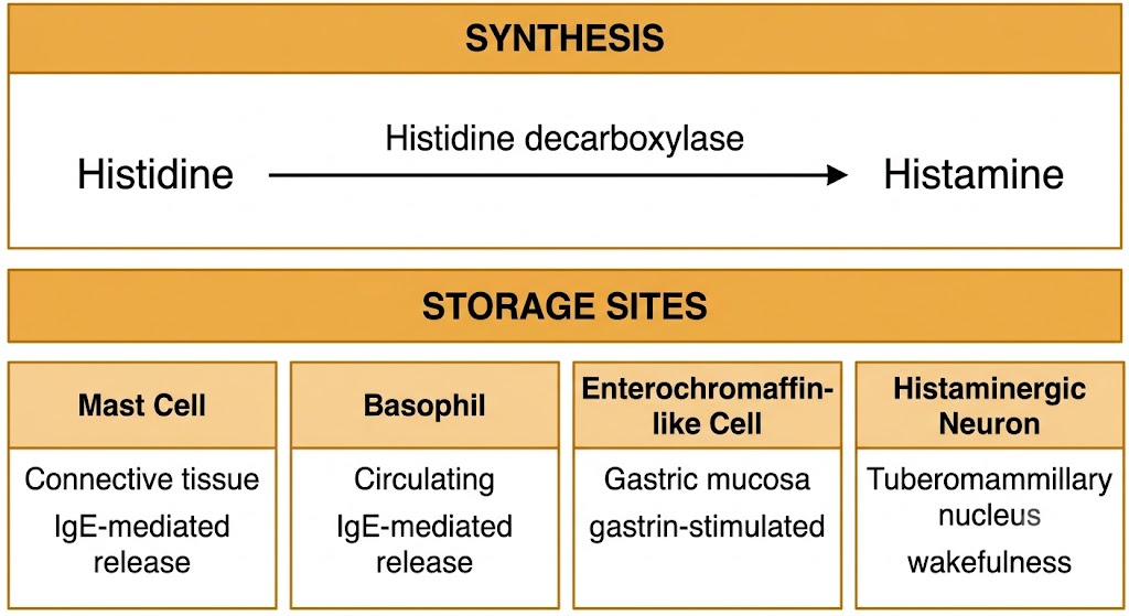
Two-zone diagram showing histamine synthesis from histidine via histidine decarboxylase (top zone) and the four storage cell types — mast cell, basophil, enterochromaffin-like cell, and histaminergic neuron — with tissue locations labeled (bottom zone).
Histamine synthesis from histidine by histidine decarboxylase and the four primary storage cell types. Figure generated by Gemini AI.

Section 2

Histamine Release Mechanisms

Immunological and non-immunological pathways of mast cell degranulation, with clinically relevant examples of each

Histamine release from mast cells and basophils can occur through immunological and non-immunological mechanisms. Only the immunological pathway requires prior sensitization; non-immunological release can occur on first drug exposure and is the basis of several well-known drug reactions that mimic allergy without involving immunoglobulin E.

Immunological Release — Immunoglobulin E-Mediated Degranulation

The canonical allergic pathway requires two exposures. On first exposure to an allergen, genetically susceptible individuals produce immunoglobulin E antibodies that bind to high-affinity immunoglobulin E receptors on mast cells and basophils, arming them. On re-exposure, the same allergen crosslinks adjacent receptor-bound immunoglobulin E molecules. This crosslinking triggers a signaling cascade that causes rapid granule-plasma membrane fusion and release of preformed histamine, tryptase, and heparin within seconds to minutes.

Because sensitization is required, true immunoglobulin E-mediated reactions cannot occur on first exposure to a drug or allergen. This is the basis of the clinical distinction between anaphylaxis (immunoglobulin E-mediated, requires prior sensitization) and anaphylactoid reactions (non-immunoglobulin E-mediated, can occur on first exposure). Both produce the same multisystem syndrome and are managed identically with epinephrine as first-line treatment.

Non-Immunological Release — Direct Mast Cell Activation

Several drug classes directly activate mast cells without involving immunoglobulin E, producing histamine release on any exposure — including the first. This mechanism underlies a set of predictable, dose- or rate-dependent adverse drug reactions that are commonly encountered in clinical practice.

Non-Immunological Histamine Releasers

Clinically Tested Drug Examples

  • Morphine and codeine: displace histamine from granule storage by direct ionic interaction, producing cutaneous flushing, whealing, and pruritus at the injection site; not immunoglobulin E-mediated
  • Vancomycin: rapid intravenous infusion causes direct mast cell activation producing Red Man Syndrome — flushing and erythema over the face, neck, and upper chest; rate-dependent, not dose-dependent in the immunological sense; managed by slowing infusion and premedication with H1 antihistamines
  • Tubocurarine and atracurium: neuromuscular blocking agents that release histamine directly from mast cells; atracurium causes more histamine release than rocuronium or vecuronium, which is clinically relevant during anesthesia induction
  • Radiocontrast media: high-osmolality ionic agents activate mast cells through osmotic and membrane effects; risk substantially reduced with modern low-osmolality non-ionic agents

Complement activation also generates non-immunological histamine release. The complement fragments C3a and C5a (anaphylatoxins) bind receptors on mast cells and basophils to trigger degranulation. This occurs in transfusion reactions, drug-induced immune complex formation, and certain infections. The clinical syndrome it produces is managed identically to immunoglobulin E-mediated anaphylaxis.


Section 3

Histamine Receptor Subtypes

H1 through H4 receptors — G protein coupling, tissue distribution, and pharmacological significance

The four histamine receptor subtypes are all G protein-coupled receptors, but they couple to distinct G proteins, activate different second messenger cascades, and are expressed in different tissues. H1 and H2 receptors are the targets of the most clinically important histamine pharmacology — antihistamines and H2 blockers respectively — while H3 and H4 receptors are lower-yield Step 1 topics.

H1 Receptor — Gq-Coupled, Vascular and Allergic Effects

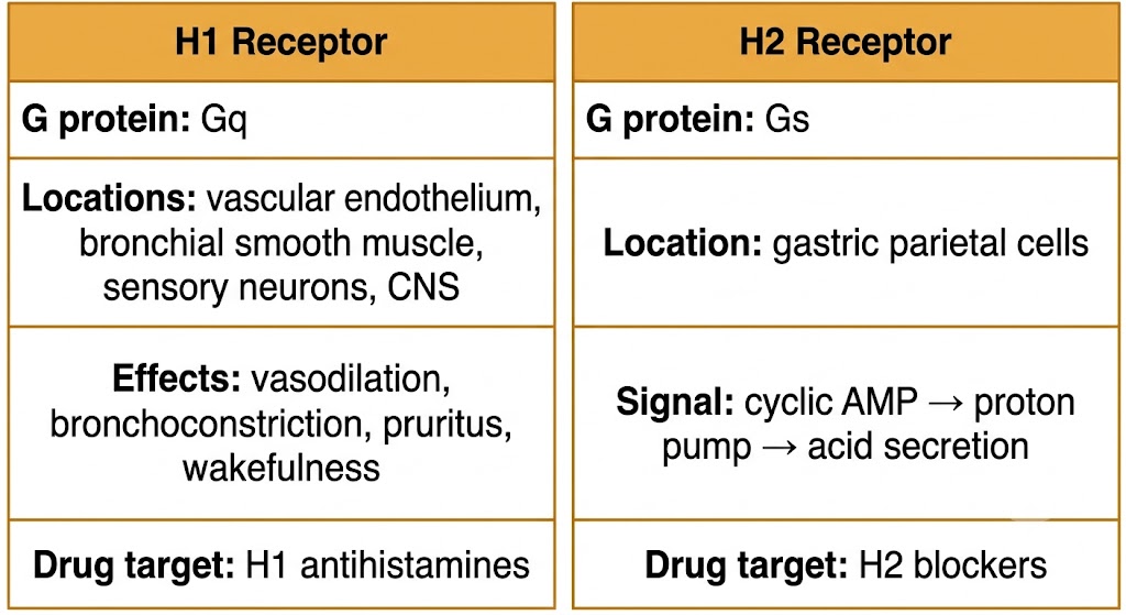
The H1 receptor couples to the Gq protein, activating phospholipase C and raising intracellular calcium. It is expressed prominently on vascular endothelium, vascular and bronchial smooth muscle, sensory neurons (particularly the C fibers that mediate pruritus), and central nervous system neurons of the tuberomammillary nucleus. The physiological consequences of H1 activation differ by tissue: in vascular endothelium it triggers nitric oxide production, producing vasodilation and increased vascular permeability (the basis of wheal formation); in bronchial smooth muscle it causes contraction and bronchoconstriction; in sensory C fibers it produces the itch sensation; and in central nervous system neurons it promotes cortical arousal and wakefulness.

H1 antihistamines are technically inverse agonists rather than simple competitive antagonists — they preferentially stabilize the inactive receptor conformation, suppressing constitutive receptor activity. For clinical purposes the practical effect is the same as competitive antagonism: blockade of histamine-mediated allergic responses.

Two-panel comparison: left panel shows H1 receptor with Gq coupling, locations including vascular endothelium and bronchial smooth muscle, and effects including vasodilation, bronchoconstriction, pruritus, and wakefulness; right panel shows H2 receptor with Gs coupling, location at gastric parietal cells, and cyclic AMP to proton pump to acid secretion signaling.
H1 and H2 histamine receptor subtypes: G protein coupling, tissue location, and downstream effects. Figure generated by Gemini AI.

H2 Receptor — Gs-Coupled, Gastric Acid Secretion

The H2 receptor couples to the Gs protein, activating adenylyl cyclase to raise cyclic adenosine monophosphate levels, which activates protein kinase A. In gastric parietal cells, protein kinase A activates the hydrogen-potassium ATPase (the proton pump), stimulating acid secretion. Enterochromaffin-like cells release histamine onto adjacent parietal cells in response to gastrin and acetylcholine, making histamine the final common amplifier of acid secretion by all three stimuli — the pharmacological rationale for H2 blocker therapy.

H2 receptors are also present on cardiac myocytes, where activation produces modest increases in heart rate and contractility via cyclic adenosine monophosphate. This contributes to the tachycardia seen in anaphylaxis alongside the dominant H1-mediated vasodilation.

H3 and H4 Receptors — Gi-Coupled, Lower-Yield Step 1 Targets

H3 receptors are Gi-coupled presynaptic autoreceptors on histaminergic nerve terminals in the central nervous system. When activated by locally released histamine, they inhibit further histamine synthesis and release, forming a classic negative feedback loop. H3 receptor inverse agonists (pitolisant) disinhibit histaminergic neurons, promoting wakefulness — the basis of pitolisant's indication for narcolepsy. H3 pharmacology is low-yield for Step 1 outside of narcolepsy drug identification.

H4 receptors are Gi-coupled and expressed predominantly on immune cells including mast cells, basophils, and eosinophils, where they modulate immune cell chemotaxis and activation. No H4-selective drug has reached major clinical use at the Step 1 level.

Receptor Subtype Summary — Step 1 Focus

H1 — Gq — vascular, bronchial, sensory neurons, central nervous system: target of H1 antihistamines (first- and second-generation).

H2 — Gs — gastric parietal cells: target of H2 blockers (cimetidine, famotidine).

H3 — Gi — presynaptic autoreceptor, central nervous system: pitolisant for narcolepsy; low Step 1 yield otherwise.

H4 — Gi — immune cells: no approved Step 1-level drug.


Section 4

Pathophysiological Roles of Histamine

The triple response, anaphylaxis, bronchoconstriction, gastric acid hypersecretion, and the limits of antihistamine therapy

Histamine dominates the early-phase acute manifestations of allergic disease — the immediate urticaria, rhinitis, bronchoconstriction, and vascular changes that appear within minutes of allergen exposure. Understanding which clinical effects are histamine-mediated directly predicts where antihistamines will succeed and where they will fall short.

The Triple Response of Lewis — Local Skin Reaction

Intradermal histamine release produces the triple response: a red spot at the site from local arteriolar vasodilation (H1 on vascular endothelium); a surrounding wheal from plasma extravasation through H1-mediated increased vascular permeability; and an irregular flare of erythema extending beyond the wheal, produced by an axon reflex that dilates surrounding arterioles. The itch accompanying this response is mediated by H1 receptor activation of sensory C fibers. The triple response is the skin correlate of urticaria and reproduces in miniature the local vascular events of acute allergy.

Anaphylaxis — Histamine Is One of Several Mediators

In systemic anaphylaxis, massive mast cell and basophil degranulation releases histamine alongside prostaglandins, leukotrienes, platelet-activating factor, and tryptase. Histamine is the primary mediator of the cutaneous manifestations (flushing, urticaria, angioedema) and contributes to cardiovascular collapse through H1-mediated vasodilation and increased vascular permeability. However, the multimediator nature of anaphylaxis is why antihistamines alone cannot reverse anaphylactic shock.

Epinephrine is the only agent that simultaneously addresses the multimediator, multisystem problem: it reverses vasodilation via alpha-1 adrenergic receptors, reverses bronchoconstriction via beta-2 adrenergic receptors, and supports cardiac output via beta-1 adrenergic receptors. H1 antihistamines reduce pruritus and urticaria as useful adjuncts but have no effect on hemodynamic collapse or bronchospasm and are never first-line treatment for anaphylaxis.

Angioedema With Versus Without Urticaria — A Critical Distinction

Histamine-mediated angioedema occurs almost always in the context of urticaria. Bradykinin-mediated angioedema — as in hereditary angioedema and angiotensin converting enzyme inhibitor-induced angioedema — occurs without urticaria. A patient with angioedema but no urticaria, or a patient on an angiotensin converting enzyme inhibitor with angioedema, should not be treated as histamine-mediated disease. These patients will not respond to antihistamines, corticosteroids, or epinephrine and require bradykinin-specific therapy. Failure to recognize the distinction can be fatal. Bradykinin pharmacology is covered fully in Module 4.

Bronchoconstriction and Gastric Acid Secretion

H1 receptor activation on bronchial smooth muscle produces bronchoconstriction, contributing to the early-phase bronchospasm in asthma after allergen exposure. This effect is significantly amplified in patients with asthma, whose airways are hyperresponsive to histamine. H1 antihistamines provide modest bronchodilatory benefit in allergic asthma but are not primary asthma therapy — the late-phase inflammatory response driven by leukotrienes and eosinophils is not histamine-mediated and is not addressed by antihistamines.

H2 receptor activation in the gastric mucosa drives acid secretion via the enterochromaffin-like cell to parietal cell paracrine pathway described in Section 3. This is the pharmacological basis of H2 blocker therapy for peptic ulcer disease and gastroesophageal reflux disease, covered in Module 3.


Visual Reference

Module 1 Visual Summary

Histamine receptors, signaling pathways, tissue distribution, and pathophysiological roles

Suggested References
Author / Organization Title Source
Simons FE, Simons KJ Histamine and H1-antihistamines: celebrating a century of progress J Allergy Clin Immunol. 2011;128(6):1139–1150
Galli SJ, Tsai M, Piliponsky AM The development of allergic inflammation Nature. 2008;454(7203):445–454
Schubert ML, Peura DA Control of gastric acid secretion in health and disease Gastroenterology. 2008;134(7):1842–1860
Haas HL, Sergeeva OA, Selbach O Histamine in the nervous system Physiol Rev. 2008;88(3):1183–1241
Sampson HA, Munoz-Furlong A, Campbell RL, et al. Second symposium on the definition and management of anaphylaxis: summary report J Allergy Clin Immunol. 2006;117(2):391–397
Zuberbier T, Maurer M Urticaria: current opinions about etiology, diagnosis and therapy Acta Derm Venereol. 2007;87(3):196–205
Leurs R, Church MK, Taglialatela M H1-antihistamines: inverse agonism, anti-inflammatory actions and cardiac effects Clin Exp Allergy. 2002;32(4):489–498
Haas H, Panula P The role of histamine and the tuberomamillary nucleus in the nervous system Nat Rev Neurosci. 2003;4(2):121–130
Thurmond RL, Gelfand EW, Dunford PJ The role of histamine H1 and H4 receptors in allergic inflammation: the search for new antihistamines Nat Rev Drug Discov. 2008;7(1):41–53
Holgate ST, Polosa R Treatment strategies for allergy and asthma Nat Rev Immunol. 2008;8(3):218–230
Dauvilliers Y, Bassetti C, Lammers GJ, et al. Pitolisant versus placebo or modafinil in patients with narcolepsy: a double-blind, randomised trial Lancet Neurol. 2013;12(11):1068–1075