Injection into cerebrospinal fluid produces a rapid, dense block with a predictable sequence of sympathetic, sensory, and motor effects
Spinal anesthesia — also called subarachnoid block — is produced by injecting local anesthetic directly into the cerebrospinal fluid in the subarachnoid space. Because the drug acts immediately on nerve roots bathed in cerebrospinal fluid, onset is rapid and the block is dense. A single-injection technique produces predictable, time-limited anesthesia used for lower abdominal, pelvic, and lower extremity surgery.
Mechanism and Drug Distribution
The subarachnoid space contains cerebrospinal fluid surrounding the spinal cord and nerve roots. Local anesthetic injected into this space distributes through the cerebrospinal fluid and contacts nerve roots as they pass through the subarachnoid space before exiting the spinal column. Because there is no barrier between the drug and the nerve roots — unlike epidural anesthesia where drug must diffuse through epidural fat and the dura — onset is fast and the block is complete at appropriate doses.
The spread of drug within the cerebrospinal fluid is influenced by the baricity of the solution — the density of the local anesthetic preparation relative to cerebrospinal fluid. Hyperbaric solutions (denser than cerebrospinal fluid, typically prepared by adding glucose) tend to sink with gravity, allowing the anesthesiologist to direct spread by positioning the patient. Hypobaric solutions rise. Isobaric solutions spread less predictably with position. This principle is used clinically to target the level of blockade.
Agents used for spinal anesthesia include bupivacaine (the most widely used for spinal anesthesia due to its reliable duration), tetracaine, and lidocaine. Lidocaine spinal anesthesia has fallen out of favor in many centers because of its association with transient neurologic symptoms, discussed in Section 3.
Sequence of Block Development
After injection into the subarachnoid space, the block develops in a predictable sequence reflecting fiber sensitivity: sympathetic fibers are blocked first, followed by sensory fibers, then motor fibers. This sequence has important clinical consequences. Sympathetic blockade — loss of vasoconstrictor tone in blood vessels below the level of the block — produces vasodilation and a drop in blood pressure. Hypotension is the most common side effect of spinal anesthesia and is managed with intravenous fluids and vasopressors such as phenylephrine (a drug that selectively activates alpha-1 receptors to constrict blood vessels) or ephedrine (a drug that activates both alpha and beta receptors).
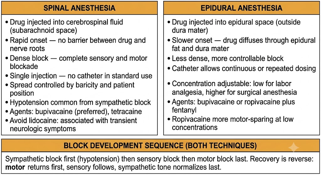
Sensory block follows sympathetic block, producing loss of pain, temperature, and touch sensation in the anesthetized dermatomes. Motor block develops last and is typically dense — patients undergoing surgery under spinal anesthesia are completely unable to move the affected limbs. Recovery follows the reverse sequence: motor function returns first, sensory block resolves, and sympathetic tone is last to normalize.
Spinal Versus Epidural — Key Distinctions
Spinal: drug injected into cerebrospinal fluid (subarachnoid space). Rapid onset. Dense block. Single injection — no catheter. Hypotension from sympathetic block is common.
Epidural: drug injected into epidural space (outside dura). Slower onset — drug must diffuse across dura. Less dense block at equivalent volumes. Catheter allows repeated dosing and titration. More controllable.
Spinal versus epidural anesthesia: site of drug deposition, onset characteristics, block density, and the shared sequence of sympathetic, sensory, then motor blockade. Figure generated by Gemini AI.
Section 2
Epidural Anesthesia
Drug deposited outside the dura diffuses to nerve roots — a catheter allows continuous dosing and precise titration
Epidural anesthesia places local anesthetic into the epidural space — the potential space between the ligamentum flavum and the dura mater surrounding the spinal cord. Drug deposited here must diffuse through epidural fat and across the dura to reach nerve roots in the subarachnoid space. This additional diffusion step produces slower onset and a less dense block than spinal anesthesia at equivalent doses, but the ability to place a catheter and dose continuously or repeatedly makes epidural anesthesia far more controllable and versatile.
Technique and Catheter Placement
A needle is advanced through the skin, subcutaneous tissue, interspinous ligaments, and ligamentum flavum until it enters the epidural space. The epidural space is identified by a characteristic loss of resistance — the ligamentum flavum offers significant resistance to injection, which disappears suddenly when the needle tip enters the epidural space. A catheter is then threaded through the needle into the epidural space, the needle is withdrawn, and the catheter remains in place, secured to the patient's back. Local anesthetic is injected through the catheter in incremental doses.
The catheter approach distinguishes epidural from spinal anesthesia. With a catheter in place, additional drug can be administered whenever the block begins to wear off, an infusion can be run continuously, and the concentration can be adjusted to target sensory blockade only (for labor analgesia) or denser surgical anesthesia (for cesarean section or major surgery).
Agents and Adjuvants
Bupivacaine and ropivacaine are the primary agents for epidural analgesia. At low concentrations — typically 0.0625% to 0.125% bupivacaine or equivalent ropivacaine — epidural infusions produce sensory blockade with minimal motor block, allowing patients to remain ambulatory during labor. At higher concentrations for surgical anesthesia, denser motor block is achieved.
Opioids are frequently added to epidural local anesthetic solutions as adjuvants. Fentanyl (a potent, short-acting opioid) added to the epidural solution acts synergistically with the local anesthetic, allowing lower concentrations of local anesthetic to be used while maintaining effective analgesia. This combination reduces motor block and local anesthetic-related side effects. Epidural morphine provides prolonged postoperative analgesia but carries a risk of delayed respiratory depression from rostral spread within the cerebrospinal fluid.
Clinical Uses
Epidural anesthesia is used for labor analgesia, cesarean section, major abdominal and thoracic surgery, and postoperative pain management. For labor, a continuous epidural infusion of dilute bupivacaine or ropivacaine with fentanyl provides effective pain relief through contractions while preserving enough motor function for the patient to push during the second stage. For cesarean section, the concentration is increased to achieve surgical-level anesthesia. For postoperative thoracic and abdominal surgery, epidural analgesia reduces opioid requirements, promotes earlier mobilization, and has been shown to reduce pulmonary complications.
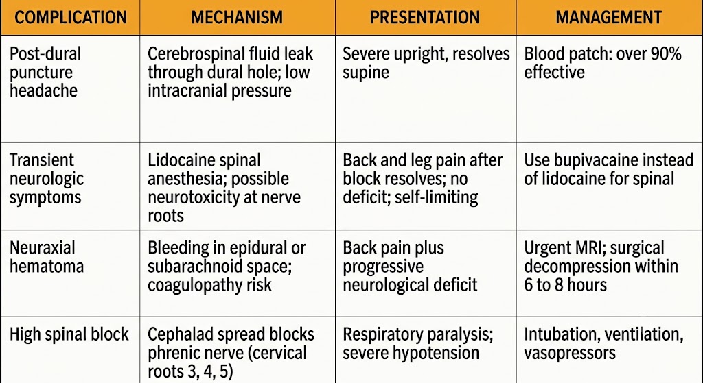
Four neuraxial anesthesia complications: mechanism, clinical presentation, and management for post-dural puncture headache, transient neurologic symptoms, neuraxial hematoma, and high spinal block. Figure generated by Gemini AI.
Section 3
Neuraxial Complications
Post-dural puncture headache, transient neurologic symptoms, neuraxial hematoma, and high spinal block — each with distinct mechanisms and management
Neuraxial anesthesia is associated with a set of specific complications that arise from the anatomy of the subarachnoid and epidural spaces, the drugs deposited there, and the needle or catheter placement itself. Recognizing these complications, understanding their mechanisms, and knowing the appropriate management are high-yield topics for pharmacology and clinical examinations.
Post-Dural Puncture Headache
Post-dural puncture headache is caused by leakage of cerebrospinal fluid through the hole made in the dura by the spinal needle. Loss of cerebrospinal fluid reduces intracranial pressure, causing traction on pain-sensitive intracranial structures — meninges, bridging veins, and cranial nerves — when the patient sits upright. The hallmark feature is postural dependency: the headache is severe in the upright position and rapidly improves or disappears when the patient lies flat. This postural component distinguishes it from other causes of headache after neuraxial anesthesia.
The risk is proportional to needle size and needle tip design. Larger needles create larger dural holes with more cerebrospinal fluid leakage. Pencil-point needles (Whitacre, Sprotte design) separate dural fibers rather than cutting them and produce dramatically lower rates of post-dural puncture headache compared to cutting-tip (Quincke) needles of the same gauge. Accidental dural puncture during epidural placement — when the epidural needle (which is much larger than a spinal needle) penetrates the dura — produces a very high incidence of severe headache.
Conservative management includes bed rest, oral hydration, caffeine (which causes cerebral vasoconstriction and raises intracranial pressure), and oral analgesics. Definitive treatment is an epidural blood patch — injection of 15 to 20 milliliters of the patient's own autologous blood into the epidural space at the level of the dural puncture. The blood clots and seals the dural hole, rapidly restoring normal cerebrospinal fluid pressure and resolving the headache in over 90% of cases. The blood patch is the most effective treatment for post-dural puncture headache that does not resolve with conservative measures within 24 to 48 hours.
Transient Neurologic Symptoms
Transient neurologic symptoms are a syndrome of back pain, buttock pain, and lower extremity pain or dysesthesia (abnormal sensation) that develop after spinal anesthesia and resolve spontaneously within a few days. The symptoms are typically bilateral, appear within hours of recovery from the block, and are not associated with any neurological deficit on examination — motor and sensory function are preserved.
The syndrome is most strongly associated with lidocaine used for spinal anesthesia, particularly at higher concentrations and in the lithotomy position (legs raised in stirrups, which is thought to stretch lumbosacral nerve roots). The incidence with lidocaine spinal anesthesia is substantially higher than with bupivacaine, which has a much lower association with this syndrome. This association has led to a marked decrease in the use of lidocaine for spinal anesthesia in many centers, replaced by small doses of bupivacaine for short procedures. The mechanism is not fully understood but may involve local neurotoxicity at high drug concentrations around nerve roots.
Neuraxial Hematoma
Neuraxial hematoma — bleeding into the epidural or subarachnoid space — is a rare but potentially catastrophic complication of neuraxial anesthesia. Blood accumulating in the confined epidural or subarachnoid space compresses the spinal cord or cauda equina, causing progressive neurological deficits. Without prompt surgical decompression, permanent paralysis results.
The primary risk factor is coagulopathy — abnormal blood clotting — at the time of needle or catheter placement or removal. Patients receiving anticoagulant or antiplatelet drugs require careful timing of neuraxial procedures relative to drug administration. Guidelines specify minimum intervals between the last dose of various anticoagulants and neuraxial needle placement or catheter removal. The American Society of Regional Anesthesia and Pain Medicine guidelines are the reference standard for these intervals.
The clinical presentation is new or progressive back pain combined with lower extremity weakness or sensory loss, or bladder or bowel dysfunction developing after neuraxial anesthesia. Any new neurological symptom after neuraxial anesthesia must be evaluated urgently. Magnetic resonance imaging confirms the diagnosis. Emergency surgical decompression within six to eight hours of symptom onset offers the best chance of neurological recovery.
High Spinal Block
High spinal block occurs when local anesthetic spreads cephalad (toward the head) within the cerebrospinal fluid, reaching the cervical spinal cord and blocking the phrenic nerve (which originates from cervical nerve roots 3, 4, and 5 and drives diaphragmatic breathing). The result is respiratory paralysis requiring immediate airway management and ventilatory support. Hypotension is also severe because sympathetic vasoconstrictor tone to the entire body is lost.
Risk factors include excessive drug dose, hyperbaric solutions used in the head-down (Trendelenburg) position, and accidental intrathecal injection of a dose intended for epidural use. Management is immediate — airway control, endotracheal intubation, ventilation, and vasopressor support until the block recedes.
Complication
Post-Dural Puncture Headache
Cause: cerebrospinal fluid leak through dural hole
Feature: severe upright, resolves supine (postural)
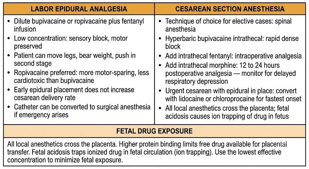
Obstetric neuraxial anesthesia: labor epidural analgesia versus cesarean section spinal anesthesia, with fetal drug exposure and ion trapping considerations. Figure generated by Gemini AI.
Section 4
Obstetric Neuraxial Anesthesia
Labor analgesia and cesarean section anesthesia — agent selection, fetal considerations, and timing
Neuraxial anesthesia is the foundation of obstetric pain management. Epidural analgesia for labor and spinal or epidural anesthesia for cesarean section are among the most commonly performed neuraxial procedures worldwide. The obstetric setting introduces additional pharmacological considerations: all local anesthetics cross the placenta, the altered physiology of pregnancy changes drug handling, and the fetal well-being must be considered alongside maternal analgesia.
Labor Epidural Analgesia
Labor pain has two components. In the first stage of labor, uterine contractions produce visceral pain transmitted by sympathetic nerve fibers entering the spinal cord at thoracic levels 10 through 12 and lumbar level 1. In the second stage, perineal pain from distension of the birth canal is transmitted by the pudendal nerve, entering the spinal cord at sacral levels 2 through 4. An epidural catheter at the lumbar level, with appropriate drug spread, covers both components when the concentration and volume are correctly chosen.
The standard approach uses a dilute epidural infusion of bupivacaine or ropivacaine combined with fentanyl. The low concentration achieves adequate sensory blockade of pain fibers while largely preserving motor function — the patient can move the legs, bear weight, and push effectively in the second stage. Ropivacaine is favored in many obstetric centers because of its greater motor-sparing properties at low concentrations and its lower cardiotoxicity profile compared to bupivacaine.
A common concern from patients is that epidural analgesia will slow labor or increase the rate of cesarean delivery. Clinical trial evidence does not support this — early epidural placement does not increase cesarean delivery rates. The epidural catheter placed for labor analgesia can also be used for cesarean section anesthesia if an emergency arises, by injecting a higher concentration of local anesthetic through the existing catheter.
Cesarean Section Anesthesia
Spinal anesthesia is the technique of choice for elective cesarean section. A single injection of hyperbaric bupivacaine into the subarachnoid space produces rapid, dense, and predictable anesthesia extending from approximately the fourth thoracic dermatome to the sacrum — sufficient for skin incision, uterine incision, delivery, and uterine closure. The addition of intrathecal opioids (fentanyl for intraoperative analgesia, morphine for prolonged postoperative analgesia lasting 12 to 24 hours) has become standard practice. Intrathecal morphine provides excellent postoperative analgesia but requires monitoring for delayed respiratory depression.
For urgent or emergency cesarean section when an epidural catheter is already in place, the epidural is converted to surgical anesthesia by injecting a larger volume of concentrated local anesthetic (typically lidocaine or chloroprocaine for fastest onset) through the existing catheter. General anesthesia is reserved for true emergencies where neuraxial anesthesia cannot be established quickly enough or is contraindicated.
Fetal Considerations
All local anesthetics cross the placenta by passive diffusion. The extent of fetal drug exposure depends on the maternal plasma concentration, the lipid solubility and protein binding of the agent, and placental blood flow. Highly protein-bound agents such as bupivacaine have less free drug available for placental transfer than less protein-bound agents at equivalent total plasma concentrations.
As discussed in Module 4, fetal acidosis causes ion trapping of local anesthetic within the fetal circulation. This is a concern particularly in the setting of fetal distress, where lower fetal pH increases the ionized fraction of local anesthetic that cannot cross back into the maternal circulation, effectively concentrating drug in the fetus. Minimizing maternal drug dose — using the lowest effective concentration — is the principal strategy for reducing fetal drug exposure.
Obstetric Neuraxial — High-Yield Summary
Labor analgesia: epidural catheter, dilute bupivacaine or ropivacaine plus fentanyl. Sensory block, motor preserved. Early placement does not increase cesarean rate.
Cesarean section: spinal anesthesia preferred for elective cases — hyperbaric bupivacaine plus intrathecal fentanyl and morphine. Epidural conversion for urgent cases if catheter in place.
Fetal exposure: all agents cross placenta. Protein binding limits transfer. Fetal acidosis causes ion trapping. Use lowest effective concentration.
Suggested References
Author / Organization
Title
Source
Butterworth JF, Mackey DC, Wasnick JD, eds.
Morgan and Mikhail's Clinical Anesthesiology. 6th ed.
New York: McGraw-Hill; 2018:937–992
Gehling M, Tryba M
Risks and side-effects of intrathecal morphine combined with spinal anesthesia: a meta-analysis
Anaesthesia. 2009;64(6):643–651
Apfel CC, Saxena A, Cakmakkaya OS, Gaiser R, George E, Radke O
Prevention of postdural puncture headache after accidental dural puncture: a quantitative systematic review
Br J Anaesth. 2010;105(3):255–263
Carvalho B, Durbin M, Drover DR, Cohen SE, Ginosar Y, Riley ET
The incidence of motor block with 0.125% versus 0.0625% bupivacaine with fentanyl for labor epidural analgesia
Anesth Analg. 2003;96(5):1468–1472
Horlocker TT, Vandermeuelen E, Kopp SL, Gogarten W, Leffert LR, Benzon HT
Regional anesthesia in the patient receiving antithrombotic or thrombolytic therapy
Reg Anesth Pain Med. 2018;43(3):263–309
Ilfeld BM
Continuous peripheral nerve blocks: a review of the published evidence
Anesth Analg. 2011;113(4):904–925
Guay J, Nishimori M, Kopp S
Epidural local anesthetics versus opioid-based analgesic regimens for postoperative gastrointestinal paralysis, vomiting and pain after abdominal surgery
Cochrane Database Syst Rev. 2016;7:CD001893
Hadzic A, ed.
Hadzic's Peripheral Nerve Blocks and Anatomy for Ultrasound-Guided Regional Anesthesia. 2nd ed.
New York: McGraw-Hill; 2011
Rosenberg PH, Veering BT, Urmey WF
Maximum recommended doses of local anesthetics: a multifactorial concept