CHAPTER 15  ·  LOCAL ANESTHETICS
Section 1

Systemic Local Anesthetic Toxicity

Central nervous system toxicity appears first; cardiovascular collapse follows at higher plasma concentrations

Systemic local anesthetic toxicity occurs when drug reaches the systemic circulation in high enough concentration to block sodium channels in the brain and heart — the same mechanism that produces therapeutic nerve blockade in peripheral nerves. The two most common causes are unintentional intravascular injection (needle tip inside a blood vessel at the time of injection) and absorption of an excessive total dose from a highly vascular injection site. Recognizing the clinical progression early is essential because intervention at the central nervous system toxicity stage prevents progression to the far more dangerous cardiovascular collapse stage.

Central Nervous System Toxicity — Early Signs

The central nervous system is more sensitive to local anesthetic toxicity than the cardiovascular system at lower plasma concentrations. The first symptoms are typically circumoral and tongue numbness — a tingling or numb sensation around the mouth and on the tongue that the patient may describe as the local anesthetic "wearing off wrong." This is followed by tinnitus (ringing in the ears), lightheadedness, and visual disturbances. A metallic taste in the mouth is another early symptom that patients sometimes report.

As plasma concentration rises, the central nervous system progresses from the initial sensory symptoms through increasing agitation, confusion, and slurred speech to muscle twitching and then generalized tonic-clonic seizures. The seizures reflect excessive excitatory activity in the central nervous system — local anesthetics at toxic concentrations preferentially block inhibitory interneurons before excitatory neurons, producing a transient state of uncontrolled excitation before global depression supervenes.

Cardiovascular Toxicity — Later Signs

At higher plasma concentrations, local anesthetics block sodium channels in the cardiac conduction system and myocardium. The electrocardiographic findings progress from prolongation of the PR interval and widening of the QRS complex through ventricular arrhythmias to myocardial depression and ultimately cardiac arrest. The cardiovascular toxicity of local anesthetics is generally more dangerous than the central nervous system toxicity because it is harder to treat, particularly in the case of bupivacaine.

Bupivacaine is particularly cardiotoxic because of its kinetics of sodium channel binding in the heart. It binds cardiac sodium channels rapidly ("fast in") but dissociates from them very slowly ("slow out") — in stark contrast to lidocaine, which both binds and dissociates rapidly. The slow dissociation means that between each heartbeat, bupivacaine does not fully release its cardiac channels, and with each successive beat more channels accumulate drug. The result is progressive deepening of cardiac conduction block that is extremely resistant to resuscitation. Ropivacaine was developed partly because it has faster cardiac channel dissociation than bupivacaine, making it significantly less cardiotoxic.

Toxicity Progression — High-Yield Sequence

Mnemonic: "Tongue to seizure to cardiac arrest"

Circumoral and tongue numbness → tinnitus, lightheadedness, metallic taste → agitation, confusion → muscle twitching → generalized seizures → cardiovascular collapse → cardiac arrest.

Central nervous system signs always precede cardiovascular signs at lower concentrations. A patient reporting tongue numbness after a nerve block is showing an early warning — stop the injection.

Early — Lower Concentration
Central Nervous System Toxicity
  • Circumoral and tongue numbness (first symptom)
  • Tinnitus, lightheadedness, metallic taste
  • Agitation, confusion, slurred speech
  • Muscle twitching
  • Generalized tonic-clonic seizures
Later — Higher Concentration
Cardiovascular Toxicity
  • PR prolongation, QRS widening
  • Ventricular arrhythmias
  • Myocardial depression
  • Cardiac arrest
  • Bupivacaine: "fast in, slow out" — resuscitation extremely difficult
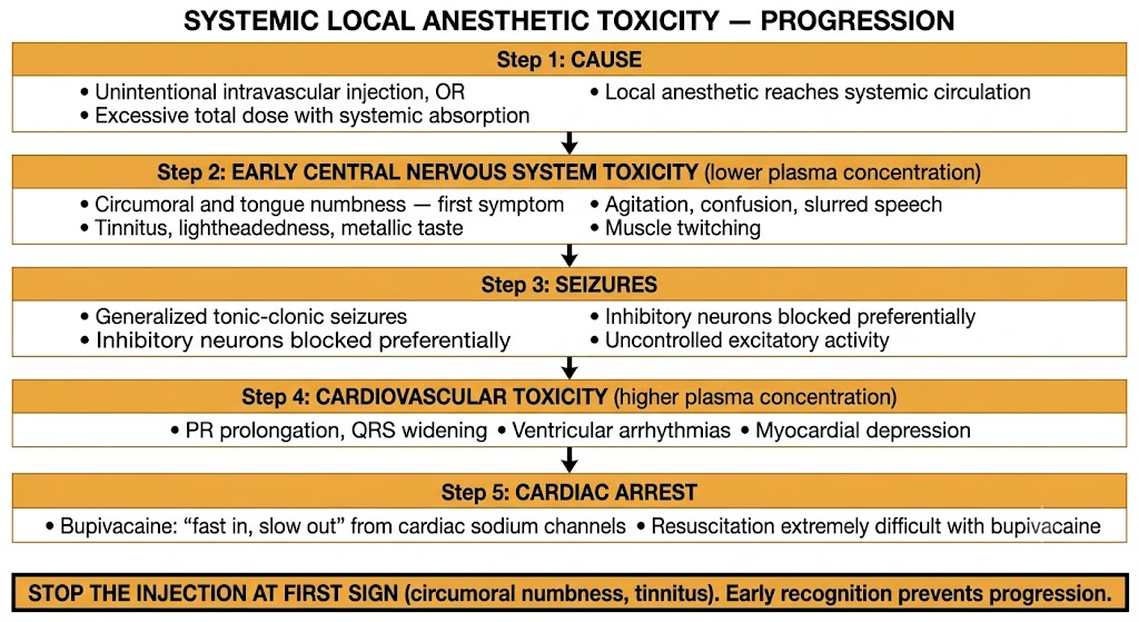
Flow diagram showing five-step progression of systemic local anesthetic toxicity from cause through early central nervous system symptoms, seizures, cardiovascular toxicity, and cardiac arrest, with a warning box to stop injection at first sign.
Systemic local anesthetic toxicity: five-step progression from cause through central nervous system symptoms, seizures, cardiovascular toxicity, and cardiac arrest. Figure generated by Gemini AI.

Section 2

Treatment of Systemic Local Anesthetic Toxicity

Airway control, seizure suppression, and intravenous lipid emulsion — with prolonged resuscitation for bupivacaine cardiac arrest

Treatment of systemic local anesthetic toxicity follows a clear sequence: stop the injection, secure the airway, manage seizures, and administer intravenous lipid emulsion for severe toxicity. The single most important advance in managing life-threatening local anesthetic toxicity — particularly bupivacaine cardiac arrest — has been the introduction of intravenous lipid emulsion therapy as a specific antidote.

Immediate Steps

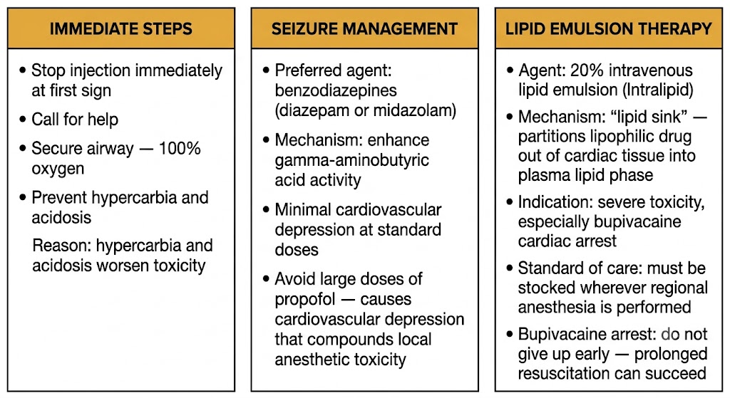
The first action is to stop the injection immediately at the first sign of toxicity. If the injection is stopped early — at the circumoral numbness or tinnitus stage — the plasma concentration may not rise further and the event may resolve without progression to seizures or cardiovascular toxicity. This is why recognizing the early central nervous system warning signs matters clinically.

Call for help immediately. Secure the airway and administer 100% oxygen. Hypercarbia (elevated blood carbon dioxide from respiratory compromise) and acidosis both worsen local anesthetic toxicity by lowering seizure threshold and increasing the fraction of ionized drug in tissues. Maintaining ventilation and preventing acidosis is a critical supportive measure throughout the resuscitation.

Seizure Management

Generalized seizures from local anesthetic toxicity are managed with benzodiazepines as the preferred agents — diazepam or midazolam given intravenously. Benzodiazepines enhance the activity of gamma-aminobutyric acid (the main inhibitory neurotransmitter in the brain), suppressing the uncontrolled excitatory activity responsible for the seizures without causing significant cardiovascular depression at standard doses.

Propofol can terminate seizures but should be used with caution in the setting of suspected cardiovascular toxicity, because propofol causes dose-dependent cardiovascular depression that compounds the myocardial effects of the local anesthetic. Small doses may be necessary if benzodiazepines are unavailable, but large doses are contraindicated in this specific context.

Intravenous Lipid Emulsion Therapy — the Lipid Sink

Intravenous lipid emulsion — commercially available as a 20% fat emulsion (Intralipid being the most recognized brand) — is the specific treatment for severe local anesthetic systemic toxicity, particularly bupivacaine-induced cardiac arrest. The mechanism is described as the "lipid sink": the infused lipid creates a new lipid phase in the blood that partitions the highly lipophilic local anesthetic drug out of cardiac and neural tissues and into the circulating lipid droplets, reducing the free drug concentration at its toxic targets.

The treatment is administered as an initial intravenous bolus followed by an infusion. It has become the standard of care for severe local anesthetic systemic toxicity and must be immediately available in any setting where regional anesthesia is performed. Its introduction has substantially improved survival from what was previously a frequently fatal complication.

For bupivacaine cardiac arrest specifically, resuscitation efforts must be sustained for an extended period — much longer than standard cardiac arrest resuscitation — because the drug dissociates from cardiac sodium channels slowly. Recovery of spontaneous circulation can occur after prolonged cardiopulmonary resuscitation when lipid emulsion therapy allows gradual redistribution of the drug away from the myocardium.

Lipid Emulsion Therapy — Key Facts

Mechanism: "lipid sink" — intravenous fat emulsion partitions lipophilic local anesthetic out of cardiac tissue into plasma lipid phase, reducing free drug at the target.

Indication: severe local anesthetic systemic toxicity — particularly bupivacaine cardiac arrest.

Agent: 20% intravenous lipid emulsion (Intralipid). Initial bolus then infusion.

Standard of care: must be immediately stocked wherever regional anesthesia is performed. Do not give up early in bupivacaine cardiac arrest — prolonged resuscitation with lipid emulsion can achieve recovery.

Three-panel diagram showing treatment of systemic local anesthetic toxicity: immediate steps (stop injection, airway, oxygen), seizure management (benzodiazepines preferred, avoid large propofol doses), and lipid emulsion therapy (20% intravenous lipid emulsion, lipid sink mechanism, standard of care for bupivacaine cardiac arrest).
Treatment of systemic local anesthetic toxicity: immediate airway management, benzodiazepines for seizures, and intravenous lipid emulsion as the specific antidote for severe toxicity and bupivacaine cardiac arrest. Figure generated by Gemini AI.

Section 3

Methemoglobinemia

Oxidation of hemoglobin iron from ferrous to ferric state — cyanosis that does not respond to oxygen

Methemoglobinemia is a potentially life-threatening complication in which hemoglobin iron is oxidized from the normal ferrous (Fe2+) state to the ferric (Fe3+) state, producing methemoglobin. Unlike normal oxyhemoglobin, methemoglobin cannot bind and transport oxygen reversibly — ferric iron does not carry oxygen. Two local anesthetics are the most clinically important causes of drug-induced methemoglobinemia: prilocaine and benzocaine.

Mechanism

Normal hemoglobin carries iron in the ferrous (Fe2+) state, which allows reversible oxygen binding. When certain oxidizing agents — including metabolites of specific drugs — donate an electron to hemoglobin iron, it is converted to the ferric (Fe3+) state. Ferric iron cannot bind oxygen, so methemoglobin is functionally useless for oxygen transport. As the proportion of methemoglobin rises, the oxygen-carrying capacity of the blood falls.

Prilocaine causes methemoglobinemia through its hepatic metabolite ortho-toluidine, which is the direct oxidizing agent acting on hemoglobin. Benzocaine causes methemoglobinemia through a similar oxidative mechanism and is a particular concern when used as a topical spray in endoscopy or intubation procedures, where the drug can be applied in large amounts to mucous membranes with high systemic absorption. Excessive spraying with benzocaine topical preparations is the most common cause of clinically significant drug-induced methemoglobinemia encountered in hospital practice.

Clinical Presentation

The characteristic presentation of methemoglobinemia is cyanosis that does not improve with supplemental oxygen administration. This failure to respond to oxygen is the key diagnostic clue — in ordinary hypoxemia from respiratory or cardiac causes, supplemental oxygen raises arterial oxygen saturation and resolves cyanosis. When cyanosis persists despite 100% oxygen delivery, methemoglobinemia must be considered.

Pulse oximetry gives a characteristic reading in methemoglobinemia: approximately 85% oxygen saturation regardless of the true oxygen saturation. This occurs because the spectrophotometric properties of methemoglobin cause the pulse oximeter to misread the signal and report a spuriously intermediate value near 85%. The actual oxygen saturation may be much lower or much higher than 85% — the pulse oximeter reading is unreliable. Confirmation requires co-oximetry, which directly measures the fraction of methemoglobin in the blood.

The blood itself has a characteristic appearance: dark chocolate-brown color rather than the bright red of well-oxygenated blood or the dark blue-red of deoxygenated blood. This dark brown discoloration of blood drawn from the patient is a highly characteristic and immediately recognizable sign.

Treatment

The treatment for symptomatic methemoglobinemia is methylene blue administered intravenously at a dose of 1 to 2 milligrams per kilogram. Methylene blue works by donating electrons through the nicotinamide adenine dinucleotide phosphate (the reduced form of nicotinamide adenine dinucleotide phosphate, abbreviated in biochemistry as NADPH) — methemoglobin reductase enzymatic pathway, reducing methemoglobin back to functional oxyhemoglobin. The response is typically rapid — cyanosis clears and oxygen saturation improves within minutes of administration.

One critical exception: patients with glucose-6-phosphate dehydrogenase deficiency cannot respond to methylene blue. Glucose-6-phosphate dehydrogenase is the enzyme responsible for generating the reduced form of nicotinamide adenine dinucleotide phosphate through the pentose phosphate pathway. Without adequate reduced nicotinamide adenine dinucleotide phosphate, the methylene blue reduction pathway cannot function, and methylene blue fails to reduce methemoglobin. In these patients, ascorbic acid (vitamin C) provides an alternative, albeit slower, electron donor for methemoglobin reduction.

Causative Agents
Prilocaine and Benzocaine
  • Prilocaine: metabolite ortho-toluidine oxidizes hemoglobin; used in EMLA cream
  • Benzocaine: topical sprays for endoscopy and intubation; excessive use causes significant methemoglobinemia
  • Both are the two local anesthetics most associated with this complication
Diagnosis
Clinical Presentation
  • Cyanosis that does not respond to supplemental oxygen
  • Pulse oximetry reads approximately 85% regardless of true saturation
  • Dark chocolate-brown blood (characteristic appearance)
  • Confirmed by co-oximetry (direct methemoglobin measurement)
Treatment
Methylene Blue
  • Methylene blue 1 to 2 mg/kg intravenously
  • Reduces methemoglobin via reduced nicotinamide adenine dinucleotide phosphate — methemoglobin reductase pathway
  • Rapid response — cyanosis clears within minutes
  • Exception: glucose-6-phosphate dehydrogenase deficiency — methylene blue fails; use ascorbic acid instead
Key Exception
Glucose-6-Phosphate Dehydrogenase Deficiency
  • Glucose-6-phosphate dehydrogenase generates reduced nicotinamide adenine dinucleotide phosphate via pentose phosphate pathway
  • Without reduced nicotinamide adenine dinucleotide phosphate, methylene blue cannot reduce methemoglobin
  • Methylene blue ineffective in these patients
  • Alternative: ascorbic acid (vitamin C) as electron donor
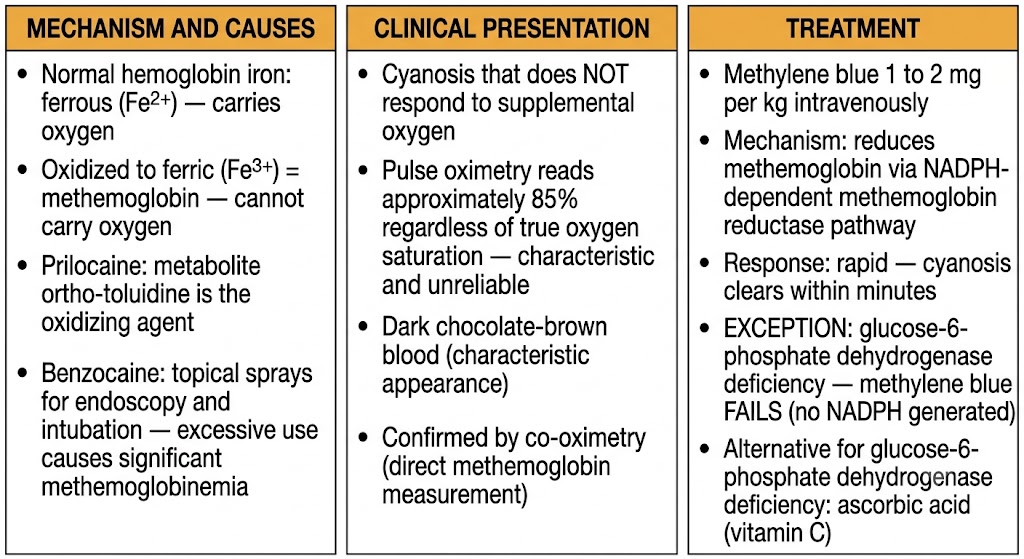
Three-panel diagram showing methemoglobinemia: mechanism and causes (ferrous to ferric hemoglobin oxidation, prilocaine metabolite ortho-toluidine, benzocaine sprays), clinical presentation (cyanosis unresponsive to oxygen, pulse oximetry reads 85%, dark chocolate-brown blood), and treatment (methylene blue, glucose-6-phosphate dehydrogenase exception requiring ascorbic acid instead).
Methemoglobinemia from local anesthetics: mechanism of hemoglobin iron oxidation, characteristic clinical presentation, and treatment with methylene blue including the glucose-6-phosphate dehydrogenase deficiency exception. Figure generated by Gemini AI.

Section 4

Special Populations and Considerations

Liver disease, pseudocholinesterase deficiency, and pregnancy alter local anesthetic pharmacokinetics and risk

Several patient populations have altered pharmacokinetics or pharmacodynamics that change the risk profile of local anesthetic administration. Recognizing these situations allows appropriate agent selection and dose adjustment.

Liver Disease

Amide local anesthetics depend on hepatic metabolism for elimination. Patients with significant liver disease — cirrhosis, severe hepatitis, or acute liver failure — have impaired amide metabolism, leading to slower drug clearance and higher plasma concentrations after a given dose. The practical implication is that amide local anesthetics should be used at reduced doses in patients with significant hepatic impairment, and the clinician should have a lower threshold for concern about accumulation with repeated doses or continuous infusions.

Ester local anesthetics, which are metabolized in the plasma by pseudocholinesterase rather than in the liver, are unaffected by hepatic disease and represent a reasonable alternative when liver function is severely impaired.

Pseudocholinesterase Deficiency

Ester local anesthetics depend on plasma pseudocholinesterase for metabolism. Patients with pseudocholinesterase deficiency — either genetic or acquired (from liver disease, malnutrition, or certain medications) — metabolize ester local anesthetics much more slowly than normal. The drug accumulates in the plasma, producing prolonged duration and elevated systemic concentrations that increase toxicity risk. In these patients, amide local anesthetics are the appropriate alternative, as their metabolism by hepatic enzymes is unaffected by pseudocholinesterase activity.

Pregnancy and Ion Trapping

Local anesthetics cross the placenta, as virtually all drugs do, and reach the fetal circulation. The fetal pH is slightly lower than maternal pH under normal conditions and becomes more acidic in the setting of fetal distress. This lower fetal pH shifts the equilibrium of the weak base local anesthetic toward the ionized form within fetal tissues — the ionized drug cannot easily cross back through the placenta into the maternal circulation, so it accumulates in the fetal compartment. This phenomenon is called ion trapping.

The clinical implication is that excessive maternal doses carry compounded risk in the fetus: not only does more drug reach the fetus, but any drug that does cross is trapped there and cannot be cleared as efficiently as in the maternal circulation. Bupivacaine and ropivacaine are the agents of choice for obstetric epidural analgesia, used at the lowest effective concentration to minimize fetal drug exposure while providing effective maternal pain relief.

Population Affected agents Clinical implication
Liver disease Amide local anesthetics (hepatic metabolism impaired) Reduce dose; consider ester agents as alternative
Pseudocholinesterase deficiency Ester local anesthetics (plasma hydrolysis impaired) Prolonged duration and increased toxicity risk; use amide agents instead
Pregnancy All local anesthetics (cross placenta; ion trapping in fetus) Use lowest effective concentration; bupivacaine and ropivacaine are agents of choice for epidural analgesia

Suggested References
Author / Organization Title Source
Neal JM, Barrington MJ, Fettiplace MR, et al. The Third American Society of Regional Anesthesia and Pain Medicine Practice Advisory on Local Anesthetic Systemic Toxicity: executive summary 2017 Reg Anesth Pain Med. 2018;43(2):113–123
Clarkson CW, Hondeghem LM Mechanism for bupivacaine depression of cardiac conduction: fast block of sodium channels during the action potential with slow recovery from block during diastole Anesthesiology. 1985;62(4):396–405
Rosenberg PH, Veering BT, Urmey WF Maximum recommended doses of local anesthetics: a multifactorial concept Reg Anesth Pain Med. 2004;29(6):564–575
Weinberg GL Lipid emulsion infusion: resuscitation for local anesthetic and other drug overdose Anesthesiology. 2012;117(1):180–187
Bhole MV, Manson AL, Seneviratne SL, Misbah SA IgE-mediated allergy to local anesthetics: separating fact from perception Br J Anaesth. 2012;108(6):903–911
Ash-Bernal R, Wise R, Wright SM Acquired methemoglobinemia: a retrospective series of 138 cases at 2 teaching hospitals Medicine (Baltimore). 2004;83(5):265–273
Freedman JM, Li DK, Drasner K, et al. Transient neurologic symptoms after spinal anesthesia: an epidemiologic study of 1,863 patients Anesthesiology. 1998;89(3):633–641
Lonnqvist PA, Morton NS Postoperative analgesia in infants and children Br J Anaesth. 2005;95(1):59–68
Gantenbein M, Attolini L, Bruguerolle B, et al. Oxidative metabolism of bupivacaine into pipecolylxylidine in humans is mainly catalyzed by CYP3A Drug Metab Dispos. 2000;28(4):383–385
Butterworth JF 4th, Strichartz GR Molecular mechanisms of local anesthesia: a review Anesthesiology. 1990;72(4):711–734