Introduction to Medical Pharmacology
Large-molecule pharmacokinetics, target-specific toxicities, and ADC payload biology
ONC2 — Module 3 of 4Section 1
FcRn recycling, target-mediated drug disposition, and immunogenicity
Monoclonal antibodies (mAbs) follow pharmacokinetic principles fundamentally different from small molecule drugs. Their large molecular size (approximately 150 kDa for a full IgG), proteinaceous nature, and specific interactions with the neonatal Fc receptor (FcRn) and their pharmacologic targets produce an ADME profile that is largely independent of the hepatic cytochrome P450 enzyme system — creating a strikingly different drug interaction landscape from the targeted small molecules covered in the preceding modules.
The vast majority of therapeutic monoclonal antibodies are administered intravenously because antibodies are degraded by gastrointestinal proteases and have negligible oral bioavailability. Subcutaneous formulations are available for selected agents and improve patient convenience, but require pharmacokinetic bridging studies to confirm therapeutic equivalence with the intravenous formulation. Full-length IgG1 antibodies have a volume of distribution of approximately 3 to 8 liters, reflecting primarily plasma and interstitial fluid distribution with very limited intracellular penetration; they do not cross the blood-brain barrier under normal conditions. This restricted distribution contrasts sharply with small molecule drugs, whose volumes of distribution in the hundreds to thousands of liters reflect extensive tissue partitioning. The limited central nervous system penetration of conventional monoclonal antibodies is a significant pharmacologic constraint in cancers with central nervous system involvement.
The characteristically long half-lives of therapeutic IgG antibodies — typically 14 to 21 days — are governed by the neonatal Fc receptor (FcRn). After pinocytosis into endosomes, FcRn binds IgG in the acidic endosomal environment, protecting it from lysosomal degradation and recycling it back into the circulation at physiological pH. This FcRn-mediated recycling is saturable: at very high antibody concentrations, FcRn becomes occupied and proteolytic degradation increases, producing nonlinear pharmacokinetics. FcRn is expressed in placental syncytiotrophoblasts, which is the mechanism by which maternal IgG — including therapeutic monoclonal antibodies — crosses the placenta after the first trimester. This has clinical implications for prescribing monoclonal antibodies during pregnancy, as the antibody can affect fetal immune development or target fetal antigens.
Many therapeutic antibodies exhibit target-mediated drug disposition (TMDD) — a pharmacokinetic phenomenon in which binding to the high-affinity pharmacologic target constitutes a significant elimination pathway, particularly at low drug concentrations before target saturation. At sub-saturating concentrations, the antibody is rapidly cleared by receptor-mediated endocytosis of the antibody-target complex; at concentrations above target saturation, clearance is governed by the slower FcRn-recycled route and effective half-life extends substantially. This explains why some monoclonal antibodies show dose-nonlinear pharmacokinetics and may require loading doses to rapidly achieve target-saturating concentrations. Cetuximab (EGFR-mediated clearance) and trastuzumab (HER2-mediated endocytosis) are classic examples.
Unlike small molecule drugs, therapeutic monoclonal antibodies are not metabolized by hepatic cytochrome P450 enzymes. They are catabolized throughout the body by ubiquitous proteolytic degradation to amino acids, which are reutilized in protein synthesis. Hepatic impairment therefore has minimal pharmacokinetic impact, and dose adjustments for liver disease are generally not required. Renal excretion of intact antibody is negligible given the large molecular size. The clinical implication is that the cytochrome P450-based drug-drug interactions that dominate small molecule oncology pharmacology are largely absent with full-length IgG monoclonal antibodies.
Because monoclonal antibodies are proteins, they can elicit immune responses generating anti-drug antibodies (ADAs) that may neutralize the therapeutic antibody, accelerate its clearance, or cause hypersensitivity reactions. Immunogenicity has been progressively reduced through antibody engineering, reflected in the International Nonproprietary Name suffix: murine antibodies (suffix -omab) have the highest immunogenicity; chimeric antibodies (suffix -ximab, approximately 25 to 35 percent human sequence) are intermediate; humanized antibodies (suffix -zumab, approximately 90 to 95 percent human) are lower; and fully human antibodies (suffix -umab, 100 percent human sequence) have the lowest immunogenicity. Even fully human antibodies can, however, elicit ADAs against idiotypic epitopes in the antigen-binding region. Pre-medication with corticosteroids, antihistamines, and acetaminophen before monoclonal antibody infusions reduces infusion-related reactions, most of which are cytokine release-driven rather than true IgE-mediated anaphylaxis.
Section 2
Cardiotoxicity monitoring, sequential anthracycline dosing, and bevacizumab's vascular toxicity profile
The anti-HER2 (human epidermal growth factor receptor 2) and anti-VEGF (vascular endothelial growth factor) antibodies represent two of the most consequential classes in oncology pharmacology. Their distinct mechanisms generate toxicity profiles and prescribing rules that are high-yield because they govern high-stakes decisions around cardiac monitoring, blood pressure management, surgical timing, and drug sequencing.
Trastuzumab is a humanized IgG1 monoclonal antibody that binds domain IV of the extracellular domain of HER2 (also designated ErbB2), a transmembrane receptor tyrosine kinase of the ErbB family. HER2 has no known direct ligand but is the preferred dimerization partner for all other ErbB family members; overexpression amplifies signaling from all ErbB heterodimers. HER2 amplification or overexpression occurs in approximately 15 to 20 percent of breast cancers and 10 to 15 percent of gastric and gastroesophageal junction cancers. Trastuzumab acts through several mechanisms: steric blockade of HER2 extracellular domain cleavage (preventing generation of the constitutively active p95-HER2 fragment), inhibition of downstream PI3K (phosphoinositide 3-kinase)/AKT (protein kinase B) and MAPK (mitogen-activated protein kinase) signaling, and antibody-dependent cellular cytotoxicity (ADCC) via Fc-region interaction with natural killer cells and macrophages.
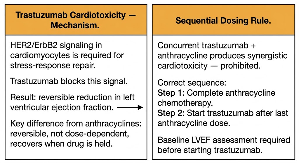
The most important toxicity of trastuzumab is cardiomyopathy with reduction in left ventricular ejection fraction (LVEF). The mechanism is fundamentally different from anthracycline cardiotoxicity: anthracycline cardiotoxicity results from irreversible free radical-mediated damage to cardiomyocyte mitochondria and DNA, is dose-dependent, cumulative, and largely irreversible. Trastuzumab cardiotoxicity results from inhibition of HER2/HER4-mediated cardiac repair signaling — ErbB2 signaling in cardiomyocytes is required for stress-response hypertrophy and protection against anthracycline-induced damage. Consequently, trastuzumab cardiotoxicity is generally not dose-dependent and is largely reversible upon drug discontinuation, in sharp contrast to anthracycline cardiotoxicity.
This mechanistic distinction has two critical clinical consequences. First, trastuzumab cardiotoxicity is managed by temporarily holding the drug and reassessing LVEF, with re-initiation possible in most cases after recovery. Second, and most important: trastuzumab must not be given concurrently with anthracyclines (doxorubicin, epirubicin). The combination produces unacceptably high rates of severe cardiomyopathy because HER2 inhibition eliminates the cardiac protective signaling that normally limits anthracycline damage. The standard practice is anthracycline-based chemotherapy completed first, then trastuzumab initiated after the last anthracycline dose. Baseline LVEF assessment by echocardiography is mandatory before starting trastuzumab, with repeat monitoring during therapy.
Pertuzumab is a humanized IgG1 monoclonal antibody that binds domain II of the HER2 extracellular domain — a different epitope from trastuzumab, which binds domain IV. This distinct binding site allows pertuzumab to inhibit HER2 dimerization with other ErbB family members, particularly the HER2-HER3 heterodimer that drives the most potent downstream oncogenic signaling. The combination of pertuzumab plus trastuzumab achieves dual HER2 blockade at distinct epitopes, producing superior outcomes compared to trastuzumab alone. Pertuzumab is used in combination with trastuzumab in HER2-positive metastatic breast cancer (CLEOPATRA trial) and in early-stage HER2-positive breast cancer in both neoadjuvant and adjuvant settings. Cardiac monitoring requirements are identical to trastuzumab. Diarrhea is a common pertuzumab-related adverse effect.
Bevacizumab is a humanized IgG1 monoclonal antibody that binds all isoforms of VEGF-A (vascular endothelial growth factor A), preventing its binding to VEGFR-1 and VEGFR-2 (VEGF receptors 1 and 2) on endothelial cells and thereby inhibiting tumor angiogenesis. It is approved in combination with chemotherapy for metastatic colorectal cancer, non-squamous non-small cell lung cancer, recurrent glioblastoma, and other solid tumors. Like all monoclonal antibodies, bevacizumab does not undergo cytochrome P450 metabolism and has no CYP-based drug interactions. Its half-life of approximately 20 days is consistent with FcRn-recycled IgG.
The toxicity profile of bevacizumab is mechanistically derived from inhibiting physiological VEGF-A signaling in normal vasculature. Hypertension occurs in approximately 23 to 35 percent of patients and results from VEGF-A withdrawal in the endothelium, reducing nitric oxide and prostacyclin synthesis and causing vasoconstriction. Proteinuria results from VEGF-A-dependent maintenance of glomerular podocyte function. Wound healing impairment is critical: VEGF-A is required for neovascularization in healing wounds, and bevacizumab must be held for at least 28 days before elective surgery and not resumed until wound healing is confirmed. Bowel perforation is a rare but potentially fatal complication resulting from compromise of tumor-adjacent bowel vasculature and impaired repair of microscopic perforations. Posterior reversible encephalopathy syndrome (PRES), presenting with headache, altered mental status, visual disturbances, and seizures, requires permanent drug discontinuation. Bevacizumab is also contraindicated with squamous cell histology in non-small cell lung cancer due to risk of life-threatening hemoptysis from tumor cavitation.
Trastuzumab + Anthracycline: Sequential, Not Concurrent
Concurrent administration of trastuzumab with anthracyclines is contraindicated. HER2 signaling in cardiomyocytes provides the protective response to anthracycline-induced stress; blocking it with trastuzumab during anthracycline therapy produces a synergistic cardiotoxic effect. The rule is absolute: complete anthracycline-based chemotherapy first, then initiate trastuzumab. Pertuzumab shares this requirement.
Section 3
RAS mutation testing, hepatitis B reactivation, blood bank interference, and osteonecrosis prevention
Four mechanistically distinct monoclonal antibody targets carry distinct companion diagnostic requirements, toxicity profiles, and clinical decision rules that appear frequently in high-order clinical reasoning: EGFR in colorectal cancer, CD20 (cluster of differentiation 20) in B-cell lymphomas, CD38 (cyclic ADP-ribose hydrolase) in multiple myeloma, and RANKL (receptor activator of NF-kB ligand) in bone metastases.
Cetuximab is a chimeric IgG1 monoclonal antibody and panitumumab is a fully human IgG2 monoclonal antibody; both bind the extracellular domain of EGFR (epidermal growth factor receptor), blocking ligand binding and receptor activation. They are approved for EGFR-expressing metastatic colorectal cancer and head and neck squamous cell carcinoma (HNSCC). A prerequisite for anti-EGFR therapy in metastatic colorectal cancer is wild-type RAS (rat sarcoma viral proto-oncogene) status: patients with any RAS mutation — KRAS (Kirsten rat sarcoma viral proto-oncogene) or NRAS (neuroblastoma RAS viral proto-oncogene) in exons 2, 3, or 4 — derive no benefit from anti-EGFR therapy because constitutively activated RAS bypasses EGFR signaling and renders EGFR blockade futile. Extended RAS testing is mandatory before prescribing. Additionally, anti-EGFR antibodies are effective only in left-sided colorectal cancers (splenic flexure to rectum); right-sided tumors have higher rates of RAS and BRAF mutations and substantially lower benefit regardless of mutation status.
Acneiform rash occurs in approximately 80 percent of patients and, as with EGFR tyrosine kinase inhibitors, its severity positively correlates with treatment response. A distinctive toxicity not shared by EGFR small molecule inhibitors is hypomagnesemia, resulting from EGFR-dependent magnesium reabsorption in the distal convoluted tubule via the TRPM6 (transient receptor potential melastatin 6) magnesium channel; EGFR blockade reduces TRPM6 expression, causing renal magnesium wasting. Cetuximab, produced in a murine cell line, carries a risk of severe IgE-mediated hypersensitivity reactions in patients with pre-existing antibodies to galactose-alpha-1,3-galactose (alpha-gal), a sugar epitope present on the cetuximab Fc region; this reaction clusters geographically in areas where lone-star tick bites sensitize individuals to alpha-gal. Panitumumab, produced in a non-murine cell line, lacks this epitope and has a lower severe infusion reaction rate.
Rituximab is a chimeric IgG1 monoclonal antibody targeting CD20, a B-cell surface antigen expressed from the pre-B cell stage through mature B cells but absent on plasma cells — which explains why plasma cell-derived immunoglobulin production is preserved after rituximab treatment. CD20 is expressed on approximately 95 percent of B-cell non-Hodgkin lymphomas and chronic lymphocytic leukemia (CLL). Rituximab depletes B cells through antibody-dependent cellular cytotoxicity (ADCC), complement-dependent cytotoxicity (CDC), and direct induction of apoptosis. It is approved for B-cell lymphomas, chronic lymphocytic leukemia, rheumatoid arthritis, and other autoimmune conditions. B-cell depletion persists for 6 to 9 months after a standard course.
Two serious infectious complications define rituximab's safety profile. Hepatitis B virus (HBV) reactivation is a potentially fatal complication: B-cell depletion removes immune control of occult HBV, allowing viral replication to surge in patients who are hepatitis B surface antigen (HBsAg)-positive or who have evidence of prior resolved infection (hepatitis B core antibody, HBcAb-positive). All patients must be screened for HBsAg and HBcAb before rituximab initiation; HBsAg-positive patients require prophylactic antiviral therapy started before rituximab and continued well after completion. Progressive multifocal leukoencephalopathy (PML), caused by JC virus reactivation in the central nervous system, is a rare but devastating complication presenting as progressive neurological deficits; brain MRI shows characteristic white matter lesions without mass effect; cerebrospinal fluid JC virus PCR confirms diagnosis; no proven treatment exists and mortality is high.
Obinutuzumab is a glycoengineered type II anti-CD20 IgG1 monoclonal antibody with enhanced ADCC compared to rituximab (through Fc modifications that increase affinity for Fc gamma receptors on natural killer cells and macrophages) and stronger direct cell death induction. It is approved for chronic lymphocytic leukemia and follicular lymphoma. Infusion-related reactions are more frequent and severe with obinutuzumab than rituximab, particularly with the first infusion.
Daratumumab is a fully human IgG1 monoclonal antibody targeting CD38, a transmembrane glycoprotein expressed at high levels on myeloma cells and at lower levels on normal hematopoietic cells including T cells, B cells, natural killer cells, and red blood cells. Daratumumab depletes myeloma cells through ADCC, CDC, antibody-dependent cellular phagocytosis (ADCP), and direct apoptosis induction, and additionally depletes CD38-expressing regulatory T cells (Tregs) and myeloid-derived suppressor cells (MDSCs), potentially enhancing anti-tumor immunity.
A clinically critical consequence of daratumumab binding to CD38 on red blood cells is interference with pre-transfusion testing: the drug coats patient red blood cells, causing pan-reactive false-positive indirect antiglobulin tests (indirect Coombs tests) that can mask alloantibodies against donor blood antigens. Before initiating daratumumab, blood typing and antibody screening must be performed and the results stored for reference; blood bank laboratories must be notified of daratumumab use so that special techniques — dithiothreitol (DTT) treatment of reagent red blood cells or genotyping-based crossmatch — are used for all future compatibility testing throughout therapy and for months after the last dose.
Denosumab is a fully human IgG2 monoclonal antibody that binds RANKL (receptor activator of NF-kB ligand), preventing it from binding its receptor RANK (receptor activator of NF-kB) on osteoclast precursors and mature osteoclasts. By blocking the RANKL-RANK interaction, denosumab suppresses osteoclast differentiation, activation, and survival, reducing bone resorption and skeletal-related events in patients with bone metastases. It is administered subcutaneously and, unlike bisphosphonates, does not undergo renal elimination, making it usable in patients with significant renal impairment where bisphosphonates are contraindicated. Hypocalcemia and osteonecrosis of the jaw are the principal toxicities; calcium and vitamin D supplementation throughout therapy is mandatory to prevent hypocalcemia. A dental evaluation with completion of invasive dental procedures before starting denosumab is recommended to reduce osteonecrosis of the jaw risk.
Daratumumab Blood Bank Alert
Daratumumab binds CD38 on red blood cells, causing pan-reactive false-positive indirect antiglobulin tests that can persist for months after the last dose, masking clinically significant alloantibodies and potentially compromising safe blood transfusion. The blood bank must be notified before the first dose of daratumumab. Special crossmatch techniques are required for all future blood compatibility testing. Failure to notify the blood bank before initiating daratumumab is a prescribing safety error.
Section 4
Linker chemistry, bystander killing, interstitial lung disease risk, and the HER2-low paradigm
Antibody-drug conjugates (ADCs) combine the target selectivity of monoclonal antibodies with the cytotoxic potency of small molecule drugs, delivering a chemotherapy payload directly to tumor cells expressing the target antigen. The clinical development of HER2-directed ADCs has introduced pharmacologic concepts — including HER2-low as a distinct therapeutic category and bystander killing as a mechanism of action — that are now central to understanding modern oncology pharmacology.
An ADC consists of three components: a targeting monoclonal antibody, a chemical linker, and a cytotoxic payload (the warhead). The antibody confers target specificity and distributes the ADC with large-molecule pharmacokinetics — no cytochrome P450 metabolism, no CYP-based interactions — while also mediating ADCC as an additional anti-tumor mechanism. The linker determines the ADC's stability in circulation and its release mechanism: cleavable linkers (hydrazone, disulfide, or peptide-based) release payload in the tumor microenvironment or within lysosomes after internalization; non-cleavable linkers release payload only after complete proteolytic degradation of the antibody inside lysosomes, requiring tumor cell internalization for activity. The drug-to-antibody ratio (DAR) defines the average number of payload molecules per antibody molecule — traditional ADCs carry DARs of approximately 3 to 4, while newer agents carry DARs of approximately 8. Higher DARs increase potency but can reduce antibody stability and increase off-target toxicity.
Bystander killing is the ability of a released payload to diffuse across cell membranes into neighboring tumor cells that may not express the target antigen. This property is most significant with membrane-permeable payloads and contributes to efficacy in tumors with heterogeneous target expression — an important pharmacologic advantage when tumor antigen expression is not uniform throughout the tumor mass.
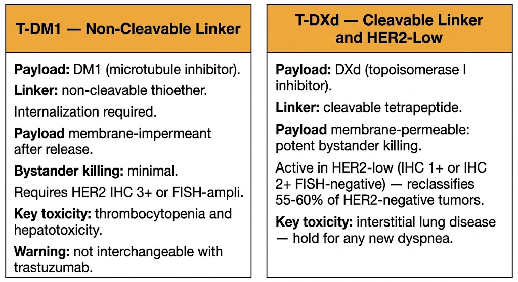
Ado-trastuzumab emtansine (T-DM1) links trastuzumab to emtansine (DM1), a potent microtubule polymerization inhibitor of the maytansinoid class, via a non-cleavable thioether linker with a DAR of approximately 3.5. After HER2-mediated internalization, lysosomal catabolism releases the DM1-containing metabolite, which disrupts microtubule dynamics in the target cell. Because the linker is non-cleavable and the released metabolite is charged and membrane-impermeant, bystander killing is minimal — activity is restricted primarily to HER2-expressing cells. T-DM1 is approved for HER2-positive metastatic breast cancer after prior trastuzumab and taxane therapy, and as adjuvant therapy for HER2-positive early breast cancer with residual disease after neoadjuvant chemotherapy (KATHERINE trial).
T-DM1 must not be substituted for trastuzumab; they are distinct drugs with distinct mechanisms, indications, and toxicity profiles, and confusion between them has caused fatal medication errors. The primary toxicities of T-DM1 are thrombocytopenia (the DM1 payload disrupts platelet microtubule dynamics required for proplatelet formation in megakaryocytes) and hepatotoxicity with elevated liver enzymes. T-DM1 does not cause the alopecia, nausea, or mucositis typical of conventional microtubule agents because payload delivery is largely restricted to HER2-expressing tumor cells.
Fam-trastuzumab deruxtecan (T-DXd) links trastuzumab to deruxtecan, a topoisomerase I inhibitor payload derived from the camptothecin class, via a tetrapeptide-based cleavable linker, with a high DAR of approximately 8. After HER2-mediated internalization, lysosomal cathepsins cleave the linker, releasing a membrane-permeable topoisomerase I inhibitor payload that can diffuse into neighboring cells, producing potent bystander killing. The high DAR combined with membrane-permeable payload and bystander killing activity produces efficacy even at low levels of HER2 expression — a property that gave rise to the concept of HER2-low breast cancer.
HER2-low is defined as immunohistochemistry (IHC) 1+ or IHC 2+/fluorescence in situ hybridization (FISH)-negative — tumors that were previously classified as HER2-negative and treated with standard chemotherapy. The DESTINY-Breast04 trial demonstrated that T-DXd produces superior progression-free and overall survival compared to chemotherapy in HER2-low breast cancer, establishing T-DXd as the standard of care in this newly defined population and reclassifying approximately 55 to 60 percent of previously HER2-negative breast cancers as potentially eligible for HER2-directed therapy.
The most serious toxicity of T-DXd is interstitial lung disease (ILD)/pneumonitis, thought to result from bystander killing of pulmonary epithelial cells by the membrane-permeable payload combined with inflammatory mechanisms. Any new respiratory symptoms in a patient receiving T-DXd must prompt evaluation for ILD; the drug is held for any grade of ILD and corticosteroids are initiated for symptomatic disease. Grade 3 or higher ILD requires permanent discontinuation.
T-DM1 vs. Trastuzumab — These Are Not Interchangeable
Trastuzumab (Herceptin) and T-DM1 (Kadcyla; ado-trastuzumab emtansine) are distinct drugs with different mechanisms, dosing, toxicity profiles, and indications. T-DM1 contains a cytotoxic payload. They must never be substituted for each other. Pharmacy verification of the exact drug name — not just the trastuzumab prefix — is mandatory at every dispensing step.
Section 5
CD30, CD79b, and TROP-2 targeting — MMAE neuropathy, UGT1A1 pharmacogenomics, and payload-specific toxicities
Three additional ADCs have transformed the treatment of specific lymphoma subtypes and triple-negative breast cancer, each exploiting a distinct tumor surface antigen and delivering a distinct cytotoxic payload whose mechanism determines the toxicity profile.
Brentuximab vedotin links an anti-CD30 (cluster of differentiation 30, a TNF receptor superfamily member) IgG1 antibody to MMAE (monomethyl auristatin E), a potent microtubule polymerization inhibitor, via a protease-cleavable valine-citrulline dipeptide linker. CD30 is highly expressed on Reed-Sternberg cells in classical Hodgkin lymphoma and on tumor cells in anaplastic large cell lymphoma (ALCL), making it an ideal ADC target. After CD30-mediated endocytosis, lysosomal cathepsin B cleaves the linker, releasing membrane-permeable MMAE that disrupts microtubule polymerization in the target cell and — through bystander killing — in surrounding tumor microenvironment cells. Brentuximab vedotin is approved in combination with AVD (doxorubicin, vinblastine, dacarbazine) as first-line therapy for advanced Hodgkin lymphoma and in relapsed or refractory Hodgkin lymphoma and ALCL.
The dose-limiting toxicity is peripheral neuropathy, driven by MMAE disruption of axonal microtubule dynamics in dorsal root ganglion neurons. Dose modification and discontinuation criteria parallel those for bortezomib. MMAE is metabolized by cytochrome P450 3A4; strong inhibitors increase MMAE exposure and strong inducers reduce it. Progressive multifocal leukoencephalopathy (PML) has been reported with brentuximab vedotin and requires evaluation when new neurological symptoms develop.
Polatuzumab vedotin links an antibody against CD79b (cluster of differentiation 79b, immunoglobulin-associated beta — a signaling subunit of the B-cell receptor complex expressed on mature B cells and most B-cell non-Hodgkin lymphomas) to MMAE via the same protease-cleavable valine-citrulline linker used in brentuximab vedotin. Because the payload is identical MMAE, the toxicity profile mirrors brentuximab vedotin — peripheral neuropathy from axonal microtubule disruption, myelosuppression, and the same cytochrome P450 3A4-based drug interactions govern MMAE release. Polatuzumab vedotin is approved in combination with bendamustine and rituximab for relapsed or refractory diffuse large B-cell lymphoma (DLBCL), and with R-CHP (rituximab, cyclophosphamide, doxorubicin, prednisone) as first-line therapy for previously untreated DLBCL (POLARIX trial).
Sacituzumab govitecan links an anti-TROP-2 (trophoblast cell surface antigen 2, a cell surface glycoprotein overexpressed in multiple solid tumors including triple-negative breast cancer, urothelial carcinoma, and non-small cell lung cancer) IgG1 antibody to SN-38 — the active metabolite of irinotecan, a topoisomerase I inhibitor — via a pH-sensitive cleavable linker with a high DAR of approximately 7.6. The high DAR and cleavable linker enable substantial bystander killing. Sacituzumab govitecan is approved for metastatic triple-negative breast cancer (ASCENT trial), HER2-negative hormone receptor-positive metastatic breast cancer, and urothelial carcinoma after platinum-based and PD-1/PD-L1 inhibitor therapy.
Sacituzumab govitecan carries a pharmacogenomic drug interaction of high clinical importance. SN-38 is detoxified by glucuronidation via UGT1A1 (UDP-glucuronosyltransferase 1A1). Patients with the UGT1A1*28 homozygous genotype — a promoter polymorphism that reduces UGT1A1 enzyme expression, occurring in approximately 10 percent of patients — cannot efficiently glucuronidate and eliminate SN-38, placing them at substantially higher risk of severe neutropenia and diarrhea. The same pharmacogenomic risk applies to irinotecan, and the principle is pharmacologically identical: reduced UGT1A1 activity increases SN-38 accumulation and toxicity. UGT1A1 genotyping is recommended before initiating sacituzumab govitecan. The dominant toxicities reflect the SN-38 payload: neutropenia and diarrhea in the majority of patients.
ADC Payload and Toxicity
ADC Class Safety Rules
Section 6
HER2 testing interpretation, bevacizumab perioperative rules, infusion reaction management, and companion diagnostic requirements
Monoclonal antibodies and ADCs are encountered across inpatient and outpatient settings, and their toxicities, drug interaction profiles, and prescribing rules generate high-stakes clinical decisions that require systematic pharmacologic reasoning.
HER2 overexpression or amplification is the biomarker gating eligibility for trastuzumab, pertuzumab, T-DM1, and T-DXd in HER2-positive disease. Standard testing uses immunohistochemistry (IHC) as the initial screen: IHC 3+ (uniform strong staining in more than 10 percent of invasive tumor cells) is HER2-positive; IHC 0 or 1+ has historically been HER2-negative; IHC 2+ requires FISH (fluorescence in situ hybridization) confirmation. The T-DXd approval for HER2-low disease created a fourth category: IHC 1+ or IHC 2+/FISH-negative now constitutes HER2-low, which is a distinct therapeutic biomarker for T-DXd eligibility. This reclassification means that approximately 55 to 60 percent of previously HER2-negative breast cancers are now HER2-low and potentially eligible for T-DXd — a major expansion of the treatable population that requires updated IHC reporting practices.
RAS wild-type status is a companion diagnostic mandate before prescribing cetuximab or panitumumab in colorectal cancer. Any KRAS or NRAS mutation in exons 2, 3, or 4 predicts lack of benefit and mandates exclusion from anti-EGFR therapy. Prescribing anti-EGFR antibodies in RAS-mutant colorectal cancer exposes patients to significant toxicity without clinical benefit. Left-sided primary tumor location (splenic flexure to rectum) is also required for anti-EGFR benefit; right-sided tumors have lower benefit regardless of RAS status, and bevacizumab-based regimens are preferred in that setting.
The 28-day pre- and post-operative hold for bevacizumab is the most important perioperative pharmacology rule for monoclonal antibodies in oncology. Bevacizumab must be held at least 28 days before elective surgery and not resumed until complete wound healing — at least 28 days after the procedure. Bevacizumab is also contraindicated in squamous cell non-small cell lung cancer because the drug impairs tumor vasculature repair, creating risk of catastrophic hemoptysis from tumor cavitation. Patients with recent arterial thromboembolic events (myocardial infarction, stroke) within 6 months should not receive bevacizumab due to arterial thromboembolism risk.
Infusion-related reactions are the most common acute complication of monoclonal antibody and ADC administration, occurring most frequently on the first infusion. The vast majority are cytokine release reactions — fever, chills, hypotension, dyspnea — rather than true IgE-mediated anaphylaxis; they are managed by stopping or slowing the infusion, administering intravenous corticosteroids and antihistamines, and resuming at a slower rate after symptom resolution. Pre-medication protocols are agent-specific: cetuximab requires diphenhydramine given the alpha-gal IgE hypersensitivity risk; daratumumab requires dexamethasone, antihistamines, acetaminophen, and a leukotriene modifier for the first infusions.
Hepatitis B virus screening with HBsAg and HBcAb is mandatory before all anti-CD20 (rituximab, obinutuzumab) and anti-CD38 (daratumumab) therapies, and before any biologic that produces significant B-cell depletion. Inactivated vaccines should be given before rituximab therapy if possible, as B-cell depletion abolishes vaccine responses for 6 to 9 months. Live vaccines are absolutely contraindicated in patients receiving rituximab — the attenuated vaccine strain can cause disseminated infection in B-cell-depleted patients.
High-Stakes Clinical Rules
Companion Diagnostics Required
| Author / Organization | Title | Source |
|---|---|---|
| Ryman JT, Meibohm B | Pharmacokinetics of monoclonal antibodies | CPT Pharmacometrics Syst Pharmacol. 2017;6(9):576-588 |
| Hansel TT, Kropshofer H, Singer T, et al. | The safety and side effects of monoclonal antibodies | Nat Rev Drug Discov. 2010;9(4):325-338 |
| Slamon DJ, Leyland-Jones B, Shak S, et al. | Use of chemotherapy plus a monoclonal antibody against HER2 for metastatic breast cancer that overexpresses HER2 | N Engl J Med. 2001;344(11):783-792 |
| Hurwitz H, Fehrenbacher L, Novotny W, et al. | Bevacizumab plus irinotecan, fluorouracil, and leucovorin for metastatic colorectal cancer | N Engl J Med. 2004;350(23):2335-2342 |
| Van Cutsem E, Kohne CH, Hitre E, et al. | Cetuximab and chemotherapy as initial treatment for metastatic colorectal cancer | N Engl J Med. 2009;360(14):1408-1417 |
| Coiffier B, Lepage E, Briere J, et al. | CHOP chemotherapy plus rituximab compared with CHOP alone in elderly patients with diffuse large-B-cell lymphoma | N Engl J Med. 2002;346(4):235-242 |
| Lonial S, Weiss BM, Usmani SZ, et al. | Daratumumab monotherapy in patients with treatment-refractory multiple myeloma (SIRIUS) | Lancet. 2016;387(10027):1551-1560 |
| Stopeck AT, Lipton A, Body JJ, et al. | Denosumab compared with zoledronic acid for the treatment of bone metastases in patients with advanced breast cancer | J Clin Oncol. 2010;28(35):5132-5139 |
| Verma S, Miles D, Gianni L, et al. | Trastuzumab emtansine for HER2-positive advanced breast cancer | N Engl J Med. 2012;367(19):1783-1791 |
| Modi S, Saura C, Yamashita T, et al. | Trastuzumab deruxtecan in previously treated HER2-positive breast cancer | N Engl J Med. 2020;382(7):610-621 |
| Younes A, Gopal AK, Smith SE, et al. | Results of a pivotal phase II study of brentuximab vedotin for patients with relapsed or refractory Hodgkin lymphoma | J Clin Oncol. 2012;30(18):2183-2189 |
| Bardia A, Hurvitz SA, Tolaney SM, et al. | Sacituzumab govitecan in metastatic triple-negative breast cancer | N Engl J Med. 2021;384(16):1529-1541 |
| Modi S, Jacot W, Yamashita T, et al. | Trastuzumab deruxtecan in previously treated HER2-low advanced breast cancer | N Engl J Med. 2022;387(1):9-20 |