CHAPTER 33  ·  ANTI-CANCER DRUGS — PART 1
Section 1

Cell Cycle Kinetics and the Basis of Chemotherapy

Log-kill hypothesis, Gompertzian growth, and cycle-specific versus cycle-nonspecific agents

Cancer pharmacology begins with a deceptively simple question: why does a drug that kills cancer cells in a dish so often fail to cure cancer in a patient? The answer lies in tumor growth kinetics, the heterogeneity of cell cycle position within a tumor mass, and the relationship between drug exposure, tumor burden, and residual viable cells.

The Cell Cycle and Drug Targeting

Every dividing cell traverses four phases: G1 (biosynthetic preparation), S (deoxyribonucleic acid synthesis), G2 (premitotic preparation), and M (mitosis). A fifth state, G0, represents reversible quiescence outside the active cycle. Cells in G0 are metabolically active but not proliferating, and they represent a critical reservoir of drug-resistant cells within a tumor mass because many cycle-specific agents require active deoxyribonucleic acid synthesis or mitotic spindle assembly to exert their lethal effects.

Cycle-specific agents kill only cells traversing a particular phase at the time of drug exposure. Antimetabolites such as methotrexate and 5-fluorouracil are S-phase specific; vinca alkaloids are M-phase specific. Because their cytotoxic effect is self-limiting once cells in the sensitive phase have been killed, cycle-specific agents exhibit a plateau on the dose-response curve. Beyond the dose that kills all cycling cells in the sensitive phase, further dose escalation does not increase cell kill. This plateau has profound scheduling implications: cycle-specific agents are best administered as prolonged infusions or repeated dosing to expose cells that enter the sensitive phase after the initial dose.

Cycle-nonspecific agents, principally the alkylating agents and platinum compounds, kill cells regardless of cell cycle position, including G0 cells. Their dose-response relationship is more nearly linear over a wider dose range, making dose escalation a meaningful strategy up to the limits imposed by organ toxicity.

The Log-Kill Hypothesis

The log-kill hypothesis states that a given dose of a chemotherapeutic agent kills a constant fraction, not a constant number, of tumor cells. If a drug kills 99.9% of cells (a 3-log kill) and a patient harbors 10 billion tumor cells at treatment initiation, 10 million viable cells remain after the first course. This arithmetic predicts that cure requires reducing the tumor burden to less than one viable cell, and that early treatment maximizes the probability of cure because the initial burden is lowest. The log-kill model also predicts why relapse occurs after apparently complete responses: if the initial tumor burden is very large, even repeated courses achieving 3-log kills will not eradicate the population before resistance emerges.

Gompertzian Growth and the Growth Fraction

Real tumors do not grow exponentially throughout their natural history. Early in tumor development, when the mass is small and vascularity is adequate, growth is approximately exponential. As tumor size increases, the growth fraction (the proportion of cells actively cycling) decreases and the doubling time lengthens because nutrient and oxygen supply cannot keep pace with the expanding cell population.

The clinical consequence is that small tumors have the highest growth fraction and are, paradoxically, the most chemosensitive. This is the biological rationale for adjuvant chemotherapy after surgical resection: even when imaging detects no residual disease, microscopic metastatic deposits are growing with a high growth fraction and are more vulnerable to cell cycle-active drugs than the bulky primary tumor was. The Norton-Simon hypothesis extends this reasoning, predicting that dose-dense chemotherapy (the same total dose delivered over a shorter interval) should be more effective than standard scheduling because it prevents tumor regrowth between cycles during the exponential phase of residual disease kinetics.

Scheduling Cycle-Specific Agents: The Cytarabine Example

For S-phase-specific drugs, the standard approach uses continuous intravenous infusion to expose cells as they enter S phase. Cytarabine in acute myeloid leukemia induction is administered as a 7-day continuous infusion precisely because at any given moment only about 20 to 40% of leukemic blast cells are in S phase. A bolus dose would kill this fraction but miss cells that enter S phase in the hours following drug clearance. Continuous infusion maintains cytotoxic concentrations throughout the entire duration of S phase for every dividing cell, explaining why this scheduling principle produces deeper remissions than equivalent bolus dosing.
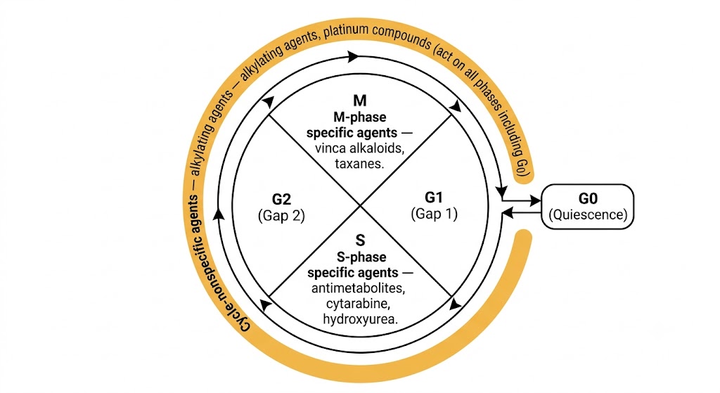
Cell cycle diagram showing G1, S, G2, and M phases in a circle with G0 quiescence connected externally. S-phase specific agents (antimetabolites, cytarabine, hydroxyurea) labeled in S segment; M-phase specific agents (vinca alkaloids, taxanes) labeled in M segment; cycle-nonspecific agents (alkylating agents, platinum compounds) shown encircling all phases including G0.
Cell cycle phases and chemotherapy drug targeting. Cycle-specific agents act only during S or M phase; cycle-nonspecific agents kill cells regardless of cell cycle position, including G0 quiescent cells. Generated with Gemini AI.

Section 2

Drug Resistance Mechanisms

Efflux transporters, metabolic inactivation, target alteration, and apoptosis defects

Drug resistance is the central unsolved problem of clinical oncology. Most patients who relapse after initial chemotherapy do so with disease that is resistant not only to the agents used in initial treatment but also to structurally unrelated drugs, a phenomenon called multidrug resistance.

P-Glycoprotein and Efflux Transporters

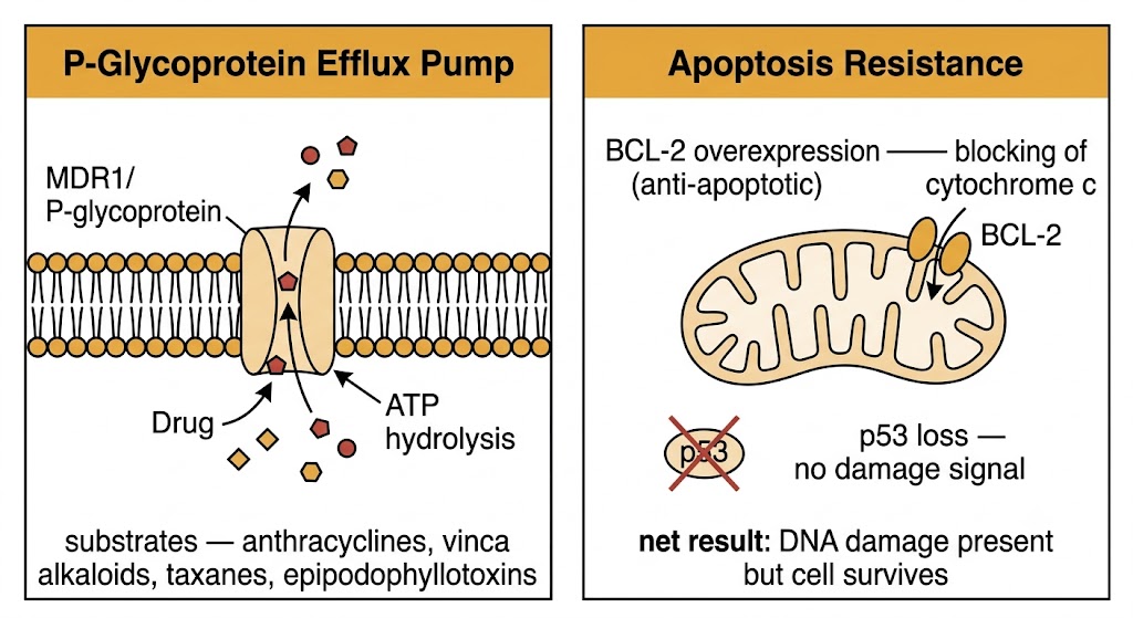
The most extensively characterized resistance mechanism is overexpression of P-glycoprotein, an energy-dependent efflux pump embedded in the plasma membrane that uses adenosine triphosphate hydrolysis to actively transport a broad range of structurally diverse hydrophobic compounds out of the cell, reducing intracellular drug concentrations below cytotoxic thresholds. Its substrate range includes anthracyclines, vinca alkaloids, taxanes, epipodophyllotoxins, and many tyrosine kinase inhibitors. Tumor cells acquire P-glycoprotein overexpression through gene amplification, promoter hypomethylation, or selection of pre-existing P-glycoprotein-high clones during chemotherapy exposure.

Additional adenosine triphosphate-binding cassette family transporters contribute to multidrug resistance. The multidrug resistance-associated protein 1 transports organic anions and glutathione conjugates and is particularly relevant to resistance against anthracyclines, vinca alkaloids, and methotrexate. The breast cancer resistance protein is highly expressed in hematopoietic stem cells and confers resistance to mitoxantrone, camptothecin derivatives, and several oral kinase inhibitors.

Altered Drug Metabolism and Target Changes

For prodrugs that require intracellular activation, reduced expression of the activating enzyme confers resistance. Cytarabine resistance in acute myeloid leukemia frequently involves downregulation of deoxycytidine kinase, the enzyme responsible for phosphorylating cytarabine to its active triphosphate form. 6-mercaptopurine resistance involves loss of hypoxanthine-guanine phosphoribosyltransferase, the enzyme that converts 6-mercaptopurine to its active nucleotide form.

Target amplification or mutation directly reduces drug binding. Dihydrofolate reductase gene amplification is the prototypic mechanism of methotrexate resistance: the tumor cell produces so much dihydrofolate reductase enzyme that therapeutic methotrexate concentrations cannot inhibit all of it, and the residual uninhibited fraction is sufficient to maintain folate metabolism and deoxyribonucleic acid synthesis.

Defective Apoptosis Signaling

Most cytotoxic drugs kill cells by inducing apoptosis. Loss of functional p53 is present in approximately 50% of human cancers and eliminates the primary transcription factor responsible for upregulating pro-apoptotic genes in response to deoxyribonucleic acid damage. Without p53-mediated apoptotic signaling, cells with drug-induced deoxyribonucleic acid damage arrest but do not die, allowing time for deoxyribonucleic acid repair and eventual resumption of proliferation.

Overexpression of BCL-2 (B-cell lymphoma 2 protein) and its relatives tilts the balance at the mitochondrial outer membrane toward cell survival by sequestering pro-apoptotic proteins. BCL-2 overexpression was first identified in follicular lymphoma through the translocation that places the BCL-2 gene under immunoglobulin heavy chain promoter control; it subsequently became a therapeutic target addressed by venetoclax, a selective BCL-2 inhibitor discussed in Chapter 34.

Resistance Category
Reduced Drug Accumulation
  • P-glycoprotein efflux (anthracyclines, vinca alkaloids, taxanes)
  • Multidrug resistance-associated protein 1 efflux
  • Breast cancer resistance protein efflux
  • Reduced drug uptake transporter expression
Resistance Category
Metabolic and Target Changes
  • Loss of activating enzyme (deoxycytidine kinase, hypoxanthine-guanine phosphoribosyltransferase)
  • Target amplification (dihydrofolate reductase)
  • Target mutation (topoisomerase II)
  • Increased catabolism (dihydropyrimidine dehydrogenase)
Two-panel diagram. Left panel: P-Glycoprotein Efflux Pump showing MDR1/P-glycoprotein spanning the cell membrane, pumping drug molecules out using ATP hydrolysis; substrates listed as anthracyclines, vinca alkaloids, taxanes, epipodophyllotoxins. Right panel: Apoptosis Resistance showing BCL-2 overexpression on mitochondrial outer membrane blocking cytochrome c release, and p53 crossed out indicating loss of damage signal; net result is DNA damage present but cell survives.
Two major drug resistance mechanisms. Left: P-glycoprotein efflux reduces intracellular drug concentration. Right: BCL-2 overexpression and p53 loss prevent apoptosis despite DNA damage. Generated with Gemini AI.

Section 3

Combination Chemotherapy

Non-overlapping toxicity, dose intensity, dose density, and the design of curative regimens

The era of curative chemotherapy began with combination regimens. The individual drugs in MOPP (mechlorethamine, vincristine, procarbazine, prednisone), CHOP (cyclophosphamide, doxorubicin, vincristine, prednisone), and BEP (bleomycin, etoposide, cisplatin) were each inadequate as monotherapy for the diseases they cure; it was their combination, guided by the principles of non-overlapping toxicity and non-cross-resistant mechanisms, that transformed Hodgkin lymphoma, aggressive B-cell lymphoma, and testicular germ cell tumors from uniformly fatal diseases into predominantly curable ones.

Principles of Rational Combination Design

The first principle of rational combination chemotherapy design is that each agent should have demonstrated single-agent activity against the target tumor type. Adding an ineffective drug to a regimen does not improve outcomes and adds toxicity. The second principle is non-overlapping dose-limiting toxicity: the drugs in the combination should have different organs at risk for their most severe adverse effects, allowing each to be given at or near its full single-agent dose.

CHOP exemplifies this principle. Cyclophosphamide dose-limits on myelosuppression; doxorubicin dose-limits on myelosuppression and cumulative cardiotoxicity; vincristine dose-limits on peripheral neurotoxicity with minimal myelosuppression; prednisone contributes anti-lymphoma activity with essentially no myelosuppression. The four drugs together deliver four independent mechanisms of action with partially non-overlapping toxicity profiles.

The mathematical rationale for combination chemotherapy also addresses resistance: if the frequency of resistance to drug A is one in a million cells and to drug B is one in a million cells, the probability of simultaneous resistance to both is approximately one in a trillion, below the tumor cell number present even in microscopic disease. This multiplicative probability argument predicts that regimens with more mechanistically distinct components should theoretically suppress resistance emergence more effectively, within the limits imposed by combined toxicity.

Dose Intensity and Dose Density

Dose intensity, defined as the amount of drug delivered per unit time, is a critical determinant of outcome in responsive tumors. Retrospective analyses of breast cancer adjuvant trials demonstrated that patients who received less than 85% of their planned dose intensity had significantly worse disease-free and overall survival than those who received full-dose therapy. This finding motivated the development of granulocyte colony-stimulating factor support to allow full-dose delivery by accelerating neutrophil recovery after myelosuppressive chemotherapy.

Dose-dense chemotherapy compresses the interval between cycles from three weeks to two weeks, maintaining the same per-cycle dose but increasing dose intensity by reducing the recovery time during which residual tumor cells can proliferate. Clinical evidence has demonstrated that dose-dense doxorubicin, cyclophosphamide, and paclitaxel with granulocyte colony-stimulating factor support improved disease-free and overall survival compared with the same regimen given on a standard three-week schedule in node-positive breast cancer. Dose-dense scheduling requires granulocyte colony-stimulating factor support to be practical and safe; without it, the shortened interval would produce cumulative myelosuppression that would force delay or dose reduction.

Canonical Curative Regimens: BEP and CHOP

BEP for testicular germ cell tumors illustrates that even metastatic solid tumors can be curable with combination chemotherapy if the biology is favorable. Cisplatin provides backbone alkylating-like activity through platinum-deoxyribonucleic acid adduct formation; etoposide adds topoisomerase II inhibition; bleomycin contributes deoxyribonucleic acid strand-break induction. The combination achieves cure rates exceeding 80% even in disseminated disease. BEP's limitations are defined by cisplatin nephrotoxicity and ototoxicity, and by the strict bleomycin cumulative dose limit and the avoidance of supplemental high-concentration oxygen in the perioperative period for any patient who has received bleomycin.


Section 4

Supportive Care Pharmacology

Febrile neutropenia, granulocyte colony-stimulating factors, antiemetics, and tumor lysis syndrome

Supportive care pharmacology determines whether a patient can complete planned chemotherapy at full dose intensity, whether febrile neutropenia is managed safely, and whether tumor lysis syndrome is recognized and treated before it produces life-threatening metabolic consequences.

Febrile Neutropenia and Granulocyte Colony-Stimulating Factors

Febrile neutropenia is defined as a temperature above 38.3 degrees Celsius as a single measurement, or above 38.0 degrees Celsius sustained over one hour, in a patient with an absolute neutrophil count below 500 cells per microliter or below 1,000 cells per microliter and expected to fall below 500 within 48 hours. It carries a mortality risk of 5 to 10% in unselected oncology patients. Management requires prompt broad-spectrum antibacterial coverage initiated within one hour of presentation. For low-risk febrile neutropenia (solid tumors, absolute neutrophil count expected to recover within 7 days, no significant comorbidities), oral fluoroquinolone plus amoxicillin-clavulanate outpatient management is appropriate. High-risk febrile neutropenia requires hospital admission and intravenous anti-pseudomonal beta-lactam therapy, most commonly piperacillin-tazobactam as first-line.

Granulocyte colony-stimulating factors stimulate neutrophil precursor proliferation and differentiation in the bone marrow, shortening the duration of chemotherapy-induced neutropenia by three to five days and reducing the incidence of febrile neutropenia by approximately 50% in high-risk regimens. Filgrastim is a non-glycosylated recombinant human granulocyte colony-stimulating factor requiring daily subcutaneous injection beginning 24 to 72 hours after the last dose of chemotherapy. Pegfilgrastim is filgrastim conjugated to polyethylene glycol, which dramatically reduces renal clearance and prolongs half-life, allowing single-injection per-cycle dosing. Granulocyte colony-stimulating factor should not be started within 24 hours of chemotherapy because it mobilizes proliferating neutrophil precursors that become vulnerable to cycle-specific cytotoxic agents if drug levels are still elevated.

Antiemetics: Emetogenic Classification and Drug Selection

Chemotherapy-induced nausea and vomiting is classified by its temporal relationship to chemotherapy administration and the emetogenic potential of the regimen. Acute nausea and vomiting, occurring within 24 hours of chemotherapy, is mediated primarily by serotonin release from enterochromaffin cells stimulating vagal afferent serotonin type 3 receptors. Delayed nausea and vomiting, occurring 24 to 120 hours after chemotherapy, is mediated predominantly by substance P binding to neurokinin-1 receptors in the brainstem. Anticipatory nausea and vomiting is a conditioned response best prevented by achieving optimal control from the first cycle.

Highly emetogenic chemotherapy, including cisplatin, cyclophosphamide at doses above 1,500 mg per square meter, and dacarbazine, requires triple-drug antiemetic prophylaxis with a serotonin type 3 receptor antagonist (ondansetron, granisetron, or palonosetron), a neurokinin-1 receptor antagonist (aprepitant or fosaprepitant), and dexamethasone. Moderately emetogenic chemotherapy such as carboplatin, doxorubicin, and irinotecan requires at minimum a serotonin type 3 antagonist plus dexamethasone.

Tumor Lysis Syndrome

Tumor lysis syndrome results from the massive release of intracellular contents into the systemic circulation when large numbers of tumor cells die simultaneously after initiation of cytotoxic therapy. The metabolic consequences are hyperuricemia from nucleic acid catabolism, hyperkalemia from intracellular potassium release, hyperphosphatemia from intracellular phosphate release, and secondary hypocalcemia from calcium-phosphate precipitation. Clinical consequences include acute uric acid nephropathy, life-threatening cardiac arrhythmias from hyperkalemia, and hypocalcemic tetany or seizures. The highest-risk tumors are those with large cell mass and high proliferative rate that are highly chemosensitive: Burkitt lymphoma, acute lymphoblastic leukemia with high white blood cell counts, and acute myeloid leukemia with blast counts above 100,000 cells per microliter.

Tumor lysis syndrome prophylaxis begins before chemotherapy with aggressive intravenous hydration, allopurinol (a xanthine oxidase inhibitor that blocks new uric acid production, initiated 24 to 48 hours before chemotherapy), and correction of pre-existing electrolyte abnormalities. For patients at high tumor lysis syndrome risk, rasburicase (recombinant urate oxidase) is preferred over allopurinol because it rapidly degrades existing uric acid to allantoin, a highly soluble product readily excreted. Allopurinol only prevents new uric acid formation and does not lower the pre-existing uric acid burden. Rasburicase is absolutely contraindicated in patients with glucose-6-phosphate dehydrogenase deficiency because the hydrogen peroxide generated by the rasburicase reaction causes severe hemolytic anemia in glucose-6-phosphate dehydrogenase-deficient erythrocytes.

Rasburicase and Glucose-6-Phosphate Dehydrogenase Deficiency: A Mandatory Pre-Treatment Check

Rasburicase cannot be given safely without knowing the patient's glucose-6-phosphate dehydrogenase status. Glucose-6-phosphate dehydrogenase deficiency affects approximately 10% of African-American males and 5% of Mediterranean-ancestry males. In deficient patients, the hydrogen peroxide generated by the rasburicase-catalyzed oxidation of uric acid to allantoin causes acute intravascular hemolysis that can be severe and life-threatening. Glucose-6-phosphate dehydrogenase screening should be performed before rasburicase administration in any patient with ancestry from regions where the deficiency is prevalent. In urgent situations where testing is not yet available, allopurinol plus aggressive hydration is the safer default while results are pending.


Suggested References
Author / Organization Title Source
Chabner BA, Roberts TG Jr Chemotherapy and the war on cancer Nat Rev Cancer. 2005;5(1):65–72
Skipper HE, Schabel FM Jr, Wilcox WS Experimental evaluation of potential anticancer agents. XIII. On the criteria and kinetics associated with curability of experimental leukemia Cancer Chemother Rep. 1964;35:1–111
Norton L Gompertzian growth as a central principle of tumor response to therapy J Clin Oncol. 2014;32(4):312–314
Calvert AH, Newell DR, Gumbrell LA, et al Carboplatin dosage: prospective evaluation of a simple formula based on renal function J Clin Oncol. 1989;7(11):1748–1756
Bleyer WA The clinical pharmacology of methotrexate: new applications of an old drug Cancer. 1978;41(1):36–51
Gottesman MM, Fojo T, Bates SE Multidrug resistance in cancer: role of ATP-dependent transporters Nat Rev Cancer. 2002;2(1):48–58
Longley DB, Johnston PG Molecular mechanisms of drug resistance J Pathol. 2005;205(2):275–292
DeVita VT Jr, Chu E A history of cancer chemotherapy Cancer Res. 2008;68(21):8643–8653
Citron ML, Berry DA, Cirrincione C, et al Randomized trial of dose-dense versus conventionally scheduled and sequential versus concurrent combination chemotherapy as postoperative adjuvant treatment of node-positive primary breast cancer J Clin Oncol. 2003;21(8):1431–1439
Perez Fidalgo JA, Garcia Fabregat L, Cervantes A, et al Management of chemotherapy extravasation: ESMO-EONS clinical practice guidelines Ann Oncol. 2012;23(Suppl 7):vii167–vii173
Aapro MS, Bohlius J, Cameron DA, et al 2010 update of EORTC guidelines for the use of granulocyte-colony stimulating factor to reduce the incidence of chemotherapy-induced febrile neutropenia in adult patients Eur J Cancer. 2011;47(1):8–32
Cairo MS, Bishop M Tumour lysis syndrome: new therapeutic strategies and classification Br J Haematol. 2004;127(1):3–11
Chipuk JE, Moldoveanu T, Llambi F, Parsons MJ, Green DR The BCL-2 family reunion Mol Cell. 2010;37(3):299–310
National Institute for Occupational Safety and Health NIOSH List of Antineoplastic and Other Hazardous Drugs in Healthcare Settings, 2016 DHHS (NIOSH) Publication No. 2016-161
Freifeld AG, Bow EJ, Sepkowitz KA, et al Clinical practice guideline for the use of antimicrobial agents in neutropenic patients with cancer: 2010 update by the Infectious Diseases Society of America Clin Infect Dis. 2011;52(4):e56–e93
Hesketh PJ, Kris MG, Basch E, et al Antiemetics: American Society of Clinical Oncology clinical practice guideline update J Clin Oncol. 2017;35(28):3240–3261