How topoisomerases resolve deoxyribonucleic acid supercoiling and how drug-stabilized cleavable complexes generate lethal strand breaks
The topoisomerases are nuclear enzymes that relieve the torsional stress created ahead of advancing replication forks and transcription complexes. Topoisomerase inhibitors exploit this catalytic mechanism by trapping the enzyme in a covalent intermediate with deoxyribonucleic acid, converting a necessary cellular enzyme into a lethal deoxyribonucleic acid-damaging agent.
Topoisomerase I relieves deoxyribonucleic acid supercoiling by making a transient single-strand nick, forming a covalent 3-prime-phosphotyrosine bond between an active-site tyrosine and the cleaved strand. This creates a swivel point around which the intact complementary strand rotates to relieve torsional stress, after which topoisomerase I religates the strand and dissociates. The intermediate in which the enzyme is covalently bound to deoxyribonucleic acid is called the cleavable complex.
Camptothecin-class drugs bind at the interface between topoisomerase I and deoxyribonucleic acid at the cleavable complex, stabilizing it and preventing religation. The stabilized cleavable complex is not inherently lethal — it becomes lethal when a replication fork collides with it, converting the reversible nick into an irreversible double-strand break that triggers apoptosis. This collision-dependent lethality explains why camptothecins are S-phase-specific.
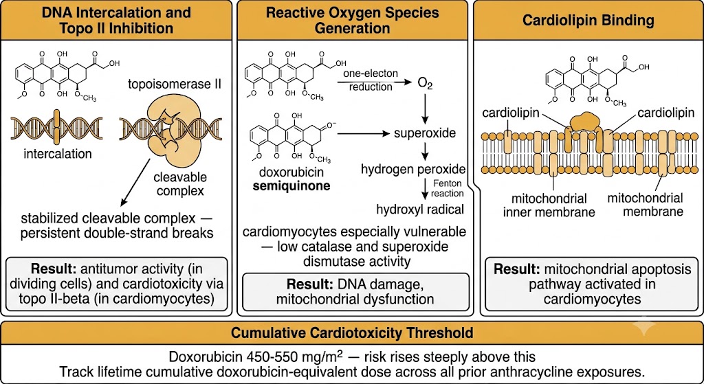
Topoisomerase II resolves both positive and negative supercoiling and decatenates sister chromatid pairs after replication by making transient double-strand breaks. Two isoforms are clinically relevant: topoisomerase II-alpha, which is cell cycle regulated and expressed predominantly in dividing cells, and topoisomerase II-beta, which is expressed in postmitotic cells including cardiomyocytes. Epipodophyllotoxins (etoposide, teniposide) and anthracyclines (doxorubicin, epirubicin, daunorubicin) stabilize the topoisomerase II cleavable complex, generating persistent double-strand breaks. The activity of anthracyclines at topoisomerase II-beta in cardiomyocytes contributes to cardiotoxicity.
Topoisomerase I inhibitors (camptothecins) produce single-strand breaks stabilized into lethal double-strand breaks only at replication forks: they are S-phase-specific and do not cause the secondary leukemias or cumulative cardiotoxicity associated with topoisomerase II inhibitors. Topoisomerase II inhibitors (anthracyclines, epipodophyllotoxins) produce double-strand breaks in any phase of the cell cycle and carry risks of treatment-related acute myeloid leukemia and, for anthracyclines, cumulative cardiomyopathy. Etoposide-related secondary acute myeloid leukemia has a latency of 1 to 3 years — shorter than alkylating agent-related acute myeloid leukemia (5 to 7 years). This difference influences drug selection in young patients receiving curative-intent therapy.
Irinotecan prodrug activation, dual diarrhea mechanisms, and UGT1A1*28 pharmacogenomics
Irinotecan and topotecan are semisynthetic derivatives of the plant alkaloid camptothecin, both acting as topoisomerase I inhibitors through the cleavable complex mechanism. Their clinical profiles differ substantially: irinotecan is used primarily in colorectal and gastrointestinal cancers and has a complex prodrug pharmacology governed by a clinically significant pharmacogenomic variant, while topotecan is used in small cell lung cancer and ovarian cancer.
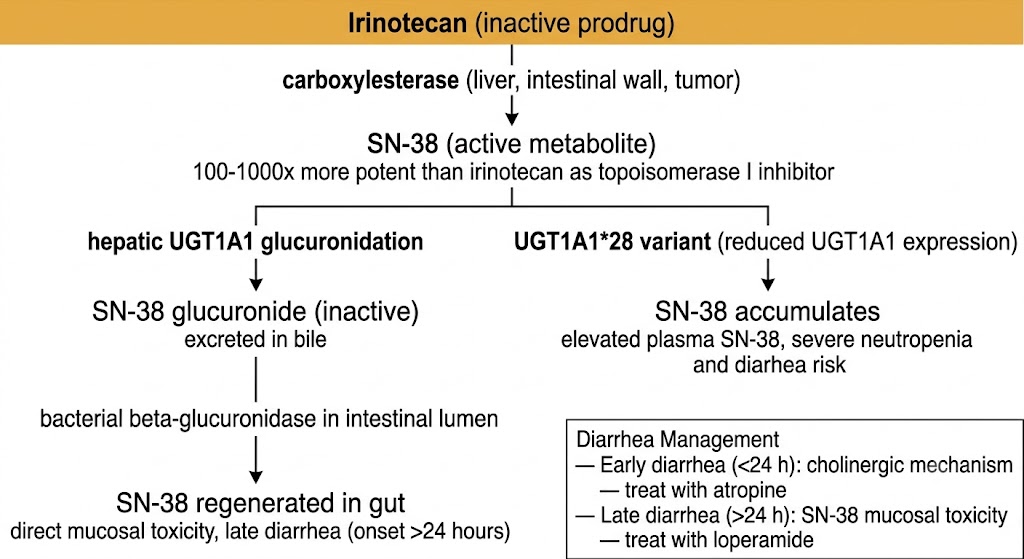
Irinotecan is an inactive prodrug requiring hepatic enzymatic conversion to the active metabolite SN-38, which is substantially more potent than the parent drug as a topoisomerase I inhibitor. SN-38 is inactivated by glucuronidation by uridine diphosphate-glucuronosyltransferase isoform 1A1 in the liver to SN-38 glucuronide, which is excreted in bile. In the intestinal lumen, bacterial beta-glucuronidases deconjugate SN-38 glucuronide back to active SN-38, which causes direct mucosal toxicity and drives late diarrhea.
The two distinct diarrhea syndromes require mechanistically different treatments. Early diarrhea, occurring within 24 hours of irinotecan infusion, is cholinergic in mechanism — irinotecan inhibits acetylcholinesterase, producing abdominal cramping, diaphoresis, flushing, lacrimation, and early-onset diarrhea. Treatment is atropine 0.25 to 1 mg intravenously or subcutaneously. Late diarrhea, onset more than 24 hours after infusion, is caused by intraluminal SN-38 mucosal toxicity. Treatment is high-dose loperamide: 4 mg at the first loose stool, then 2 mg every 2 hours until diarrhea-free for at least 12 hours. Loperamide is not effective for early cholinergic diarrhea; atropine is not effective for late diarrhea. Confusing the two syndromes leads to treatment failure and potentially fatal dehydration.
The UGT1A1*28 variant reduces uridine diphosphate-glucuronosyltransferase isoform 1A1 enzyme expression by approximately 30 to 70%. Patients homozygous for UGT1A1*28 (present in approximately 10% of North Americans of European ancestry) are at substantially increased risk of severe neutropenia and grade 3 to 4 diarrhea at standard irinotecan doses because SN-38 accumulates to higher plasma concentrations due to impaired glucuronidation. The Food and Drug Administration label for irinotecan recommends considering a dose reduction for UGT1A1*28 homozygous patients. Genotyping is increasingly performed before high-dose irinotecan regimens such as FOLFIRI (fluorouracil, leucovorin, irinotecan) or FOLFIRINOX (fluorouracil, leucovorin, irinotecan, oxaliplatin) to guide starting dose selection.
Every patient starting irinotecan must receive written instructions distinguishing early from late diarrhea with the correct treatment for each. Early cholinergic diarrhea (within 24 hours): treat with atropine. Late diarrhea (after 24 hours): treat with loperamide 4 mg at first loose stool, then 2 mg every 2 hours. Hospitalization is required for any patient who develops grade 3 or 4 late diarrhea with concurrent neutropenia — this combination is potentially fatal without prompt intravenous hydration and antibiotics.
Topotecan does not require prodrug activation and is active as the parent compound. It is a substrate for the breast cancer resistance protein, which limits oral absorption and mediates cellular efflux resistance. Topotecan is primarily renally excreted, and dose reduction is required when creatinine clearance falls below 40 mL per minute. The dose-limiting toxicity is myelosuppression, predominantly neutropenia. Topotecan is used in relapsed small cell lung cancer, recurrent ovarian cancer, and relapsed cervical cancer.
Multiple cytotoxic mechanisms, cumulative dose limits, liposomal formulations, and etoposide secondary leukemia risk
The anthracyclines are among the most effective antitumor agents in clinical oncology, with activity across breast cancer, sarcoma, lymphoma, leukemia, and gastric cancer. Their clinical use is inextricably linked to the risk of dose-dependent cumulative cardiomyopathy, which imposes absolute lifetime dose limits and requires active cardiotoxicity surveillance.
Doxorubicin exerts antitumor activity through at least three simultaneous mechanisms. First, it intercalates into double-stranded deoxyribonucleic acid and stabilizes the topoisomerase II-alpha cleavable complex, generating persistent double-strand breaks. Second, doxorubicin generates reactive oxygen species in cardiac tissue, causing oxidative damage to deoxyribonucleic acid, proteins, and lipid membranes. Third, doxorubicin disrupts mitochondrial function in cardiomyocytes, contributing to the cumulative irreversible cardiomyopathy. The cardiomyopathy mechanism involves reactive oxygen species generation combined with topoisomerase II-beta-mediated double-strand breaks in postmitotic cardiomyocyte nuclei.
Doxorubicin undergoes extensive hepatic metabolism and biliary excretion. Hepatic dose modification is required: for bilirubin 1.2 to 3.0 mg per deciliter, give 50% of the dose; for bilirubin above 3.0 mg per deciliter, give 25% of the dose. The cumulative dose-dependent cardiotoxicity threshold for conventional doxorubicin is 450 to 550 mg per square meter; the incidence of clinical heart failure rises steeply with each additional 100 mg per square meter above this threshold.
Epirubicin is the 4-prime-epimer of doxorubicin, with faster glucuronidation and biliary clearance and approximately 20% lower cardiotoxicity per unit dose, allowing a higher per-cycle dose. The cumulative dose limit for epirubicin is approximately 900 mg per square meter. It is used in breast cancer (fluorouracil, epirubicin, cyclophosphamide regimen) and gastric cancer. Daunorubicin and idarubicin are anthracyclines used in acute myeloid leukemia induction; idarubicin is more lipophilic with greater central nervous system penetration and is preferred by many centers for acute myeloid leukemia induction.
Pegylated liposomal doxorubicin encapsulates doxorubicin in polyethylene glycol-coated liposomes that exploit the enhanced permeability and retention effect — the disordered, leaky vasculature of tumors allows preferential extravasation of nano-scale liposomal particles while normal capillaries do not. The result is a dramatically different toxicity profile: cardiotoxicity is substantially reduced, myelosuppression is reduced, and alopecia is significantly less. The new dose-limiting toxicities are palmar-plantar erythrodysesthesia (hand-foot syndrome) and mucositis. Pegylated liposomal doxorubicin is approved for ovarian cancer, AIDS-related Kaposi sarcoma, and multiple myeloma.
Etoposide is incorporated into multiple curative-intent regimens including BEP (bleomycin, etoposide, cisplatin) for testicular germ cell tumors, BEACOPP (bleomycin, etoposide, doxorubicin, cyclophosphamide, vincristine, procarbazine, prednisone) for high-risk Hodgkin lymphoma, and etoposide plus cisplatin for small cell lung cancer. It stabilizes the topoisomerase II-alpha-deoxyribonucleic acid cleavable complex during the G2 phase and G2/M boundary, generating persistent double-strand breaks and triggering apoptosis.
The most serious long-term toxicity of etoposide is treatment-related secondary acute myeloid leukemia, characterized by balanced chromosomal translocations, with a latency of 1 to 3 years and de novo acute myeloid leukemia presentation without a prior myelodysplastic syndrome phase. The cumulative risk is approximately 1 to 2% in testicular cancer survivors treated with BEP and approximately 3 to 5% in Hodgkin lymphoma patients treated with BEACOPP. These risks must be disclosed during informed consent for curative-intent regimens containing etoposide.
Oxidative deoxyribonucleic acid strand scission, pulmonary toxicity, anesthesia oxygen risk, and ribonucleic acid polymerase inhibition
Bleomycin and actinomycin D are antitumor antibiotics with distinct mechanisms of deoxyribonucleic acid damage and toxicity profiles that differ markedly from the topoisomerase inhibitors. Both are used in curative-intent regimens and carry severe organ-specific toxicities requiring active monitoring.
Bleomycin is a glycopeptide antibiotic that kills cells by generating reactive oxygen species that directly cleave deoxyribonucleic acid strands. The mechanism involves bleomycin chelating iron in the nucleus to form a bleomycin-iron complex that, in the presence of molecular oxygen, generates hydroxyl radicals cleaving deoxyribonucleic acid strands preferentially at 5-prime-guanine-cytosine and 5-prime-guanine-thymine sequences. The lung and skin have low capacity to inactivate bleomycin compared with other tissues such as the liver, which explains why these organs are selectively vulnerable to bleomycin-induced toxicity. Bleomycin is cell cycle specific with maximal cytotoxicity in G2 phase and M phase.
The dose-limiting and potentially fatal toxicity of bleomycin is pulmonary toxicity, manifesting as bleomycin-induced pneumonitis progressing in severe cases to pulmonary fibrosis. Pulmonary toxicity occurs in approximately 10% of patients receiving standard BEP doses and is associated with cumulative lifetime bleomycin doses above 400 units, age above 40 years, prior thoracic radiation, renal impairment (bleomycin is renally eliminated), and high inspired oxygen concentration. Serial diffusing capacity for carbon monoxide measurements are used for monitoring. Treatment is bleomycin discontinuation; corticosteroids are used in moderate to severe cases.
High inspired oxygen fractions during general anesthesia can trigger acute respiratory distress syndrome in patients with prior bleomycin exposure, with reported mortality rates exceeding 50% in severe cases. The inspired oxygen fraction must be kept at the lowest level consistent with adequate oxygen saturation (target 93 to 95%) during and after anesthesia in any patient with a history of bleomycin therapy. This requirement does not expire with time — there is no established safe interval after bleomycin beyond which normal inspired oxygen fractions can be used. Bleomycin history must be prominently documented in the patient chart and communicated to the anesthesia team before any procedure requiring general anesthesia.
Actinomycin D intercalates into double-stranded deoxyribonucleic acid by inserting its flat phenoxazinone chromophore between guanine-cytosine base pairs, physically blocking ribonucleic acid polymerase movement along the deoxyribonucleic acid template and inhibiting transcription of all ribonucleic acid species without direct inhibition of deoxyribonucleic acid replication at clinical concentrations. Actinomycin D is a potent radiation sensitizer because it inhibits transcription of deoxyribonucleic acid repair enzymes required after radiation-induced deoxyribonucleic acid damage. It is used in Wilms tumor (nephroblastoma) in children, gestational trophoblastic disease, Ewing sarcoma, and rhabdomyosarcoma. The dose-limiting toxicities are myelosuppression and mucositis; actinomycin D is also an extreme vesicant requiring scrupulous administration technique.
| Author / Organization | Title | Source |
|---|---|---|
| Pommier Y | Topoisomerase I inhibitors: camptothecins and beyond | Nat Rev Cancer. 2006;6(10):789–802 |
| Pommier Y, Sun Y, Huang SN, Nitiss JL | Roles of eukaryotic topoisomerases in transcription, replication and genomic stability | Nat Rev Mol Cell Biol. 2016;17(11):703–721 |
| Pizzolato JF, Saltz LB | The camptothecins | Lancet. 2003;361(9376):2235–2242 |
| Iyer L, Das S, Janisch L, et al | UGT1A1*28 polymorphism as a determinant of irinotecan disposition and toxicity | Pharmacogenomics J. 2002;2(1):43–47 |
| Hande KR | Etoposide: four decades of development of a topoisomerase II inhibitor | Eur J Cancer. 1998;34(10):1514–1521 |
| Minotti G, Menna P, Salvatorelli E, Cairo G, Gianni L | Anthracyclines: molecular advances and pharmacologic developments in antitumor activity and cardiotoxicity | Pharmacol Rev. 2004;56(2):185–229 |
| Weiss RB | The anthracyclines: will we ever find a better doxorubicin? | Semin Oncol. 1992;19(6):670–686 |
| Swain SM, Whaley FS, Ewer MS | Congestive heart failure in patients treated with doxorubicin: a retrospective analysis of three trials | Cancer. 2003;97(11):2869–2879 |
| Chen J, Stubbe J | Bleomycins: towards better therapeutics | Nat Rev Cancer. 2005;5(2):102–112 |
| de Wit R, Roberts JT, Wilkinson PM, et al | Equivalence of three or four cycles of bleomycin, etoposide, and cisplatin chemotherapy and of a 3- or 5-day schedule in good-prognosis germ cell cancer | J Clin Oncol. 2001;19(6):1629–1640 |
| Lipshultz SE, Lipsitz SR, Mone SM, et al | Female sex and drug dose as risk factors for late cardiotoxic effects of doxorubicin therapy for childhood cancer | N Engl J Med. 1995;332(26):1738–1743 |
| Swain SM, Whaley FS, Gerber MC, et al | Cardioprotection with dexrazoxane for doxorubicin-containing therapy in advanced breast cancer: a randomized trial | J Clin Oncol. 1997;15(4):1318–1332 |