CHAPTER 2 · ADME

Section 1

Renal Drug Elimination — Glomerular Filtration

How the kidney filters drugs from plasma and why renal function determines drug clearance

The kidneys are the principal organ for eliminating drugs and their metabolites from the body. Renal drug elimination involves three distinct processes: glomerular filtration, tubular secretion, and tubular reabsorption. Understanding each process and how they interact determines not only the rate of drug elimination but also how disease states and urine pH manipulations can be exploited to treat drug toxicity.
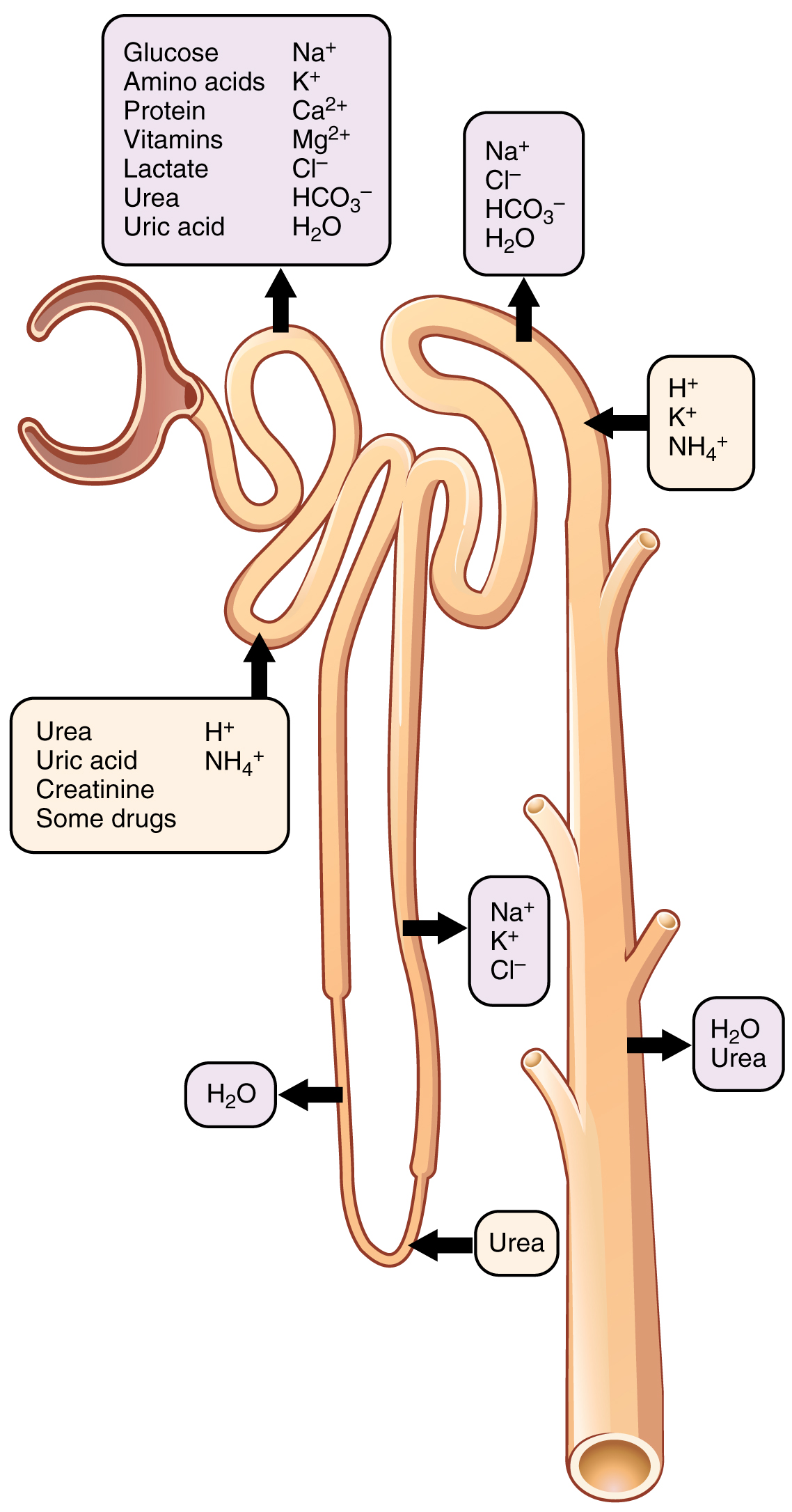
Diagram of a nephron showing the locations of secretion and reabsorption of substances along the renal tubule. Arrows indicate tubular secretion of urea, uric acid, creatinine, hydrogen ions, ammonium, and some drugs from the peritubular blood into the proximal tubule lumen, and reabsorption of glucose, amino acids, sodium, potassium, bicarbonate, and water from the tubular fluid back into the bloodstream at the proximal convoluted tubule and distal segments.
OpenStax College. Schematic showing locations of secretion and reabsorption of substances in the renal nephron. Source: Wikimedia Commons. License: CC BY 3.0.
Glomerular Filtration and the Role of Plasma Protein Binding

Each kidney contains approximately one million nephrons, the functional filtration units. Blood enters the glomerulus — a tuft of fenestrated capillaries enclosed within Bowman's capsule — at high hydrostatic pressure. The high pressure forces unbound (free) drug molecules and other small solutes through the glomerular filtration membrane into the tubular fluid. Protein-bound drug cannot pass through this barrier: only the free drug fraction is filtered, because plasma proteins are too large to cross the glomerular membrane.

This selectivity for free drug has an important implication that is often overlooked: glomerular filtration rate does not equal drug filtration rate. For a drug that is 99 percent bound to albumin, only 1 percent of the plasma drug concentration is free and available for filtration. The filtered load of drug equals the product of glomerular filtration rate, the free drug fraction in plasma, and the plasma drug concentration.

Glomerular Filtration Rate as the Measure of Kidney Function

Glomerular filtration rate is the volume of plasma filtered by the kidneys per unit time, normally approximately 100 to 125 milliliters per minute in a healthy adult. Because it cannot be measured directly, it is estimated from the plasma concentration of creatinine — a muscle metabolism byproduct that is freely filtered at the glomerulus, minimally secreted, and not reabsorbed. When the kidneys filter less effectively, plasma creatinine rises and the estimated glomerular filtration rate falls proportionally.

For drugs whose elimination is primarily through glomerular filtration (or filtration plus tubular secretion), the elimination rate is directly proportional to renal function. When renal function falls to 50 percent of normal, the filtration-dependent clearance of such a drug falls by approximately 50 percent, causing plasma drug concentrations to approximately double at the same dose. This proportionality is the pharmacokinetic rationale for dose adjustment in renal impairment: reducing the dose or extending the dosing interval in proportion to the reduction in glomerular filtration rate maintains the same average plasma exposure as in a patient with normal renal function.

Renal Drug Elimination — Three Processes

Glomerular filtration: passive, pressure-driven; only free (unbound) drug filtered; rate proportional to glomerular filtration rate. Tubular secretion: active transport from peritubular capillaries into tubular lumen; adds to filtered drug; requires specific transporters (organic anion and organic cation transporters). Tubular reabsorption: passive diffusion of un-ionized, lipophilic drug back from tubular lumen into blood; reduces net excretion; manipulable by urine pH. Net renal excretion = filtered + secreted − reabsorbed.

Section 2

Tubular Secretion and Reabsorption

Active transport into the tubule, passive reabsorption, and how urine pH manipulation treats drug overdose

Beyond simple filtration, the renal tubule actively secretes drugs into the tubular lumen and passively reabsorbs them back into the bloodstream. These two processes substantially modify the net excretion of many drugs and can be clinically manipulated to accelerate elimination in overdose situations.

Tubular Secretion

Tubular secretion is an active, carrier-mediated process that transports drug from the peritubular capillaries into the tubular lumen, adding drug to the tubular fluid beyond what glomerular filtration alone delivers. This process is so efficient for some drugs that the tubule can secrete drug even when the drug is highly protein-bound — because the transporter removes free drug from plasma rapidly enough to pull drug off the binding protein, driving net transport from capillary to tubule.

Two major sets of transport proteins mediate tubular secretion. Organic anion transporters, located on the basolateral membrane of proximal tubular cells, take up organic acid drugs (including penicillins, cephalosporins, methotrexate, furosemide, and probenecid itself) from peritubular plasma and deposit them into the tubular lumen. Organic cation transporters handle basic drugs and cationic compounds (including metformin, ranitidine, and creatinine). The two systems are distinct and are not competitive with each other.

The probenecid-penicillin interaction is the paradigmatic clinical example of tubular secretion blockade. Probenecid competitively inhibits the organic anion transporter, reducing the tubular secretion of penicillin and extending its plasma half-life from approximately 30 minutes to several hours. This interaction was actively exploited during World War Two, when penicillin was scarce, to make each dose last longer. Today, probenecid continues to be used clinically to block the tubular secretion of antiviral agents such as cidofovir, reducing its renal toxicity by decreasing the tubular concentration of this nephrotoxic drug.

Tubular Reabsorption and Ion Trapping

As the tubular fluid flows through the nephron, lipophilic, un-ionized drug molecules passively diffuse back across the tubular epithelium into the peritubular capillaries — a process called tubular reabsorption. This reduces net drug excretion. The extent of reabsorption depends on the drug's lipophilicity and its ionization state in the tubular fluid, which in turn depends on urine pH.

Weak acids (such as aspirin and phenobarbital) are more un-ionized and therefore more reabsorbable in acidic urine, and more ionized (and therefore less reabsorbable, more trapped in the tubular lumen) in alkaline urine. Alkalinizing the urine with sodium bicarbonate therefore increases the excretion of weak acid drugs — a principle exploited in the treatment of aspirin overdose and phenobarbital overdose. Alkaline urine traps the ionized form of these drugs in the tubular lumen, preventing reabsorption and accelerating elimination.

The opposite applies to weak bases (such as amphetamines and phencyclidine): acidifying the urine with ammonium chloride increases their ionization in the tubular fluid, trapping them and increasing excretion. Historically, urine acidification was used to increase amphetamine excretion, though this approach has fallen out of favor because systemic acidification carries risks. Understanding ion trapping is an examination favorite because it links urine pH manipulation directly to drug toxicokinetics and allows prediction of the direction of the intervention needed.

Urine pH Manipulation — Ion Trapping Summary

Alkalinize urine (sodium bicarbonate): ionizes weak acids in tubular fluid → traps ionized form → reduces reabsorption → increases excretion. Use for: aspirin overdose, phenobarbital overdose, methotrexate elimination enhancement. Acidify urine (ammonium chloride): ionizes weak bases → traps in tubular lumen → increases excretion. Use for: amphetamine overdose (historical), phencyclidine (theoretical). Probenecid: blocks organic anion transporter → reduces penicillin and cidofovir tubular secretion.


Section 3

Dose Adjustment in Renal Impairment

Which drugs require adjustment, how to adjust them, and why renal failure changes more than just filtration

Renal impairment reduces the elimination of drugs that are primarily excreted by the kidneys, causing accumulation and toxicity if doses are not adjusted. Knowing which drugs require adjustment and how to approach that adjustment is one of the most practically important applications of pharmacokinetics in clinical medicine.

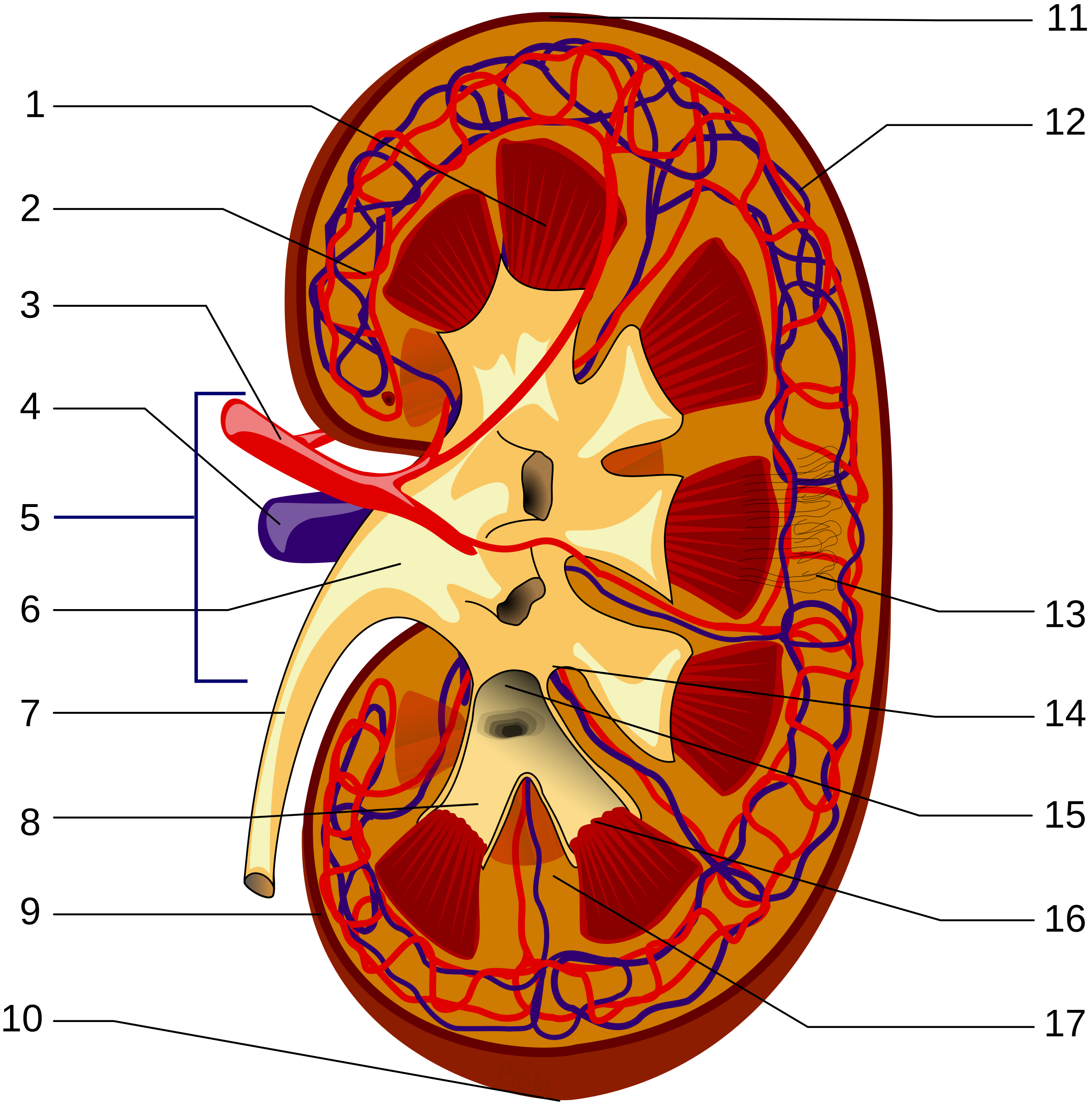
Anatomical diagram of a cross-sectioned kidney showing the major structural components including the renal pyramid, interlobular arteries, renal artery and vein, renal hilum, renal pelvis, ureter, minor and major calyces, renal capsule, interlobar vein, nephrons, renal papilla, and renal columns, illustrating the organizational context within which drug elimination by glomerular filtration and tubular secretion takes place.
Piotr Michał Jaworski (PioM). Kidney anatomy diagram. Source: Wikimedia Commons. License: CC BY-SA 3.0. 1. Renal pyramid10. Inferior renal capsule 2. Interlobular artery11. Superior renal capsule 3. Renal artery12. Interlobar vein 4. Renal vein13. Nephron 5. Renal hilum14. Minor calyx 6. Renal pelvis15. Major calyx 7. Ureter16. Renal papilla 8. Minor calyx17. Renal column 9. Renal capsule
Which Drugs Require Renal Dose Adjustment

Drugs that require dose adjustment in renal impairment share two characteristics: they are substantially excreted by the kidneys (typically more than 30 to 50 percent of unchanged drug eliminated renally), and they have a narrow therapeutic index where drug accumulation produces toxicity. Drugs that are primarily eliminated by hepatic metabolism and have wide therapeutic indexes generally do not require renal dose adjustment, because the accumulated drug concentration, though higher than in patients with normal renal function, remains within a safe range.

The most clinically important drugs requiring careful renal dose adjustment include aminoglycoside antibiotics (gentamicin, tobramycin, amikacin), vancomycin, beta-lactam antibiotics at high doses, digoxin, lithium, metformin, enoxaparin and other low-molecular-weight heparins, direct oral anticoagulants (apixaban, rivaroxaban, dabigatran have different degrees of renal dependence), gabapentin and pregabalin, allopurinol, and many antiretroviral agents. Conversely, azithromycin, doxycycline, linezolid, clindamycin, and most macrolide antibiotics do not require renal dose adjustment because they are primarily eliminated by non-renal routes.

How to Adjust Doses

The two fundamental approaches to renal dose adjustment are dose reduction (administering a smaller dose at the normal dosing interval) and interval extension (administering the normal dose at a longer dosing interval). These produce different concentration-time profiles: dose reduction lowers both the peak and trough concentrations, while interval extension maintains the same peak but allows the trough to fall lower before the next dose.

Which approach is appropriate depends on the pharmacodynamic properties of the drug. For drugs where efficacy correlates with time above a minimum effective concentration (most beta-lactam antibiotics), maintaining a consistent concentration by reducing the dose and keeping the interval the same is preferred. For drugs where efficacy correlates with peak concentration (aminoglycosides — concentration-dependent bactericidal activity), maintaining a high peak while extending the interval to allow drug to clear is the preferred approach, and once-daily dosing becomes even more rational in renal impairment. For narrow-therapeutic-index drugs, the dose and interval are typically both adjusted empirically based on published guidelines with therapeutic drug monitoring to verify that concentrations are within range.

Renal Failure Changes More Than Filtration

Renal impairment affects drug pharmacokinetics through multiple mechanisms beyond reduced filtration. Protein binding changes: uremia generates endogenous organic acids that compete with drugs for albumin binding sites, increasing the free fraction of acidic drugs including phenytoin and some nonsteroidal anti-inflammatory drugs. Volume of distribution changes: fluid retention associated with renal failure increases the effective volume of distribution for hydrophilic drugs, requiring higher loading doses. Absorption may be altered: uremia-induced nausea, gastrointestinal dysmotility, and gastroparesis reduce the reliability of oral drug absorption. Metabolism is modestly impaired: some cytochrome P450 enzyme activities are reduced in advanced renal failure, though the magnitude is less than in severe hepatic disease. For drugs with active or toxic metabolites that are renally eliminated (such as morphine-6-glucuronide from morphine, or normeperidine from meperidine), renal failure causes accumulation of the metabolite as well as the parent drug — a consideration that makes meperidine particularly dangerous in renal failure due to normeperidine-induced seizures.

Category Approach Examples Key concern
Avoid in severe renal failure Use alternative Metformin (lactic acidosis), meperidine (normeperidine seizures), nitrofurantoin (inadequate urinary concentration), NSAIDs (further renal injury) Drug or metabolite causes harm in renal failure regardless of dose
Reduce dose Lower dose, same interval Digoxin, beta-lactams, gabapentin, many antivirals Maintains steady-state exposure; prevents toxicity from accumulation
Extend interval Normal dose, longer interval Aminoglycosides, vancomycin Preserves peak concentration for efficacy; reduces trough toxicity
Monitor levels Dose by measured concentration Vancomycin, aminoglycosides, digoxin, lithium, phenytoin (with albumin correction) Variable pharmacokinetics in renal failure; therapeutic drug monitoring guides dosing
No adjustment needed Standard dosing Azithromycin, doxycycline, linezolid, clindamycin, most macrolides Primarily non-renal elimination; accumulation not clinically significant

Section 4

Hepatic and Biliary Elimination

Biliary excretion as a complementary elimination route and its molecular weight threshold

While renal excretion is the dominant route for most drugs and their water-soluble metabolites, biliary excretion provides a complementary pathway through which the liver secretes drugs directly into bile, delivering them to the intestinal lumen for fecal elimination. The relative importance of renal versus biliary excretion depends primarily on molecular size and chemical characteristics.

Molecular Weight Threshold and Biliary Excretion

Biliary excretion is preferentially used for larger, more polar molecules that are not efficiently filtered at the glomerulus. As a rough rule, drugs with molecular weights above approximately 500 to 600 daltons tend to be excreted in bile rather than urine, while smaller molecules are more effectively renally eliminated. This threshold reflects the capacity of hepatocyte canalicular transporters, which preferentially handle larger conjugated metabolites and polar compounds.

Active transport proteins on the canalicular membrane of hepatocytes pump drug molecules and their conjugates from hepatocytes into bile. The resulting bile contains conjugated drug metabolites — primarily glucuronide and sulfate conjugates generated by phase two metabolism — at high concentrations. Bile is then stored in the gallbladder and released into the duodenum during meals, delivering the conjugated drug into the intestinal lumen.

Drugs Prominently Excreted in Bile

Rifampin is one of the most prominent examples of biliary drug excretion: after absorption, it is rapidly taken up by hepatocytes, undergoes deacetylation in the intestine (not in the liver), and is excreted in bile. Its characteristic red-orange discoloration of urine, feces, tears, and sweat reflects this excretion pattern across multiple routes. Erythromycin and other macrolide antibiotics are substantially excreted in bile, accounting for their relatively high fecal concentrations and their efficacy against biliary tract pathogens. Digoxin and its glucuronide conjugates are partially excreted in bile, contributing to the enterohepatic recirculation discussed in Section 5. Irinotecan (a cancer chemotherapy drug) undergoes biliary excretion of its toxic metabolite SN-38 glucuronide, which is deconjugated in the intestine by bacterial beta-glucuronidase to release the highly toxic parent SN-38 compound — the mechanism underlying irinotecan-associated severe diarrhea.


Section 5

Enterohepatic Recirculation

How biliary excretion becomes reabsorption, and the clinical consequences of interrupting the cycle

For drugs that are both excreted in bile and able to be reabsorbed from the intestine, elimination is not a single pass through the liver but a recurring cycle of hepatic secretion, intestinal delivery, and reabsorption. This enterohepatic recirculation substantially extends drug half-life beyond what would be predicted from hepatic and renal clearance alone.

The Mechanism of Enterohepatic Recirculation

The cycle proceeds as follows: the drug or its conjugated metabolite is secreted from hepatocytes into bile, stored in the gallbladder, and released into the small intestine. In the colon, bacteria express beta-glucuronidase and sulfatase enzymes that cleave the glucuronide or sulfate conjugate, liberating the free (de-conjugated) drug. The free drug, which is now lipophilic enough to be absorbed across the colonic mucosa, is reabsorbed into the portal circulation and returns to the liver, where it re-enters the hepatic processing cycle. Each cycle delays drug elimination and extends the effective half-life.

This recycling is physiologically important for bile salts, which undergo enterohepatic recirculation approximately 6 to 10 times per day, with only a small fraction lost in feces. For drugs, enterohepatic recirculation is clinically relevant when it significantly prolongs drug exposure or when interruption of the cycle is used therapeutically.

Clinical Consequences and Therapeutic Interruption

Digoxin, morphine, chloramphenicol, ethinyl estradiol (in oral contraceptives), and numerous other drugs undergo clinically meaningful enterohepatic recirculation. For digoxin, the recirculation contributes to its long effective half-life (36 to 48 hours) and means that elimination continues for days after the drug is stopped. For oral contraceptives containing ethinyl estradiol, the estrogen component undergoes hepatic glucuronidation, biliary excretion, and colonic deconjugation before reabsorption — a cycle that was hypothesized to be disrupted by broad-spectrum antibiotics that kill the colonic bacteria responsible for deconjugation.

The antibiotic-oral contraceptive interaction has been a longstanding clinical concern and teaching point. The theoretical mechanism is plausible: if antibiotics substantially deplete the colonic bacteria that deconjugate estrogen glucuronide, less free estrogen would be reabsorbed, potentially reducing the circulating estrogen concentration and diminishing contraceptive efficacy. However, careful pharmacokinetic studies have generally found that most commonly used antibiotics (amoxicillin, tetracyclines) do not substantially reduce ethinyl estradiol plasma concentrations or ovarian suppression. Rifampin is the important exception: it profoundly reduces oral contraceptive efficacy through enzyme induction (cytochrome P450 3A4) rather than enterohepatic mechanism, and alternative or additional contraception is strongly recommended during rifampin use.

Cholestyramine, a bile acid sequestrant resin, actively interrupts enterohepatic recirculation for several drugs by binding them in the intestinal lumen and preventing reabsorption. This is therapeutically useful for accelerating the elimination of digoxin in toxicity (cholestyramine interrupts digoxin's enterohepatic recirculation, accelerating fecal elimination), and it is the mechanism by which cholestyramine and colesevelam lower cholesterol (by interrupting bile salt recirculation, forcing the liver to synthesize new bile salts from cholesterol).

Enterohepatic Recirculation — Clinical Pearls

Mechanism: hepatic conjugation → biliary excretion → intestinal bacterial deconjugation → reabsorption → prolonged half-life. Cholestyramine interrupts recirculation of digoxin, warfarin, thyroxine — useful in toxicity but separates administration from these drugs by at least 4 hours to avoid impaired absorption. Rifampin reduces oral contraceptive efficacy via enzyme induction (not enterohepatic mechanism) — strong recommendation for alternative contraception. Irinotecan: intestinal bacterial beta-glucuronidase releases toxic SN-38 from bile conjugate — mechanism of severe diarrhea; inhibiting intestinal beta-glucuronidase is a pharmacological strategy to reduce toxicity.


Section 6

Other Routes of Elimination

Pulmonary elimination, mammary and salivary excretion, and practical implications for breastfeeding

While renal and biliary elimination account for the clearance of most drugs, several minor routes of elimination are clinically significant for specific drug classes or patient situations.

Pulmonary Elimination

Volatile and gaseous substances are eliminated through the lungs. Inhaled general anesthetics — including isoflurane, sevoflurane, and desflurane — are primarily eliminated by exhalation. This mirrors their administration: the same routes used for delivery are used for removal. The elimination rate by the pulmonary route depends on the drug's blood-gas partition coefficient, which determines how readily the drug moves from blood into exhaled air. Anesthetics with low blood-gas partition coefficients (desflurane, sevoflurane) are eliminated more rapidly than those with higher coefficients (isoflurane, halothane), which is one pharmacokinetic basis for the faster recovery from desflurane and sevoflurane anesthesia. Ethanol is also partially eliminated by the pulmonary route, which is the pharmacokinetic basis for breathalyzer testing: the exhaled ethanol concentration is proportional to the blood ethanol concentration.

Mammary, Salivary, and Skin Excretion

Breast milk is slightly acidic relative to plasma (pH approximately 6.8 to 7.1 versus plasma pH 7.4), which influences the ion trapping of drugs in milk. Weak bases (such as opioids, most psychotropic drugs, and many antibiotics) are ionized to a greater extent in the slightly acidic milk than in plasma, accumulating in breast milk by ion trapping. Lipophilic drugs also distribute into the fat-rich milk compartment. The practical clinical consequence is that any drug taken by a breastfeeding parent may be present in breast milk, and the infant receives a dose proportional to the milk drug concentration and volume consumed.

Several drugs are specifically contraindicated during breastfeeding because of demonstrated or theoretical infant harm: most chemotherapy agents, amiodarone (iodine-rich, thyroid effects), lithium, radioactive iodine, codeine in mothers who are ultra-rapid cytochrome P450 2D6 metabolizers (as discussed in Module 3), and several others. For many other drugs, the infant's exposure through breast milk is low and clinically acceptable, but each situation warrants case-by-case review using resources such as the Lactation Risk Classification system.

Salivary excretion is minor for most drugs but forms the basis for saliva-based therapeutic drug monitoring, which exploits the correlation between salivary and plasma drug concentrations for several drugs including phenytoin, carbamazepine, and some antiretrovirals. Skin excretion via sweat contributes to the characteristic color changes seen with rifampin (orange) and is one route of elimination for heavy metals.

Pulmonary Elimination

Volatile drugs exhaled via lungs

  • Inhaled anesthetics: elimination mirrors administration via exhalation
  • Low blood-gas partition coefficient → faster elimination (desflurane, sevoflurane)
  • High partition coefficient → slower elimination (isoflurane, halothane)
  • Ethanol: partial pulmonary elimination → basis for breathalyzer testing

Mammary Excretion

Breastfeeding drug safety considerations

  • Breast milk pH ~6.8–7.1: ion-traps weak bases from plasma
  • Lipophilic drugs accumulate in fat-rich milk
  • Contraindicated: chemotherapy, amiodarone, lithium, radioactive iodine
  • Codeine: FDA black box warning for ultra-rapid CYP2D6 metabolizer mothers
  • Consult lactation resources for each drug; most drugs are low-risk

Suggested References
Author / Organization Title Source
Rowland M, Tozer TN Clinical Pharmacokinetics and Pharmacodynamics: Concepts and Applications, 4th ed. Lippincott Williams & Wilkins, 2011
Brunton LL, Hilal-Dandan R, Knollmann BC, eds. Goodman & Gilman’s: The Pharmacological Basis of Therapeutics, 13th ed. McGraw-Hill, 2018
Aronoff GR et al. Drug Prescribing in Renal Failure: Dosing Guidelines for Adults and Children, 5th ed. American College of Physicians, 2007
Nolin TD et al. ESRD-associated alterations in drug disposition: mechanisms and clinical implications Semin Dial. 2010;23(3):250-257
Giacomini KM et al. Membrane transporters in drug development Nat Rev Drug Discov. 2010;9(3):215-236
Klaassen CD, Aleksunes LM Xenobiotic, bile acid, and cholesterol transporters: function and regulation Pharmacol Rev. 2010;62(1):1-96
Hale TW Medications and Mothers’ Milk, 19th ed. Springer, 2021
Sager JE et al. Physiologically based pharmacokinetic (PBPK) modeling and simulation approaches: a systematic review of published models, applications, and model verification Drug Metab Pharmacokinet. 2015;30(1):3-25