![]()
![]()
|
|
|
Anesthesia Pharmacology Chapter 15: Cardiac Anesthesiology
![]()
![]()
|
|
|
Special Topics:
Halothane-induced Myocardial Sensitization to Dysrhythmogenic Effect of Epinephrine
Inhalational Agents and Baroreflex Control & the Sympathetic System
Differential Effects of Propofol and Sevoflurane on Heart Rate Variability
Effect of Sevoflurane on QT Interval in a Patient with Congenital Long QT Syndrome
1Introduction: Most inhalational anesthetics are myocardial depressants. Depression in myocardial contractility may be well-tolerated and even beneficial in some circumstances.
Assessment of effects of inhalational agents on myocardial and cardiovascular performance depends on initially evaluating different means of assessing myocardial contractility.
Isolated papillary muscle preparations have been historically used to provide information about contractility with specified pre and afterload conditions; however, in the intact heart it becomes much more difficult to measure contractility.
Representation of contractility and has been inferred following determination (invasively) of some myocardial functional parameters. These parameters include circumferential fiber shortening velocity (Vcf), isovolumic phase indices, maximal rates of ventricular pressurize (dP/dt), maximal ratio of dP/dt at a given ventricular pressure (Vpm) and the ratio of dP/dt relative to a given isometric pressure, usually 40 mm Hg.
Ejection indices tend to be afterload-dependent; isovolumic phase indices usually are preload-dependent as well as dependent on myocardial inotropic state (myocardial contractility)
Echocardiography-determined measurements of left ventricular inotropic state (contractility) tend to be load-dependent. These measurements include: circumferential fiber shortening velocity heart-rate corrected (Vcfc), stroke volume, fractional shortening, ejection fraction, and dP/dt.
A preload-independent measure of systolic function is left ventricular end-systolic wall tension. The relationship between left ventricular end-systolic wall stress and Vcfc appears to be a load-independent, sensitive measure of contractility. Electrocardiographic-based systolic functional assessment uses ejection fraction measurements.
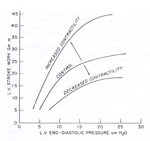
Frank-Starling curve analysis as an index of contractility is generally appreciated.
Cardiac performance could be estimated as a point on the Frank-Starling curve using measurement techniques relating flow (cardiac output) and associated left ventricular filling pressures. Cardiac output might be estimated from thermal dilution methods and left ventricular filling pressures estimated using pulmonary artery catheters. Frank-Starling curves do not define contractile status as such but relate pumping efficacy to load dependency.
The Frank-Starling relationship is indicated above. Application to the clinical environment dependent on the use of balloon-guided pulmonary artery catheters with thermistors allowing cardiac output measurement by thermal dilution with the occlusive balloon permitting estimation of pulmonary capillary wedge pressure. The lack of the linear relationship between ventricular end diastolic volume and pressure has been viewed as a limitation, at least in terms of quantifying pump function in terms of a specific number.
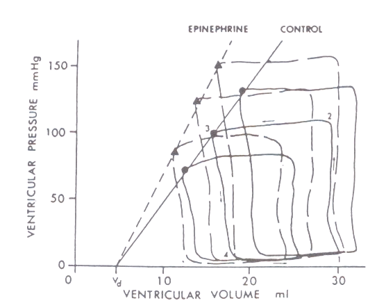
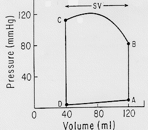
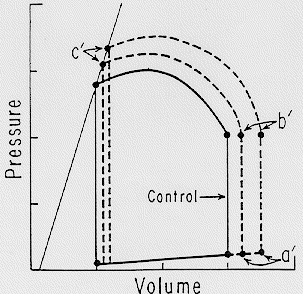
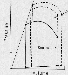
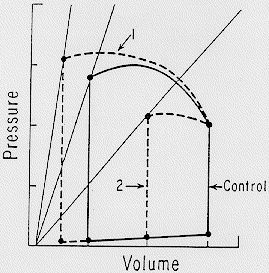
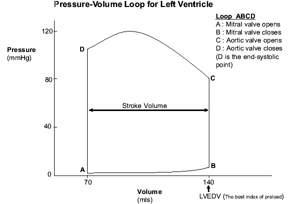
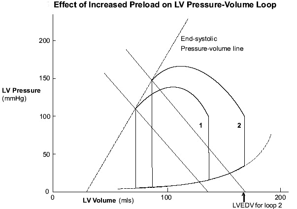
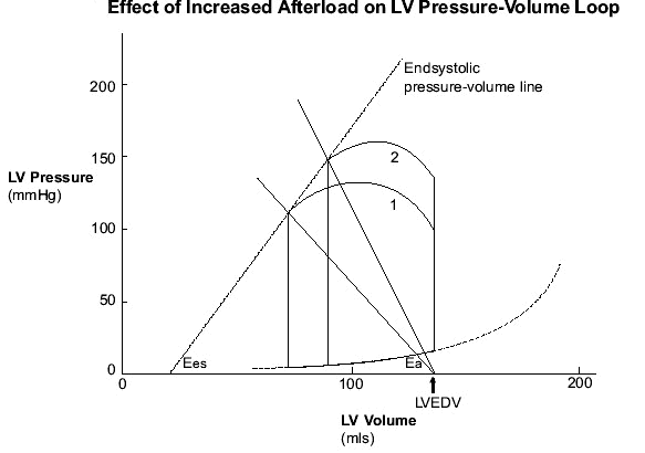
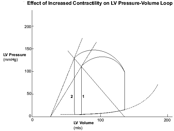
Linearity is associated with relationships between ventricular pressure and ventricular volume, as noted by graphing upper left corners of pressure-volume loops of ejecting contractions. As a result, the end-systolic pressure-volume relationship can be viewed as representing a given contractile state. This linearity in the end-systolic pressure-volume relationship shifts left and upward with a enhancement in the inotropic state and conversely to the right and downward with negative intropism. Furthermore, these changes appear independent of preload or afterload
1,2Changes in contractile state and their effects on pressure-volume lops, end-systolic pressure-volume lines and the volume axis intercept Vd (Figure 16-3 from Ref. 1)
Volume Pressure Loop Annimation attribution: D. Penny, Ph.D. Wayne State University, School of Medicine Virtual Classroom "My Physiology0
|
|
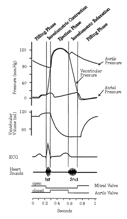
"4The cardiac cycle refers to all of the events associated with beat-to-beat activity of the heart including electrical and contractile events, ventricular volume changes and heart sounds. The graphical representation of these events as a function of time, is referred to as the Wiggers Diagram.... The events illustrated in the Wiggers Diagram are causally related and thus can be derived from "first principles". The parameters of function measured and displayed in the Wiggers Diagram include: aortic pressure (obtained by placing a catheter and associated pressure transducer in the aorta); left ventricular pressure (obtained by placing a catheter and associated pressure transducer in the left ventricular chamber - through the aortic valve); left atrial pressure (obtained as a "wedge pressure" from a catheter inserted into the pulmonary artery from the right side of the heart); aortic blood flow (obtained by one of a variety of techniques including doppler); ventricular volume (obtained from indicator dilution techniques); heart sounds (obtained by auscultation); venous pulse (obtained by placement of a catheter and associated pressure transducer in the inferior or superior vena cava); and electrocardiogram (obtained from placement of surface electrodes)."
|
0
0
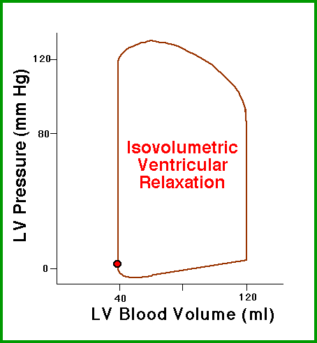
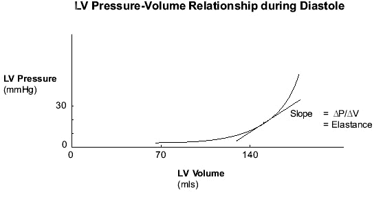
0 Questions and Answers Concerning Cardiac Pressure-Volume Curves6 (Some of these curves are similar to the curves above, but with additional explanations in some cases)
6Brandis, K., Anesthesia Self-Education Modules, Queensland Anaesthesia, Chapter 30
0
0
0
|
1Methods to estimate ESPVR have included contrast ventriculography, scintigraphy, echocardiography, etc. Echocardiography has a limitation due to limited dimensional assessment.
More current technology including three-dimensional reconstructions of echocardiographic data may facilitate more accurate volume determinations.
Even with conventional echocardiography, reasonable methods have been developed to assess ESPVR in patients.
Femoral artery pressure is used to as an estimate ventricular pressure and left ventricular cross-sectional area is used instead of left ventricular volume estimates. With these approximations, rapid estimates of left ventricular contractility may be made.
A limitation in this process is related to the two-dimensional data rather than a full 3-D reconstruction of the ventricle which can be done using echocardiography.
Without the complete surface representation of the ventricle, anomalies due to myocardial ischemia such as akinetic regions or poorly contracting regions may lead to incorrect volume estimates. ESPVR as a measure of contractility itself has been challenged from the point of view that the relationship could be affected by altered preload and afterload.
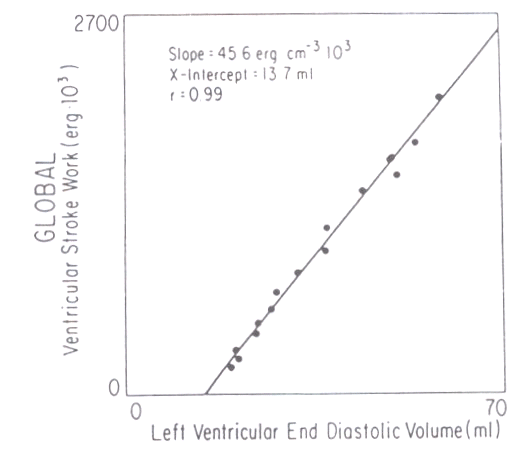
If filling pressures are used as representing preload, then Frank-Starling relationships are curvilinear; however, Frank-Starling relationships turn out to be rectilinear if end diastolic volumes are used to represent preload (see below).
The slope of this relationship ("preload recruitable stroke work or PRSW") can indicate changes in myocardial contractile state.
The authors of this relationship indicated later than the area under the line was a more sensitive indicator of myocardial contractility and was, at least within physiological ranges, probably insensitive to preload and afterload changes.
|
|
1Using the indices of contractility described above, myocardial effects on anesthetics may be evaluated. For example, using papillary muscle preparations, inhalational anesthetic-induced dose-dependent decreases in maximal fiber shortening velocity, mean maximal developed force, and dP/dt have been described. Considering dP/dt, the percentage decrease appeared comparable in considering isoflurane, enflurane and halothane.
High doses of isoflurane were not associated with increased left ventricular filling pressures, by contrast to that observed with enflurane or halothane.
As a result, isoflurane may be somewhat less of a myocardial depressant than enflurane or halothane in this assay system.
Another approach utilized the slope of the ESPVR in assessing contractility.
This method was applied to both intact, chronically instrumented, unpremedicated dogs and patients following cardiopulmonary bypass.
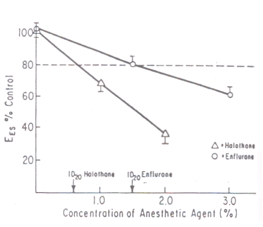
Halothane and enflurane depressed, to comparable extent, myocardial contractility (at equianesthetic levels). 0.7 MAC for both agents was needed to reduce end systolic elastance (slope of end-systolic pressure-diameter relationship) by 20%.
|
|
"Comparison of effects of halothane versus enflurane on myocardial depression, measured as the anesthetic concentration that inhibits inotropic state by 20 percent of the control value (ID20 ). ID20 for both anesthetics is 0.7 MAC. EES = slope of the end-systolic pressure-diameter relation"1,5
![]()
|
|
|
![]()
![]()
1Park, KW, Haering, JM, Reiz, S, Lowenstein, E Effects of Inhalation Anesthetics on Systemic Hemodynamics and the Coronary Circulation in Cardiac Anesthsia, Fourth Edition (Kaplan, JA, ed; Reich, Dl and Konstadt, SN, Assoc eds) W.B. Saunders Co. A Divsion of Harcourt Bract & Company, Philadelphia, 1999. This chapter is the primary reference for all above material, except as noted.
2Suga, H, Sagawa, K, Shoukas, AA: Load independence of instantaneous pressure-volume ratio of the canine left ventricle and effects of epinephrine and heart rate on the ratio. Circ Res 32: 314, 1973. -second sourced from reference 1.
5Van Trigt, P, Christian, CC, Fagraeus, L, et al: "Myocardial depression by anesthetic agents [halothane, enflurane, and nitrous oxide]: Quantitation based on end-systolic pressure-dimension relations" Am J Cardiol 53: 243, 1984---second sourced from reference 1.
3Tam, J, CV notes, Cardiac Performance I, University of Manitoba, Dept. of Medicine, Cardiology
4Gore, RW, The Heart as a Pump: Lecture 10, Cardiovascular Physiology 485, University of Arizona
5Glower, DD, Spratt, JA, Snow, ND, et al: Linearity of the Frank-Starling relationship in the intact heart: The concept of preload recruitable stroke work. Circulation 71: 994, 1985.--second sourced from reference 1.
6Brandis, K., Anesthesia Self-Education Modules, Chapter 3, Queensland Anaesthesia