|
|
|
|
Overview: Patient History Issues
Assessment of coronary vascular disease
History recent myocardial infarction
Resuscitation/hospitalization associated with myocardial arrhythmias
Cardiac drug use
Symptomatology Associated with Severity of CoronaryVascular Disease
Patient history following myocardial infarction (range of severity)
Better prognosis: short hospitalization; uneventful recovery
Poorer prognosis: if patient required
Intra-aortic balloon pump following myocardial infarction
Defibrillation
Assessment of patients cardiovascular status based on post-infarction drug regimen (chronic)
Normal/hyperdynamic cardiac function: ß-adrenoceptor blocking drugs (e.g.,metoprolol (Lopressor); propranolol (Inderal))
Severe left ventricular dysfunction (hypotensive state): balloon support; inotropic agents, e.g. dopamine (Intropin) and dobutamine (Dobutrex), nitrate
|
|
(Adapted from Pasternak, Braunwald and Sobel: Acute myocardial infarction: In Braunwald, editor: Hard Disease,ed 4, Philadelphia, WB Saunders, 1992, p 1250 (also, Table 68-2 from Ross, AF, Gomez, MN. and Tinker, JH Anesthesia for Adult Cardiac Procedures in Principles and Practice of Anesthesiology (Longnecker, D.E., Tinker, J.H. Morgan, Jr., G. E., eds) Mosby, St. Louis, Mo., pp. 201-218, 1998.)

Primary Reference: Shanewise, JS and Hug, Jr., CC, Anesthesia for Adult Cardiac Surgery, in Anesthesia, 5th edition,vol 2, (Miller, R.D, editor; consulting editors, Cucchiara, RF, Miller, Jr.,ED, Reves, JG, Roizen, MF and Savarese, JJ) Churchill Livingston, a Division of Harcourt Brace and Company, Philadelphia, pp. 1753-1799, 2000.
 |
|
|
|
|
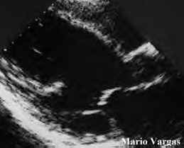
Echocardiography may be helpful in assessing:
Overall ventricular function +/-stress challenge (e.g. dobutamine (Dobutrex) challenge) with assessment of ejection fraction.
Ventricular wall motion anomalies
Mitral valve regurgitation, secondary to left ventricular dilatation or papillary muscle rupture/dysfunction
Notation of heart rate and
Observe which ECG leads indicate ischemic change -- these ECG leads may be the most helpful leads to monitor during anesthesia
Ventricular dysfunction unmasked during exercise-- (e.g.,premature ventricular contractions (PVCs), hypotension, rales)
Reason the possibility of potential intraoperative problems which would influence monitoring, anesthetic approaches and therapeutic responses
Coronary Angiography and Left Ventriculography
Atherosclerotic lesion location may be suggestive of:
The most appropriate electrocardiographic leads to monitor
Potential complications, e.g. ventricular dysfunction (hypokinesis), cardiac arrhythmias
Patency status of previous bypass grafts (internal mammary artery/saphenous vein); also:
Identifies the previous percutaneous transluminal coronary angioplasty (PTCA)
Adequacy of previous PTCA
Characteristics of atherosclerotic coronary disease, diffuseness of disease; and assessment of distal portions of coronary arteries
Cardiopulmonary bypass complications: related to revascularization completeness
May reveal coronary vasospastic disorder (Prinzmetal's angina)
Evidence of ventricular dysfunction:
Hypokinesis (poor ventricular contraction (regional))
Dyskinesia
Akinesis (absence of ventricular contraction in a region)
Aneurysm
High left ventricular end diastolic pressures (>15 mm Hg); ejection fraction < 0.5; and left ventricular and diastolic pressures increasing by > 5 mm mm Hg following contrast.
 |
 |
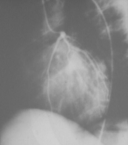
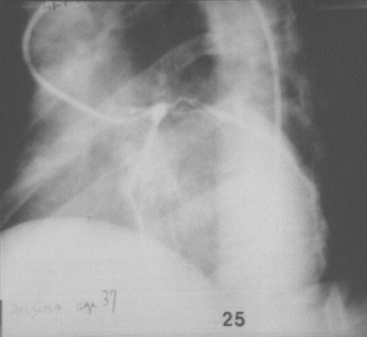
"Normal left coronary angiogram.
Left anterior oblique view (45 degrees) (Left); Left coronary angiogram.
Left anterior oblique view.
Narrow area from disease proximal end of circumflex and top of anterior descending.
Male age 37. Severe angina not controlled by medical treatment": courtesy of SouthBank University, London; used with permission
Radionuclide Angiocardiography
Adequacy of myocardial perfusion (thallium uptake study) +/-stress [exercise, dobutamine (Dobutrex) challenge]
Myocardial infarction identification: (technetium 99 uptake)
Evaluation ventricular function (equilibrium-gated blood pool imaging)
Estimation of ejection fraction
Uncovering inducible myocardial ischemia (stress thallium scan indicates reperfusion to ischemic regions)
Thallium Scans
|
|
|